Inhibitory effect of sodium metabisulphite and chlorine on growth of Aspergillus spp. and Penicillium spp. strains...
Ciência Rural, v.43, n.9, set, 2013.
1721
Inhibitory effect of sodium metabisulphite and chlorine on growth of Aspergillus spp.
and Penicillium spp. strains isolated from marine shrimp
Efeito inibitório do metabissulfi to de sódio e do cloro sobre o crescimento de cepas de
Aspergillus spp. e Penicilium spp. isoladas de camarão marinho
Ygor Flávio de Moraes SantosI,III* Átyla Peeter Batista VelosoI Rodrigo Maciel CalvetI,III
Maria Marlúcia Gomes PereiraI Carina Maricel PereyraII Ana Maria DalceroII
Adriana Mabel TorresII Maria Christina Sanches MuratoriI
ISSN 0103-8478
Received 10.14.11 Approved 04.30.13 Returned by the author 08.05.13
ABSTRACT
The sodium metabisulphite (SMB) is used in shrimp farming to prevent melanosis and the 5.0 ppm chlorine (CL) concentration used in the shrimp processing is effi cient as a bactericide, but there is no evidence of the effectiveness of these chemical compounds as fungicides. Therefore, the aim of this study was to evaluate the in vitro effect of sodium metabisulphite (SMB) and chlorine (CL) on the growth of Aspergillus and
Penicillium species isolated from marine shrimp in different stages of processing. The samples were collected from a frozen shrimp processing industry, located in Piauí State, Brazil. The total fungi and occurrence of Aspergillus and Penicillium species were evaluated. For in vitro sensibility test using the diffusion disk in agar method, fi ve concentrations of SMB (0%, 1%, 3%, 5% and 10%) and six of CL (0, 1, 2, 3, 4 and 5 μg mL-1) were used. The
fungal counts in the different processing stages ranged from 1.74 to 3.38 CFU g-1. Twenty-nine Aspergillus strains were isolated,
prevailing A. versicolor (59.3%) and twenty-two of Penicillium, prevailing P. citrinum (74%). One strain of A. fl avus was AFB1
producer. All the isolated strains of P. citrinum produced citrinin. All tested species were in vitro sensitive to 3% of SMB, except the
A. fl avus. The 10% concentration of SMB inhibited the in vitro
growth of all strains. The CL concentrations tested did not inhibit the studied species growth and SMB concentrations above 3.0% inhibited in vitro the growth of the tested strains.
Key words:Aspergillus fl avus, Litopenaeus vannamei, Penicillium citrinum, shrimp culture, conservation.
RESUMO
O metabissulfito de sódio (SMB) é usado na
carcinicultura para evitar a melanose e a concentração de 5,0ppm de cloro (CL), utilizada no benefi ciamento do camarão, é efi ciente como bactericida, porém não há comprovação da efi cácia destes compostos químicos como fungicida. Desse modo, o objetivo deste
estudo foi avaliar o efeito in vitro do metabissulfi to de sódio (SMB) e cloro (CL) sobre o crescimento de espécies de Aspergillus e
Penicillium isolados de camarão marinho em diferentes estágios de processamento. As amostras foram coletadas de uma indústria de processamento de camarão congelado, localizada no Estado do Piauí, Brasil. Fungos totais e ocorrência das espécies de
Aspergillus e Penicillium foram avaliados. Para o teste in vitro de sensibilidade pelo método disco-difusão em ágar, foram utilizadas cinco concentrações de SMB (0%, 1%, 3%, 5% e 10%) e seis de CL (0, 1, 2, 3, 4 e 5μg mL-1). As contagens fúngicas nos diferentes
estágios de processamento variaram de 1,74 a 3,38UFC g-1. Foram
isoladas 29 cepas de Aspergillus, prevalecendo o A. versicolor
(59,3%) e 22 de Penicillium, prevalecendo o P. citrinum (74%). Uma cepa de A. fl avus era produtora de AFB1. Todas as cepas de P. citrinum isoladas produziram citrinina. Todas as espécies testadas foram sensíveis in vitro a 3% do SMB, com exceção do A. fl avus. A concentração de 10% do SMB inibiu in vitro o crescimento de todas as cepas. As concentrações de CL testadas não inibiram o crescimento das espécies isoladas e concentrações de SMB acima de 3,0% inibiram in vitro o crescimento das linhagens testadas.
Palavras-chave: Aspergillus fl avus, Litopenaeus vannamei,
Penicillium citrinum, carcinicultura, conservação.
INTRODUCTION
The shrimp culture is an important activity practiced in the coast of the Piauí State, Brazil, which generates foreign exchange and is an important source of employment for the local people. In 2009, Brazilian production was 65.000 tons of shrimp, of which 843 tons of shrimp were from the State of Piauí (ABCCam, 2011).
IDepartamento de Morfo
fi siología Veterinária, Centro de Ciências Agrárias (CCA), Universidade Federal do Piauí (UFPI), Teresina, PI, Brasil. IIDepartamento de Microbiología e Inmunología, Universidad Nacional de Río Cuarto, Río Cuarto, Córdoba, Argentina.
IIIPrograma de Pós-graduação em Ciência Animal, CCA,UFPI, 64049-550, Teresina, PI, Brasil. E-mail: ygor
Fungi are widely distributed in nature and because of this can be used as parameters for determining microbiological hygienic conditions of the culture environment. Some fungi are capable of producing secondary toxic metabolites and sometimes carcinogenic to animals and humans. Its biosynthesis is related to environmental conditions such as temperature, humidity and rainfall during the growing season, harvesting, post harvest and storage of agricultural products (PITT & HOCKING, 1999; SAMSOM et al., 2001). Aspergillusspp., Penicillium
spp. and Fusarium spp. are the most commonly
fi lamentous moulds found in stored cereal grains and feeds. Thus, the hot and humid climate is conducive to growth of fungi in stored foods and therefore conducive to mycotoxin production (BINTVIHOK et al., 2003).
Due to the importance of the shrimp to the brazilian economy and the increasingly sharp demands of importing countries, and the quality of fi nal product, it is necessary to develop and enhancement techniques for postharvest processing of shrimp. Sodium metabisulphite – SMB (Na2S2O5.
H2O) is used in shrimp culture, in order to avoid
“black spot” or melanosis on post-harvest shrimp. This is a preservative for greater stability and which has the highest amount of sulfur dioxide (SO2), when
diluted with water (SILVA, 1988). For this reason, sulphites are multifunctional agents and are capable of controlling microbial growth in foods (LAURILA et al., 1998). The fi sh processing industries use 5.0μg mL-1 of chlorine (CL) concentration during all
processing steps in order to avoid bacterial growth (JAY, 2005).
There are several studies about SMB action on bacteria such as Salmonella spp., Clostridium
spp., Pseudomonas spp., Vibrio spp., Staphylococcus
spp., Bacillus spp., (VIEIRA, 2004). However have no evidence of the effectiveness of these chemical compounds as a fungicide on post-harvest shrimp.
The objective was to evaluate the effect
in vitro of sodium metabisulphite and chlorine on growth and survival of Aspergillus and Penicillium
species in cultured marine shrimp.
MATERIALS AND METHODS
Source of samples
The samples were collected from a shrimp processing industry located in the coast area of Piauí State, Brazil (region enclosed between 2º 55’ 51,39’’S - 2º 58’ 04,31’’S a 41º 20’ 09,35’’W - 41º 26’ 33,52’’W). A total of 36 samples with 100g of shrimp
was obtained from six different points classifi ed as “A” (time of harvest before the addition of SMB), point “B” (after the addition of SMB), point “C” (reception industry after washing the shrimp amounts with chlorinated water to 5.0μg mL-1); point “D”(for
the handling and removal of the carapace in the wake of processing); point “E” (after exoskeleton removal); and point “F” (at the exit of the freezing tunnel at -35ºC). After collection, the samples were placed in plastic sterile sealed bags Nasco Whirl-Pak®, identi
fi ed and transported in containers containing ice packs to the Laboratory of Microbiological Control of Food, of Centre of Agrarian Sciences of Federal University of Piauí, Brazil.
Mycological determination and identifi cation of
Aspergillus and Penicillium species
Total fungal counts from samples were performed into Dichloran Rose Bengal Chloranphenicol Agar (DRBC), recommended by PITT & HOCKING (1999). Quantitative enumeration was done using the surface-spread method.
Twenty-fi ve grams of each sample were homogenized in 225mL 0.1% peptone water solution for 30min in an orbital shaker. Serial dilutions (10-2 to 10-3) were made
and 0.1mL aliquots were inoculated in duplicates onto the culture media. Plates were incubated at 25º C for 7-10 days in darkness. Only plates containing 10-100 colony-forming units (CFU) were used for counting. The results were expressed as CFU per gram of sample (CFU g-1). Representative colonies of Aspergillusand
Penicillium spp. were transferred for sub-culturing to tubes containing Malt Extract Agar (MEA). Identifi cation of Aspergillus and Penicillium species
was performed according to taxonomic keys (KLICH, 2002; SAMSON & FRISVAD, 2004; PITT, 2004).
Afl atoxin B1 production by Aspergillusfl avus
All Aspergillus fl avus strains isolated from shrimp were assayed for afl atoxin B1 (AFB1) production. The strains were grown on MEA plates at 28ºC for 7 days. The mycelium was transferred to an Eppendorf tube and 1000μL chloroform was added. The mixture was shaken for 20min at room temperature, the mycelium was removed and the chloroform extract evaporated to dryness under N2 fl ow. The residue was redissolved in 200μL de
chloroform. The extracts were analyzed by Thin Layer Chromatography (TLC) on silica gel 60 F254 TLC aluminum sheets (20x20cm, 250mm thick, Merk, Germany). The eluent used was chloroform: acetone (90:10, v v-1). The chromatographic run was
Inhibitory effect of sodium metabisulphite and chlorine on growth of spp. and spp. strains... 1723
The observation was carried out under the plates with ultraviolet light of wavelength 360nm and the concentration of AFs method was determined by visual comparison of the retention factors and the
fl uorescence intensity. Detection limit of the used method was 5g kg-1 (GEISEN, 1996).
Citrinin production by Penicilliumcitrinum
Penicillium citrinum strains isolated were assayed for citrinin production, respectively. The detection of toxins was performed following the methodology proposed by CRUZ et al. (1992). They were preserved on MEA at 4ºC until use. The strains were grown in media Coconut Agar (CAM) at 28ºC for 7 days. Three agar plugs were removed from the central area of the colony, weighed and introduced into a small vial. A volume of 1mL of chloroform was added to each vial, the sample-solvent mixture was centrifuged for 10min at 1400rpm. The plugs were removed and the chloroform extract was evaporated to dryness under N2fl ow. The residue was re-dissolved
in 100μL chloroform. Extracts were analyzed by TLC on silica gel 60 F254 TLC aluminum sheets (20x20cm, 250μm thickness, Merck, Germany). The developing solvent was toluene: acetate of ethyl: chloroform: formic acid (70:50:50:10). The plates were previously impregnated with 10% oxalic acid in ethanol.
Sensitivity test in vitro of Aspergillus spp. and Penicillium spp.
Four strains of Aspergillus (A. fl avus,A. versicolor, A. terreus and A. niger aggregate) and
fi ve of Penicillium (P. citrinum, P. implicatum,P. citreonigrum,P. purpurogenumandP. decumbens) isolated from shrimp samples collected at different points (“A”, “B”, “C”, “D”, “E” and “F”) were in vitro tested for sensibility to SMB and CL by the diffusion disk in agar method (NCCLS / CLSI, 2004) using four concentrations of SMB (0, 1, 3, 5 and 10%) and fi ve concentrations of CL (0, 1, 2, 3, 4, 5μg ml-1). All tests were performed in duplicate with
four replicates each. The fi lter paper discs (20mm of diameter) were sterilized and subsequently immersed in concentrations of SMB and CL.
To prepare the inoculums, the recommendations of ESPINEL-INGROFF et al. (1997) and the NCCLS / CLSI (2009) were followed, as follows: conidial suspensions were prepared using one to fi ve Aspergillus and Penicillium colonies (<5mm of diameter) grown in MEA for seven days at 25°C. The colonies were covered with 1.0mL of sterile saline solution at 0.85% and were homogenized with
a pipette preparing a suspension. One drop of Tween 20 was added to facilitate the inoculum preparation. The resulting mixture of conidia and hypha fragments was removed and transferred to a sterile test tube. After the particles sedimentation (3 to 5 minutes), the supernatant was transferred to another sterile tube and homogenized in a mixer for 15 seconds. Each fungal suspension was adjusted to contain 1x10⁶ to 5,0x10⁶CFU mL-1,
fi rst, by comparison the turbidity with the 0.5 McFarland scale tube.
The densities of the conidial suspensions were read and adjusted to an optical density that ranged from 0.09 to 0.11 (80-82% of transmittance) in a spectrophotometer at 530nm, hemocytometer counting and counting of colony forming units (CFUs) in MEA. Subsequently, the suspensions were poured into Petri dishes with MEA where paper discs were added on three equidistant points. The plates were incubated at 25°C for seven days. After the incubation period, the fungal growth and sensibility to the tested compounds was observed.
Statistical analysis
Data analyses were performed by variance analysis. Total fungal counts data were transformed using a logarithmical function log10(x+1) before
applying the variance analysis.
RESULTS AND DISCUSSION
Table 1 shows fungal total medium counts on the DRBC from different stages of shrimp processing ranged from 1.74 to 3.38log10 CFU g-1. There were no
differences between the different stages of processing (P<0.05). However, some shrimp samples of point “C” had fungal counts above the recommended limit (3.0log10 CFU g-1). This results are similar with others
authors who worked with shrimp feed, water and nurseries substrate of the same industry, indicating that fungal were found in the environment culture in constant amounts after post harvest and processing industrial (CALVET et al., 2007). Fungal counts of the stage that shrimp amounts were washed with chlorinated water, was the only stage with counts above the recommended limit (3.0log10 CFU g-1). REIS et al. (2004) analyzed
shrimp samples trade collected in Sao Paulo, whose values ranged between 3.04 at 7.36log10 CFU g-1 if kept
under refrigeration. These authors say that the incorrect management after the expedition may advantage the fungal growth pre-existing during marketing.
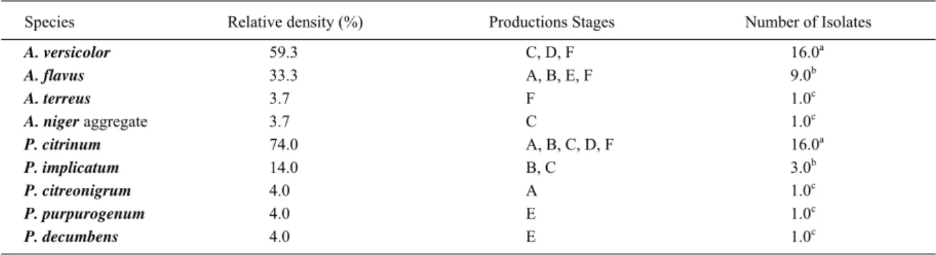
Table 2 shows relative density (%) of
Aspergillus spp. and Penicillium spp. isolated from
Aspergillus spp. strains were isolated. Aspergillus versicolor was the predominant species (59.3%) followed by A. fl avus (33.3%). Aspergillus terreus
and A. nigeraggregate were isolated at low frequency (3.7%). Twenty two Penicillium spp. strains were isolated from shrimp. Penicillium citrinum was the predominant species (74.0%) followed by P. implicatum (14.0%). Penicillium citreonigrum, P. purpurogenum and P. decumbens were isolated at a relative density from 4.0%.
Several studies have shown that shrimp have species from Aspergillus and Penicillium genera as predominant mycofl ora (LEAÑO et al., 2005; CALVET, 2008). The dominance of fi lamentous fungi indicates that these fungi can proliferate very well in this substrate. Among the fi lamentous fungi, the dominant genera observed (Penicillium and
Aspergillus spp.) are toxins producers and pathogenic to shrimps (e.g. Aspergillus and Fusarium spp.) (LEAÑO, 2001).
From the nine A. fl avus isolated, one strain (11.1%) of stage “F” was AFB1 producer. YOUSEFI et al. (2009) isolated A. fl avus strains producing of AFB1 as mycofl ora from cultured green tiger shrimps
(Penaeus semisulcatus) and water of their ponds. The presence of these fungal species isolated from shrimp at different stages of processing may be due to environmental contamination or feed contaminated with these species in the culture environment (CALVET, 2008; YOUSEFI et al., 2009).
In this study, P. citrinum was the most prevalent species isolated from shrimp. All strains of P. citrinum were positive for citrinin production. Although the citrinin is not carcinogenic it has nephrotoxic effects (PITT, 2004). These results are similar to those obtained by CALVET (2008), who reported the presence of toxigenic P. citrinum in storage sheds containing shrimp feed.
The inhibitory effect of SMB on Aspergillus
spp. and Penicillium spp. strains growth by diffusion agar method shown in table 3. Aspergillus terreus,
P. citrinum, P. implicatumand P. decumbens were sensitive to 1% SMB concentrations. Aspergillus fl avus was the only species resistant to 3% SMB concentrations. All studied strains were sensitive to 5 and 10% SMB concentrations. Chlorine concentrations studied were not effective in inhibiting the growth of the tested strains (data not shown).
Table 1 - Fungal counts (CFU g-1) from shrimp at different productions stages.
Different productions stages Fungal counts (CFU g-1 in log 10) Media
Before the addition of SMB (A) 2.81ª
After the addition of SMB (B) 2.50ª
Washing the shrimp excessive amounts of chlorine (C) 3.11ª Handling and removal of the carapace (D) 2.56ª After removal of the exoskeleton (E) 2.45ª Exit of the tunnel of freezing at -35º C (F) 2.64ª
CFU= colony-forming units; SMB= Sodium metabisulphite; Maximum recommended level: 3.0CFU g-1 in log10.
Table 2 -Relative density (%) of Aspergillus spp. and Penicillium spp. isolated fromshrimp at different productions stages.
Species Relative density (%) Productions Stages Number of Isolates
A. versicolor 59.3 C, D, F 16.0a
A. flavus 33.3 A, B, E, F 9.0b
A. terreus 3.7 F 1.0c
A. niger aggregate 3.7 C 1.0c
P. citrinum 74.0 A, B, C, D, F 16.0a
P. implicatum 14.0 B, C 3.0b
P. citreonigrum 4.0 A 1.0c
P. purpurogenum 4.0 E 1.0c
P. decumbens 4.0 E 1.0c
a,b,c= Letters are similar results in the same column. χ2=30,96 (P<0,05) A = Before the addition of Sodium metabisulphite; B = After the
Inhibitory effect of sodium metabisulphite and chlorine on growth of spp. and spp. strains... 1725
The industry investigated using 6% SMB solutions for shrimp immersion for 15 to 20 min, after post collect and so prevents the occurrence of melanose and bacterial growth (SILVA, 1988; BARBIERI & OSTRENSKY, 2002). This concentration of SMB did not inhibit fungal growth during the processing stages, it was demonstrated by fungal counts in different stages. However, the use of 5% SMB totally inhibited strains tested in vitro. The use of SMB is known against phytopathogenic fungi and bacteria, but little is known about these storage fungi in shrimp (GÓES et al., 2006; PATSOUKIS & GEORGIOU, 2007).
The chlorine concentration used (5μg mL-1) is ef
fi cient as a bactericide but not fungistatic, due to the presence of fungi during all stages of processing and ineffi cient to inhibit the strains tested
in vitro (JAY, 2005). In general, fi lamentous fungi are more resistant than yeasts and bacteria. Studies with different chemicals were not effective to inhibit the growth of Aspergillus and Penicillium species and inadequate chemicals doses can promote fungal growth (XAVIER et al., 2007; CHAPMAN, 1998).
This is the fi rst preliminary work that shows the SMB and CL effect on growth of Aspergillus and
Penicillium species isolated from marine shrimp in different stages of processing. Future studies should be conducted to see how SMB and CL can affect the parameters of growth and toxin production.
ACKNOWLEDGMENT
This work was carried out with grants from Conselho Nacional de Desenvolvimento Científi co e Tecnológico (CNPq),
Universidade Federal do Piauí (UFPI), Brazil, Universidade Federal Rural do Rio de Janeiro (UFRRJ), Brazil and Universidad Nacional de Río Cuarto, (UNRC), Argentina.
REFERENCES
ABCCam (ASSOCIAÇÃO BRASILEIRA DE CRIADORES DE CAMARÃO). Estatísticas do setor pesqueiro e da carcinicultura
brasileira. Available from: <http://www.abccam.com.br/abcc/ images/stories/estatisticas/Estatstica_DO_SETOR_PESQUEIRO. pdf>. Online. Accessed: Ago. 25, 2011.
BARBIERE, J.R.C.; OSTRENSKY, N.A. Camarão marinho: engorda. Viçosa: Aprenda Fácil, 2002. 352p.
BINTVIHOK, A. et al. Afl atoxin contamination in shrimp feed and effects of afl atoxin addition on shrimp production. Journal of
Food Protection, v.66, n.5, p.882-885, 2003.
CALVET, R.M. et al. Contagem de fungos fi lamentosos e leveduras em camarões cultivados. Revista Higiene Alimentar, v.21, n.150, p.253-251, 2007.
CALVET, R.M. Identifi cação e isolamento de fungos toxígenos em carcinicultura marinha. 2008. 82f. Dissertação (Mestrado em Ciência Animal) – Programa de Pós-graduação em Ciência Animal, Faculdade de Veterinária. Universidade Federal do Piauí.
CHAPMAN, J.S. Characterizing bacterial resistance to preservatives and disinfectants. International Biodeterioration
and Biodegradation, v.41, p.241-245, 1998.
CRUZ, L.C.H. et al. Aplicação do Ágar-Coco como meio diferencial para o isolamento de fungos citrinogênicos. Arquivos
da Universidade Federal Rural do Rio de Janeiro, v.15, n.1, p.39-60, 1992.
ESPINEL-INGROFF, A. et al. Multicenter evaluation of proposed standardized procedure for antifungal susceptibility testing of fi lamentous fungi. Journal of Clinical Microbiology, v.35, n.1, p.139-143, 1997.
GEISEN, R. Multiplex polymerase chain reaction for the detection of potential afl atoxin and sterigmatocystin producing fungi. Journal of Applied Microbiology, v.19, p.388-392, 1996.
Table 3 -Inhibitory effect of growth in vitro of sodium metabisulphite on species Aspergillus and Penicillium.
---Sodium metabisulphite (%)---Species
0,0 1,0 3,0 5,0 10,0
A. niger agregados R R S S S
A. terreus R S S S S
A. versicolor R R S S S
A. flavus R R R S S
P. citrinum R S S S S
P. implicatum R S S S S
P. citreonigrum R R S S S
P. purpurogenum R R S S S
P. decumbens R S S S S
GÓES, L.M.N. et al. Uso do metabissulfi to de sódio no controle de microorganismos em camarões marinhos Litopenaeus vannamei
(Boone, 1931). Acta Scientiarum. Biological Sciences, v.28, n.2, p.153-157, 2006.
JAY, J.M. Microbiologia de alimentos. 6.ed. Porto Alegre: Artmed, 2005. 712p.
KLICH, M.A. A laboratory guide to the common Aspergillus
species and their teleomorphs. Australia: CSIRO - Division of Food Processing, 2002. 116p.
LAURILA, E. et al. The inhibition of enzymatic browning in minimally processed vegetables and fruits. AgBiotech News and
Information, v.9, n.4, p.53-66, 1998.
LEAÑO, E.M. Fungal diseases. In: LIO-PO, G.D. et al.
Health management in aquaculture. Philippines: Aquaculture Department, Southeast Asian Fisheries Development Center, 2001. p.45-53.
LEAÑO, E.M. et al. Mycofl ora of the ‘green water’ culture system of tiger shrimp Penaeus monodon Fabricius. Aquaculture
Research, v.36, p.1581-1587, 2005.
NCCLS/ CLSI (NATIONAL COMMITTEE FOR CLINICAL LABORATORY STANDARDS/CLINICAL LABORATORY STANDARDS INSTITUTE). Reference method for antifungal disk diffusion susceptibility testing of yeasts; Approved guideline – Second Edition. M44-A2, v.29, n.17. Wayne, 2009. 8p.
PATSOUKIS, N.; GEORGIOU, C.D. Effect of sulfi te–hydrosulfi te and nitrite on thiol redox state, oxidative stress and sclerotial differentiation of fi lamentous phytopathogenic fungi. Pesticide
Biochemistry and Physiology, v.88, n.2, p.226-235, 2007.
PITT, J.I.; HOCKING, A.D. Fungi and food spoliage. 2.ed. New York: Springer Science Business Media, LLC, 2009. 419p. PITT, J.I. Guía del laboratorio para la identifi cación de especies
comunes de Penicillium. Australia: CSIRO - Division of Food Processing, 2004. 199p.
REIS, J. et al. Estudo higiênico-sanitário dos camarões dulcícolas
Macrobrachium amazonicum e M. jelskii. Revista Higiene
Alimentar, v. v.116/117, p.18, p.58-67. 2004.
SAMSON, R.A.; FRISVAD, J.C. Penicillium subgenus.
Penicillium: new taxonomic schemes and mycotoxins and other extrolites. Netherlands: Centraalbureau voor Schimmelcultures, 2004. 243p.
SAMSON, R.A. et al. Introduction to food-and airborne fungi. Netherlends: Royal Netherlands Academy of Arts and Siences, 2001. 389p.
SILVA, R.R. Considerações sobre o uso e o mal uso de sais de sulfi to em crustáceos. In:SEMINÁRIO SOBRE CONTROLE DE QUALIDADE NA INDÚSTRIA DE PESCADO, 1988, Santos, SP.
Anais...Santos: Loyolap, 1988. p.244-259.
VIEIRA, R.H.S.F. Microbiologia, higiene e qualidade do pescado: teoria e prática. São Paulo: Varela, 2004. 380p. XAVIER, M.O. et al. Atividade in vitro de três agentes químicos frente a diferentes espécies de Aspergillus. Arquivos do Instituto
Biológico, v.74, p.49-53, 2007.
YOUSEFI, S. et al. Afl atoxin production by Aspergillus fl avus
isolates from green-tiger shrimps (Penaeus semisulcatus).