Medial Hypertrophy in Patients with Pulmonary Embolism:
Anatomopathological Study
Renato Tambellini Arnoni, Fábio Biscegli Jatene, Wanderley Marques Bernardo, Vera Demarchi Aiello, Tanas Jatene,
Rosângela Monteiro, Lea Maria Macruz Demarchi
Instituto do Coração do Hospital das Clínicas – FMUSP – São Paulo, SP - Brazil
Summary
Objective: To compare the arterial response of cases of acute and chronic embolism, seeking to associate ischemic pulmonary remodeling with progression to chronicity.
Methods: Retrospective analysis of 61 necropsies of patients who died in the Instituto do Coração (31 cases of pulmonary embolism and 30 cases of acute myocardial infarction). Slides of pulmonary tissue were obtained from all cases and analyzed qualitative and quantitatively (medial thickness measurement).
Results: Qualitative analysis enabled the diferentiation between cases of embolism and the control group, thus characterizing the two groups and deining the adequate choice of the control group. The alterations predominated in patients with embolism (alveolar inlammation and edema, infarction, vasoconstriction, concentric intimal proliferation, presence of thrombus). Quantitative analysis demonstrated higher percent medial thickness in the cases of embolism than in the control group; among the cases of embolism, no diferences in intra (acute – 19.74 and chronic – 20.04) and pre-acinar (acute – 18.85 and chronic – 18.68) arteries were observed.
Conclusion: The lack of diference among the groups with embolism and the higher values of percent medial thickness in the peripheral arteries allow the conclusion that the vascular response is more intense and starts in these arteries. (Arq Bras Cardiol 2007;88(6):584-589)
Key words: Arterial occlusive diseases; pulmonary embolism/physiopathology; thromboembolism.
Mailing address: Renato Tambellini Arnoni •
Rua Jesuíno Arruda, 318/121 – 04532-080 – São Paulo, SP - Brazil E-mail: [email protected]
Manuscript received March 12, 2006; revised manuscript received August 1, 2007; accepted September 1, 2007.
Introduction
The incidence of pulmonary thromboembolism caused by obstruction of pulmonary artery branches by emboli originating in other parts of the body is high, and is estimated at 600,000 cases/year in the United States1. Mortality of this
condition is high, with 60,000 deaths, the majority of which within the irst hour. Pulmonary embolism accounts for 0.27 to 0.40% of hospital admissions2. Because of increased
blood coagulation and other efects of gestation and birth, pulmonary embolism is the major cause of maternal death in developed countries3.
Acute embolism may result in a transient pressure increase in the pulmonary territory, which tends to resolve in the absence of the obstructive factor. However, some factors may predispose to a sustained pressure elevation: previous massive embolism, multiple previous embolic episodes, signiicant perfusion defect, young age, and embolism of undetermined cause. The incidence of pulmonary hypertension after acute embolism is 1%, 3.1%, and 3.8% at six months, one year, and two years, respectively4.
Several studies have been recently conducted with the
purpose of explaining progression to chronic embolism. Two hypotheses have been studied with greater enthusiasm: the irst considers the maintenance of the occlusive factor as fundamental for the development of a pulmonary vascular response characterized by medial hypertrophy; the second relates pulmonary hypertension to a previous initial arteriopathy in patients who had an unfavorable outcome5,6.
The histological diagnosis of embolism itself is not simple; resorption of emboli by endogenous thrombolysis, variation in embolus size, and impossibility of morphological diferentiation between thrombi embolized to the lungs and those formed in situ make this diagnosis diicult. Large thrombi in major pulmonary arteries tend to be embolic as long as no disease is observed within the arterial wall. Primary thrombi are more common in the upper lobes, whereas thromboembolic events are more common in the lower lobes6,7.
The alterations in the pulmonary vascular bed are characteristic of vascular remodeling, which consists in alterations resulting from pressure elevation both in the venous and the arterial bed, or from increased blood low. Microscopic analysis allows the veriication of endothelial injury, presence of muscle in non-muscular arteries, intimal proliferation, and medial hypertrophy8.
were prepared and stained with hematoxylin-eosin. For the case to be included in the study, at least two respective lung slides adequate for analysis were required. The histological lung sections were then also stained with Miller’s stain, thus facilitating the observation of elastic ibers. Therefore, each case resulted in four lung sections (two stained with hematoxylin-eosin and two with Miller’s stain).
All slides were blinded so that the observer did not know whether it was an embolism case or not.
With the 61 cases selected (16 acute embolism, 15 chronic embolism, and 30 control cases), an analysis could be performed with an optical microscope (Q500YW – Leica – UK) coupled to an image analyzer (Quantimet Q500YW – Leica – UK), using magniication ranging from 2.5 to 40 times.
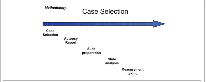
The slides obtained were analyzed both qualitative and quantitatively (Figure 1).
The hematoxylin-eosin-stained histological sections were used for the qualitative analysis, which was based on the identiication of pulmonary tissue alterations. The presence or absence of alterations was observed. The qualitative parameters of the pulmonary circulation analyzed were: permeability alterations: alveolar inlammation and edema; cell injury: infarction; vascular alterations: vasoconstriction, concentric intimal proliferation, presence of thrombus.
Quantitative analysis was performed with the image analyzer (Quantimet), which enabled the performance of measurements in the vascular walls with the purpose of determining the presence of distal arteriopathy in the pulmonary bed. Acinar and pre-acinar arteries were analyzed, and the arteries not showing clear contours (internal and external elastic laminae) were excluded.
The following vascular structures were assessed: external diameter: smallest diameter delimited by the external elastic lamina of the pulmonary arteriole; medial thickness: distance between the external and internal elastic laminae of the pulmonary arteriole. Two measurements were taken for each arteriole, and the arithmetic mean was calculated.
(not always identiiable), thrombus recanalization, capillary channel proliferation (plexiform response) more characteristic of pulmonary hypertension, and intimal hypertrophy6,9-12.
The objective of this study is to compare the arterial response of cases of acute and chronic embolism, seeking to associate ischemic pulmonary remodeling to progression to chronicity.
Methods
This study was conducted in the Laboratory of Medical Investigation, Laboratory of Research on Thoracic Surgery, and Laboratory of Pathological Anatomy of the Instituto do Coração da Faculdade de Medicina da Universidade de São Paulo.
In the present study, patients diagnosed with acute and chronic pulmonary embolism who died in the Instituto do Coração were analyzed and compared with patients who died of acute myocardial infarction, in an attempt to choose patients without previous pulmonary disease. One group was comprised of cases of pulmonary embolism, and the other, of patients who died of acute myocardial infarction. Group 1: patients who died of pulmonary embolism (31 cases); Group 2: patients who died of acute myocardial infarction (30 cases).
The study was based on the analysis of autopsies performed in the Instituto do Coração by the Service of Pathological Anatomy. Case selection was based on the Service’s database, by identifying autopsies reporting pulmonary embolism for group 1 cases and myocardial infarction for group 2 cases. In light of these results, the autopsy reports could be assessed for the correct case identiication and selection. Thus, half of the group 1 sample was deined as acute cases and half as chronic cases (of these, eight had undergone previous thromboembolectomy). Selection of cases of myocardial infarction, in turn, was made by reading the autopsy reports and excluding those mentioning chronic pulmonary disease or pulmonary infection.
Once the cases were selected, the respective lung slides
Fig. 1 - Methodology used in case selection.
Case Selection
Case Selection
Methodology
Autopsy Report
Slide preparation
Slide analysis
Percent medial thickness (%MT ): %MT = (2 x medial thickness / external diameter) x 100 (Figure 2).
Results
The analyses of the results obtained during the present study were divided into qualitative and quantitative analyses.
Qualitative analysis was performed using overall observation of the lung section with identiication of histological alterations, without considering the degree of pulmonary involvement (edema, inlammation, and infarction). Vascular alterations, however, were assessed for each of the arteries found in the lung section studied, and presented as percent involvement, that is, percentage of arteries involved within the total studied (vasoconstriction, intimal proliferation, and thrombus).
Quantitative analysis included artery measurement, as previously described, using slides stained with Miller’s stain.
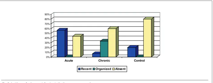
Qualitative analysis - Edema was more frequently observed in the cases of acute embolism (100%), followed by chronic cases (86.7%), and this diference was statistically signiicant. Inlammation was more frequent in the cases of both acute and chronic embolism, however a high incidence was also observed in the control group, with more than half of the cases presenting some degree of inlammation.
The incidence of pulmonary infarction was signiicantly higher in patients with acute embolism (recent infarction). Likewise, patients with chronic embolism presented alterations compatible with organized infarction in a greater number of cases. Control group patients with pulmonary infarction were those who developed some degree of associated pulmonary infarction as a result of the cardiac event, with the pulmonary infarction always being an acute event (Figure 3).
Vasoconstriction was not observed in the control group. The comparison between groups and artery types (intra-acinar and pre-(intra-acinar) showed a higher incidence of vasoconstriction in acinar vessels (6.78 greater in pre-acinar vessels – p = 0.007), and in the group of patients with acute embolism in relation to chronic cases (11.26 greater – p = 0.011) (Table 1).
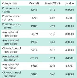
Intimal proliferation was assessed by comparing the artery types and then the groups with each other, since the estimation model used demonstrated that both factors had an inluence Thus, we could compare diferent portions of the same
lung (pre-acinar and acinar portions) and similar portions of diferent lungs, as well as analyze the pulmonary tissue lesion with local destruction of pulmonary tissue and its arterial bed, characteristic of pulmonary hypertension conditions.
Statistical analysis - All qualitative and quantitative variables were analyzed descriptively. Qualitative variables were calculated and presented as frequencies and percentages. For quantitative variables (artery diameter and percent medial thickness), the minimum and maximum values, as well as the median, mean, quartiles, standard deviation and stand errors were analyzed.
Qualitative variables were compared using the Fisher’s exact test, whereas comparisons between quantitative variables were made with generalized estimation equation models using Gamma distribution and multiple comparisons. The level of signiicance for all tests performed was 5%.
Fig. 2 - Method of artery measurement. %WT – Percent medial thickness.
WT
WT
Diameter
Fig. 3 - Incidence of pulmonary infarction in the three groups assessed. 0%
10% 20% 30% 40% 50% 60% 70% 80% 90%
Acute Chronic Control
on the incidence of this histological alteration (p = 0.006). In relation to the analysis of the type of artery afected: in patients with acute embolism and in the control group there was a higher frequency of cases of intimal proliferation in pre-acinar vessels than in intra-acinar vessels (p<0.0001). Although the incidence was higher among cases of chronic embolism, no diference was observed between the artery types.
The comparison between the diferent groups for intra-acinar arteries, in turn, demonstrated a signiicantly greater involvement in chronic cases in relation to the other two groups:
- 36.69 times in comparison to the acute embolism group, and 56.17 times in comparison to the control group.
The same was observed in the pre-acinar arteries, however with a slightly lower diference in comparison to intra-acinar arteries.
The acute embolism and control groups did not show differences as regards intimal proliferation in pre-acinar arteries; the alteration was identiied only in smaller arteries (intra-acinar) (Table 2).
Quantitative analysis - Quantitative analysis was performed in 55 cases, with measurements of 2262 arteries. Control group cases with associated pulmonary infarction were excluded.
Arteries were characterized according to their location: a) pre-acinar; b) intra-acinar.
Further, patients of each group were compared in relation to the artery types and arteries within the same group (generalized estimation equation using Gamma distribution and multiple comparisons).
No diference was observed as regards artery diameter in the three groups (Table 3).
When percent wall thickness (%WT) of intra-acinar arteries is observed in the diferent groups, we can conclude that:
values in the acute and chronic groups are higher than those of the control group, thus demonstrating medial hypertrophy in these two groups. However, no diference was observed between the groups with embolism.
In relation to %WT, the results were similar to those of the intra-acinar arteries, with values slightly lower than those of the arteries previously mentioned. Embolism groups presented greater wall thickness with higher %WT, consequently (Table 4).
Comparison of percent thickness between the groups and artery types - In the comparisons of patients of the diferent groups and artery types analyzed (pre and intra-acinar) in relation to their percent thickness, the model demonstrated that each of the parameters assessed individually presented statistical significance as for the presence of difference between patients. The joint analysis of factors established that the association of artery location and patient group did not show statistical signiicance.
Table 1 - Comparison of vasoconstriction according to vessel type and group
Comparison Mean dif Mean WT dif p-value
Pre/intra-acinar 6.7817 2.4969 0.007
Acute/chronic 11.261 4.4385 0.011
%WT – percent medial thickness; Dif - diference.
Table 2 - Comparison of incidence of intimal proliferation in the diferent groups
Comparison Mean dif Mean WT dif p-value
Pre/intra-acinar
- acute 12.46 3.12 <0.0001
Pre/intra-acinar
- chronic 1.70 5.07 0.738
Pre/intra-acinar
- control 19.86 2.94 <0.0001
Acute/chronic
- intra-acinar -36.69 7.36 <0.0001
Acute/control
- intra-acinar 19.47 4.63 <0.0001
Chronic/control
- intra-acinar 56.17 5.78 <0.0001
Acute/chronic
- pre-acinar -25.93 7.21 0.0003
Acute/control
- pre-acinar 12.07 6.31 0.056
Chronic/control
- pre-acinar 38.00 5.46 <0.0001
Dif - diference; %WT – percent medial thickness.
Table 3 - Mean artery diameter (μm)
Group Minimum Median Maximum Mean S.D. S.E.
Acute - intra-acinar 62.86 81.6 105.4 82.32 12.1 3.02
Chronic - intra-acinar 60.49 85.05 98.36 82.08 11.69 3.02
Control - intra-acinar 63.98 77.56 108.4 78.88 9.04 1.65
Acute - pre-acinar 167.62 270.42 707.43 336.65 171.69 42.92
Chronic - pre-acinar 193.99 241.41 537.26 302.12 105.2 27.16
Control - pre-acinar 121.62 244.65 367.51 243.48 62.95 11.49
Detailed analysis of this relation evidenced a constant increase of %WT in pre-acinar arteries when compared to intra-acinar arteries, always 1.09 times higher.
In the comparison between groups (acute, chronic and control) no diference was observed among patients with embolism (acute and chronic). When these two groups were individually compared with the control group, their percent thickness was shown to be approximately six times higher, with p < 0.0001 (Figure 4).
pulmonary artery, and becomes progressively thicker toward segmental arteries, and occasionally become occluded5.
Progression to chronicity results in ibrotic or cellular intimal thickening accompanied by luminal obstruction of the small arteries that may coexist with recanalized thrombi or presence of plexiform lesion.
Acute embolism cases may progress in two ways: resolution or chronicity. The latter may be associated with pulmonary hypertension. Resolution results from local ibrinolytic action with clot resorption and total renewal of the pulmonary arterial vascular bed. Usually, the embolism resolves within weeks5,13-16.
In some cases thrombus resorption does not occur and becomes an organized clot. This results from alterations in hemostasis and ibrinolysis, and from recurrent embolic events5,17.
To date, we do not know for sure which cases will become chronic; those with pulmonary artery pressure higher than 40 mmHg at the moment of the acute event are known to have a higher probability of developing chronic pulmonary involvement5,18,19. Remy-Jardin et al20 demonstrated
involvement of a larger pulmonary area in patients who did not present complete resolution of the acute episode.
Patients whose pulmonary condition progressed to chronicity had alterations characteristic of chronic arteriopathy at presentation of thromboembolism.
Chronic cases may progress without symptoms for some time, and recurrence of symptoms results from local thrombosis due to low local blood low secondary to pulmonary artery obstruction or development of arteritis in the non-obstructed vascular bed. This sequence of events also explains the progressive worsening of pulmonary hypertension in those cases where the initial embolic condition does not resolve5.
Some evidences suggest that other factors may be involved in the progression to chronic pulmonary embolism and worsening of pulmonary hypertension: recurrence of thromboembolism or in situ thrombosis, and small (distal) pulmonary artery remodeling in non-occluded areas, similar to what is observed in primary pulmonary hypertension cases.
These evidences are suppor ted by the following theories6,21-23: 1) low correlation between length of the
central obstruction and degree of pulmonary hypertension; 2) progression of pulmonary hypertension in the absence Table 4 - Mean percent thickness
Group Minimum Median Maximum Mean S.D. S.E.
Acute - intra-acinar 14.65 19.25 24.66 19.74 3.33 0.83
Chronic - intra-acinar 16.54 20.07 25.83 20.04 2.45 0.63
Control - intra-acinar 10.56 13.96 22.15 14.41 2.39 0.44
Acute - pre-acinar 12.33 18.38 24.22 18.85 3.09 0.77
Chronic - pre-acinar 13.29 18.55 24.65 18.68 3.45 0.89
Control - pre-acinar 9.32 12.85 19.08 13.16 2.49 0.45
SD – standard deviation; SE – standard error.
Fig. 4 - Percent thickness in the three groups related to the artery types. %WT – Percent medial thickness
acute chronic control
9,00
Mean % WT for intra-acinar arteries Mean % WT for pre-acinar arteries
18,00
12,00 15,00 21,00 24,00 27,00
a c control
GROUP 9.00
18.00
12.00 15.00 21.00 24.00 27.00
Discussion
Clot organization into ibrous tissue is associated with intimal disappearance and medial infiltration of arterial vessels. These lesions are partially repermeabilized at the great trunks, however they remain occluded at the ostia of the collateral branches.
References
1. PIOPED Investigators. Value of the ventilation/perfusion scan in acute pulmonary embolism: results of the prospective investigation of pulmonary embolism diagnosis (PIOPED). JAMA. 1990; 263 (20): 2753-9.
2. Stein PD, Patel KC, Kalra NK, Petrina M, Savarapu P, Furlong JW, et al. Estimated incidence of acute pulmonary embolism in a community/teaching general hospital. Chest. 2002; 121 (3): 802-5.
3. Kadlecek S, Rizi RR. New diagnostic testes for pulmonary emboli. Acad Radiol. 2005; 12 (2): 133-5.
4. Pengo V, Lensing AW, Prins MH, Marchiori A, Davidson BL, Tiozzo F, et al. Incidence of chronic thromboembolic pulmonary hypertension after pulmonary embolism. N Engl J Med. 2004; 350 (22): 2257-64.
5. Dartevelle P, Fadel E, Mussot S, Chapelier A, Herve P, de Perrot M, et. al. Chronic thromboembolic pulmonary hypertension. Eur Respir J. 2004; 23 (4): 637-48.
6. Egermayer P, Peacock AJ. Is pulmonary embolism a common cause of chronic pulmonary hypertension? Limitations of the embolic hypothesis. Eur Respir J. 2000; 15 (3): 440-8.
7. Wagenvoort CA. Pathology of pulmonary tromboembolism. Chest. 1995; 107 (Supl. 1): S10–7.
8. Riley DJ. Vascular remodeling. In: Crystal RG, West JB (eds). The Lung. New York: Raven Press Ltd.; 1991. p. 1189-98.
9. Yi ES, Kim H, Ahn H, Strother J, Morris T, Masliah E, et al. Distribution of obstructive intimal lesions and their cellular phenotypes in chronic pulmonary hypertension. Am J Respir Crit Care Med. 2000; 162: 1577-86.
10. Naeije R, Dorfmüller P. Pathophysiology of pulmonary arterial hypertension. Eur Respir Mon. 2003; 27: 191-203.
11. Fishman AP. Changing concepts of the pulmonary plexiform lesion. Physiol Res. 2000; 49(5): 485-92.
12. Ghamra ZW, Dweik RA. Primary pulmonary hypertension: an overview of epidemiology and pathogenesis. Cleve Clin J Med. 2003; 70 (Suppl 1): S2-8.
13. Tow DE, Wagner HN. Recovery of pulmonary arterial blood low in patients with pulmonary embolism. N Engl J Med. 1967; 276 (19): 1053-9.
14. Secker-Walker RH, Jackson JA, Goodwin J. Resolution of pulmonary embolism. BMJ. 1970; 4: 135-9.
15. Fred HL, Axelrad MA, Lewis JM, Alexander JK. Rapid resolution of pulmonary thromboemboli in man: an angiographic study. JAMA. 1966; 196 (13): 1137-9.
16. Wechsler BM, Karlson KE, Summers DN, Krasnow N, Garzon AA, Chait A. Pulmonary embolism: inluence of cardiac hemodynamics and natural history on selection of patients for embolectomy and inferior cava ligation. Surgery. 1969; 65 (1): 182-90.
17. Chaouat A, Weitzenblum E, Higenbottam T. The role of thrombosis in severe pulmonary embolism. Eur Respir J. 1996; 9 (2): 356-63.
18. De Soyza ND, Muphy ML. Persistant post-embolic pulmonary hipertension. Chest. 1972; 62 (6): 665-8.
19. Riedel M, Stanek V, Widimsky J, Prerovsky I. Long term follow-up of patients with pulmonary thromboembolism: late prognosis and evolution of hemodynamic and respiratory data. Chest. 1982; 81 (2): 151-8.
20. Remy-Jardin M, Louvegny S, Remy J, Artaud D, Deschildre F, Bauchart JJ, et al. Acute central thromboembolic disease: posttherapeutic follow-up with spiral CT angiography. Radiology. 1997; 203 (1): 173-80.
21. Azarian R, Wartski M, Collignon MA, Parent F, Herve P, Sors H, et al. Lung perfusion scans and hemodynamics in acute and chronic pulmonary embolism. J Nucl Med. 1997; 38 (6): 980-3.
22. Moser KM, Metersky ML, Auger WR, Fedullo PF. Resolution of vascular steal after pulmonary thromboendarterectomy. Chest. 1993; 104: 1441-4. 23. Moser KM, Bloor CM. Pulmonary vascular lesions occurring in patients with
chronic major vessel thromboembolic pulmonary hypertension. Chest. 1993; 103 (3): 685-92.
of recurrent thromboembolism; 3) evidence of pulmonary blood flow redistribution after thromboendarterectomy from non-occluded areas to endarterectomized areas as a consequence of the high resistance in the non-obstructed bed; 4) histopathological evidence of pulmonary vasculopathy with medial hypertrophy, intimal thickening and plexiform lesions; 5) persistence of pulmonary hypertension despite a satisfactory thromboendarterectomy.
In a previous study, Yi et al9 presented a percent thickness
value in cases of chronic embolism of approximately 10. This value did not vary in the diferent portions of the arteries analyzed. In this study, higher values were observed for percent medial thickness, of approximately 19 in smaller-diameter
arteries and 18.5 in larger-diameter arteries. On average, values were 1.09 times greater for intra-acinar arteries.
The lack of diference between embolism groups and the higher percent thickness values in peripheral arteries allow the conclusion that the vascular response is more intense and starts in these arteries; these may be related not to blood low obstruction, but to the ischemia resulting from this obstruction, since the response is similar in acute and chronic cases.
Potential Conlict of Interest