Arq Bras Cardiol volume 74, (nº 4), 2000
Tedoldi e Manfroi Myocardial infarction and subsequent pregnancy
3 4 7
Hospital Nossa Senhora da Conceição – Porto Alegre
Mailing address: Citânia Lúcia Tedoldi Rua Santa Cecília, 1373/502 90420-041 - Porto Alegre, RS, Brazil
Citânia Lúcia Tedoldi, W aldomiro Carlos Manfroi
Porto Alegre, RS - Brazil
Myocardial Infarction and Subsequent Pregnancy
Case Report
We report the case of a 40-year-old woman with 2 pre-vious myocardial infarctions, revascularization surgery, and an ongoing pregnancy complicated with preeclampsia and fetal hypoxia. Her follow-up performed by a multidisci-plinary team made possible the birth through cesarean sec-tion of a premature infant of the female sex with a very low birth weight, but without severe respiratory distress of the hyaline membrane disease type. Three months after the deli-very, mother and daughter were healthy.
The prevalence of coronary heart disease in women of childbearing age is low when compared with that in men of the same age group. This difference, however, decreases as age increases. Coronary heart disease accounts for 5% of the deaths in women from 35 to 44 years of age in developed countries, such as the United States of America and the United Kingdom 1. It is estimated that coronary heart disease may complicate 1/10,000 pregnancies and may be potential-ly lethal for both the mother and the fetus 2,3. In a series of published cases during the period from 1943 to 1997, 33 pregnancies in women with previous infarction were repor-ted, out of which, 16 were properly investigated 2. The risks associated with subsequent pregnancies depend on such fac-tors as the status of the coronary anatomy, the presence of myocardial ischemia, and the residual left ventricular function 4. As reports in the literature about maternal and fetal manage-ment and outcome are limited, cardiologists and obstetrici-ans feel insecure about handling these patients.
Hypertensive disorder of pregnancy is known to com-plicate 10% of gestations, causing an increase in maternal and perinatal morbidity and mortality. The incidence of that disorder is increased in patients with previous predisposing factors, such as nulliparity, multifetal gestation, hyperten-sion, and diabetes, previous or familial history of preeclam-psia, fetal hydrops, hydatidiform mole5. Maternal complica-tions associated with the hypertensive disorder of pregnan-cy include the following: premature detachment of the pla-centa, pulmonary edema, respiratory distress, disseminated intravascular coagulation, cerebral hemorrhage, hepatic fai-lure, renal failure 5. Fetal complications include the
follo-wing: prematurity, intrauterine growth retardation, fetal and neonatal death 5. The risk is higher in older women, those with preexisting vascular disease (arterial hypertension, re-nal disease, type I diabetes mellitus), in multifetal gestati-ons, and in those women with complications in previous pregnancies 6. Genetic forms of thrombophilia (tendency to the occurrence of thrombosis) are related to adverse gesta-tional results. However, the value of a routine investigation in such cases is questionable 6.
Case Report
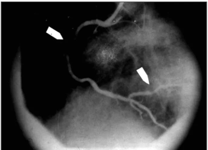
A 40-year-old pregnant white female, gravida III (GIII) para II (PII), last delivery 18 years before, with an echogra-phic gestational age of 23 weeks, sought medical care at the high-risk pregnancy unit of the Hospital Nossa Senhora da Conceição. She reported an inferior myocardial infarction at the age of 31 years and an anteroseptal myocardial infarcti-on at the age of 38 years. After the latter event, corinfarcti-onary angiography showed a proximal segmentary lesion occup-ying 95% of the lumen of the anterior descending artery, a segmentary lesion of 50% in the right coronary artery, and a segmentary lesion of 50% in the medium third of the poste-rior ventricular branch of the right coronary artery. Ventricu-lography showed inferior akinesia and anteroapical hypoki-nesia (figs. 1 to 4). Based on these findings, the patient underwent myocardial revascularization with implantation of an internal thoracic artery bypass graft to the diagonal branch and saphenous vein bypasses to the anterior des-cending and right coronary arteries.
She also reported arterial hypertension and hyperlipi-demia in the 2 preceding years, smoking for 10 years, and use of oral contraceptives until she discovered the preg-nancy. Her mother had died of a stroke and her sister had had preeclampsia. In a routine assessment, ignoring the pre-gnancy but already at the 10th week of gestation, the exer-cise test showed weak cardiorespiratory fitness, angina du-ring exercise, at the second stage, and worsening of the pre-vious alterations of the ST segment (straightening), repre-senting residual ischemia. She used captopril at a dose of 37.5mg/day and SL nitrate when necessary.
mi-3 4 8
Tedoldi e Manfroi
Myocardial infarction and subsequent pregnancy
Arq Bras Cardiol volume 74, (nº 4), 2000
nute, with diffuse pulmonary rhonchi, acyanosis, good peri-pheral perfusion, and no edema.
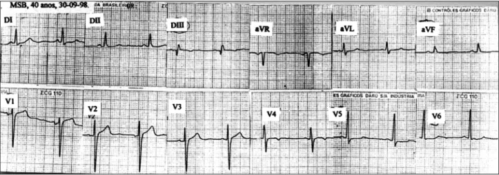
Laboratory tests were normal, except for the lipid pro-file, which showed the following values: total cholesterol of 244mg/dL; LDL-cholesterol of 142mg/dL; triglycerides of 421mg/dL. The electrocardiogram showed sinus rhythm, an inferior probably inactive area, and unspecific alterations of
ventricular repolarization (fig. 5). A maternal echocardio-gram showed a left atrium of 35mm, shortening fraction (%∆D) of 19%, ejection fraction of 45%, mild enlargement in the left ventricle, diastolic left ventricle of 56mm and systo-lic left ventricle of 45mm, segmentary contractile dysfunc-tion with inferodorsal and septoapical hypokinesia, and mild mitral reflux. The fetal echocardiogram at the 24th week of gestation was normal.
Pansinusitis was diagnosed and treated with ampicil-lin, nebulization, and respiratory physiotherapy, normali-zing the pulmonary auscultation. The therapeutical manage-ment adopted was rest, diet for hyperlipidemia, suspension of captopril, acetylsalicylic acid at a dose of 100mg/day, ate-nolol at a dose of 100 mg/day, and isosorbide mononitrate at a dose of 40mg/day through oral via.
On ambulatory assessment at the gestational age of 26 weeks, the patient was asymptomatic, with blood pressure of 135/90mmHg, and heart rate of 68 bpm. She came back at the gestational age of 30 weeks reporting sporadic uterine contractions. At that time her blood pressure measured in the right upper limb with the patient sitting was 160/100m-mHg, and the physiological decrease did not occur on left lateral decubitus. Her heart rate was 80bpm and the patient had no edema. At that time she was admitted to the hospital due to a diagnostic suspicion of preeclampsia, which was confirmed by laboratory tests (urea of 39mg/dL; creatinine of 0.9mg/dL; uric acid of 6.3mg/dL; clearance of endoge-nous creatinine of 86mL/min; hematocrit of 39.4%; hemo-globin of 13.2g/mL; platelets 163,000/mm3; fibrinogen of 530mg/dL; LDH 352 U/L). The patient denied angina and the electrocardiogram remained unchanged. At the gesta-tional age of 31 weeks, the obstetric echography showed an asymmetric retardation of the intrauterine growth. The Dop-pler blood flow test showed centralization of the fetal circu-lation. Amniocentesis was performed and fetal pulmonary immaturity was diagnosed. The patient was treated with rest in the position of left lateral decubitus, parenteral hydration, and oxygen therapy, heparin SC at the dose of 5000 U every 12 hours, reduction in atenolol to 50mg/day, association of Fig. 3 - Coronary angiography showing a 95% segmentary lesion with irregular
margins in the proximal portion of the anterior descending artery.
Fig. 4 – Coronary angiography showing a 50% segmentary ulcerated and eccentric lesion with irregular margins in the right coronary artery, and a 50% segmentary lesion in the medium third of the posterior ventricular branch of the right coronary artery. Fig. 1 - Ventriculography showing the anteroapical hypokinesia of the left ventricle.
Arq Bras Cardiol volume 74, (nº 4), 2000
Tedoldi e Manfroi Myocardial infarction and subsequent pregnancy
3 4 9
nifedipine 30mg/day orally and if necessary, betamethasone once a week. The acetylsalicylic acid and the isosorbide mo-nonitrate were maintained. The fetus was monitored throu-gh cardiotocography and fetal biophysical profile. At the gestational age of 33-34 weeks, reduction in the variability of the fetal heart rate in association with late severe decelera-tion were verified, as was a fetal biophysical profile of 2/8 wi-th absolute oligohydramnios. The pregnancy was interrup-ted with a cesarean section under peridural anesthesia and tubal ligation was performed. No maternal problems occur-red. The female newborn infant weighed 1,345g and had an Apgar score of 1 at the first minute and 6 at the fifth minute. Despite the cardiorespiratory depression, the newborn res-ponded well to the neonatal reanimating maneuvers, wi-thout progression to severe respiratory distress due to hya-line membrane disease. Pediatric gestational age (Capurro) was 36 weeks. The newborn infant remained hospitalized for treatment of septicemia and to gain weight. At the age of 3 months, the infant was healthy and weighed 4,000g. The placenta weighed 400g and had some areas of infarct. Ma-ternal hereditary thrombophilia was investigated and a deficiency (54%) in the activity of the S protein was revea-led. C protein, antithrombin III, and plasminogen were nor-mal, and no mutations in the prothrombin and in the Factor Leiden factor V were identified. Hyperhomocystinemia was not investigated. Three months after the delivery, the mo-ther was asymptomatic with blood pressure of 110/80mmHg, heart rate of 76bpm, and she was using 25mg/day of capto-pril, 100mg/day of atenolol, 200mg/day of acetylsalicylic acid, and 20mg/day of lovastatin. The patient will be reasse-sed to confirm the deficiency in the activity of the S protein.
Discussion
Even though symptomatic ischemic heart disease in pregnancy is rare, if it is identified, follow-up by a multidisci-plinary team during the gestational and puerperal periods is required, as well as counseling in regard to future pregnan-cies. The multidisciplinary team must comprise the follo-wing specialists: an obstetrician, a cardiologist, a
nutritio-nist, a social worker, a psychologist, an anesthesiologist, a neonatologist, and a gynecologist with experience in follo-wing up high-risk pregnancy.
Among the risk factors for coronary heart disease, our patient had hypertension, hyperlipidemia, and oral contra-ceptive use in association with smoking 4. As predisposing factors for the hypertensive disorders of pregnancy, she had chronic hypertension and a familial history of preeclam-psia 3. The risk of a gestation subsequent to 2 myocardial in-farctions was associated with the presence of a mild seg-mentary left ventricular dysfunction (Shorte ning fraction of 19% and ejection fraction of 45%) and residual ischemia 2, despite the previous surgical myocardial revascularization. Therefore, maternal gestational risk was increased by age and hypertension with the superposing hypertensive disor-der of pregnancy, which is characterized by an increase in vascular reactivity that may affect the coronary arterial flow 2 and the flow of other important organs. The maternal gesta-tional risk was also increased by the ischemic heart disease with sequela of 2 myocardial infarctions; mild left ventricular dysfunction accompanied by residual myocardial ischemia with the risk of decompensation due to the physiological hemodynamic overload of pregnancy.
3 5 0
Tedoldi e Manfroi
Myocardial infarction and subsequent pregnancy
Arq Bras Cardiol volume 74, (nº 4), 2000
1. Raw KT. Epidemiology of coronary heart disease in women. In: Julian DG, Wen-ger NK. Women and Heart Disease. London: Mosby, 1997: 1-20.
2. Dufour PH, Ocelli B, Puech F. Pregnancy after myocardial infarction, Int J Gynae-col Obstet 1997; 59: 251-3.
3. Rutherford JD. Coronary artery disease in chilbearing age. In: Elkayam U, Gleicher N. Cardiac Problems in Pregnancy. 3th Ed. New York: Wiley-Liss, 1998: 121-30. 4. Webber MD, Halligan RE, Schumacher JA. Acute infarction, intracoronary thrombolysis and primary PTCA in pregnancy. Cathet Cardiovasc Diagn 1997; 42: 38-43.
5. Chiari RS, Frangieh AY, Sibai BM. Hypertension during pregnancy. In: Elkayam U, Gleicher N. Cardiac Problems in Pregnancy. 3th Ed. New York: Wiley-Liss, 1998: 257-73.
6. Sibai BM. Thrombophilias and adverse outcomes of pregnancy - What should a clinician do? N Eng J Med. 1999; 340: 51-52.
7. Briggs GG, Freeman RK, Yaffe SJ. Drugs in Pregnancy and Lactation. 5th Ed. Bal-timore: Williams e Wilkins, 1998: 133c.
References
8. Moretti MM, Fairlie MF, Aki S, Khoury AD, Sibai BM. The effect of nifedipine the-rapy on fetal and placental waveforms in preeclampsia remote from term. Am J Obst Gynecol 1990; 163: 1844-8.
9. Schulman H. Doppler ultrasound in pregnancy. In: Elkayam U, Gleicher N. Car-diac Problems in Pregnancy. 3th Ed. New York: Wiley-Liss, 1998: 615-27. 10. Montella KR, Ginsberg J Thromboembolic disease in pregnancy. In: Elkayam U,
gleicher N. Cardiac Problems in Pregnancy. 3th Ed. New York: Wiley-Liss, 1998: 232.
11. Trindade CEP. Prematuridade. Em Manual de Perinatologia, 2ª edição, MEDSI 1995, Cap 40: 512-37.
12. McIntire DD, Bloom SL, et al. Birth weight in relation to morbidity and mortality among newborns infants. N Eng J Med 1999; 340: 1234-8.
13. Varella IRS, Lopes ACS, Paniz L, Silveira LML, Ferreira NF, Sandrez MM. Perfil nosológico dos recém-nascidos do berçário do Hospital Nossa Senhora da Conceição. Apresentado no XVI Congresso Brasileiro de Perinatologia, Nov 1998, Salvador, Bahia.
growth retardation, such as the use of beta-blocker 3 and previous arterial disease. We decided to reduce the dose of atenolol and to prescribe nifedipine, which in addition to providing an appropriate control of the blood pressure, does not interfere with the uterus-placenta blood flow 8.
Severe fetal hypoxia evidenced by centralization of the fetal circulation on the Doppler blood flow test creates a risk of death within 2 weeks 9. As fetal pulmonary maturity was not assured when amniocentesis was performed, betame-thasone was started and some measures to increase oxygen supply to the fetus and reduce the risks of thromboses were adopted. Severe maternal and obstetrical complications have been related to the presence of thrombophilias, mainly in white women 6. Screening for hereditary thrombophilia showed deficiency in the activity of the S protein, which should be reconfirmed because during pregnancy this acti-vity may be physiologically decreased 10. Delivery by cesa-rean section was an obstetrical indication (eminent fetal death) even though the vaginal via with analgesia is the preferred one in patients with hemodynamically stable is-chemic heart disease. Medications such as betamimetics, ergot alkaloids, bromocriptine, and prostaglandins are con-traindicated during pregnancy and puerperium because they may cause infarction or thrombosis, or both 2.
Prematurity accounts for more than 50% of the morbi-dity and mortality among newborn infants without any congenital anomalies; 90% of the premature infants wei-ghing between 1,250g and 1,500g survive 11. The major com-plications include the following: respiratory distress, which
ranges from transitory tachypnea of the newborn to hyaline membrane disease; septicemia; intraventricular hemorrha-ge; and necrotizing enterocolitis. The incidence of respira-tory distress is inversely proportional to the weight percen-tile at birth 12. Hyaline membrane consequent to pulmonary and thoracic cage immaturity and deficiency in surfactant affects 10% to 15% of the newborn infants weighing less than 2,500g 11.
Precocious neonatal mortality in the nursery of our hospital is comparable to that of the United States in the ‘80s (11%). At the Universidade Federal de Minas Gerais this mortality was reported as 31.2% in 1990, and in Latin Ameri-can countries it was reported as 16% in 1981. In the very low-weight newborn infants, precocious neonatal mortality is about 18% and in 1% of these cases this mortality is rela-ted with respiratory diseases 13.
Despite the presence of many predetermined factors that increase neonatal morbidity and mortality (prematurity, very low weight, intrauterine growth retardation, poor birth conditions, maternal age above 35 years) 13, in association with maternal diseases such as hypertension, hypertensive disorder of the pregnancy, and ischemic heart disease, our newborn infant had a good outcome.