Case Report
Introduction
Coronary artery disease (CAD) is a late complication after mediastinal irradiation. It tends to affect the coronary ostia, presumably due to their relatively central location within the radiation field. Radiotherapy (RT) may induce accelerated atherosclerosis, with adventitial fibrosis, epicardial fibrotic tissue, and little lipid material in the intimal lesion, which is different from the typical atherosclerotic lesion. We present a case of a 23-year-old male patient who developed left main coronary artery lesion following therapeutic RT for Hodgkin’s lymphoma.
Case Report
A 23 year-old-man with atypical chest pain for 2 months was admitted to our institution. He had been treated for Hodgkin’s lymphoma (HL) with mediastinal RT and chemotherapy at 5 years of age, after which complete remission was achieved. At evaluation, the treadmill exercise test was positive, and dipyridamole sestamibi scintigraphy showed transitory defects in the lateral and anterior walls, with normal left ventricular ejection fraction (Figure 1). Subsequently, the patient underwent coronary arteriography, which disclosed 80% stenosis of the left main coronary artery (Figure 2A). Afterwards, the patient underwent coronary artery
bypass grafting (CABG) from the left internal thoracic artery (ITA) to the left anterior descending coronary artery and from the right ITA to the left marginal coronary artery. Three months after surgery, the patient was admitted with atypical chest pain, and a new coronary arteriography showed occlusion of the right ITA. The patient underwent percutaneous coronary intervention (PCI) with Infinnium stent (Paclitaxel Eluting Coronary Stent) implantation at the left main coronary artery (Figure 2B) with a good outcome at 1-year follow-up.
Discussion
The treatment of HL consists of RT and chemotherapy. In the 1950s, high-voltage RT was used, which was more efficacious, but resulted in greater collateral damage. The involvement of the heart is related to the dosage and quantity of RT, RT on the left side of the chest, as well as the place and size of the tumor. Some authors report that cardiac risk increases when the dose of RT is more than 30 Gy and the heart is involved in the radioactive mantle field1,2. In several long-term series of patients who underwent RT, deaths resulting from second malignancies and heart disease exceeded those resulting from HL3. RT affects the whole heart, but mainly the pericardium, leading to constrictive pericarditis; it also affects the valvular apparatus, myocardium, conduction system and coronary artery4,5. The incidence of CAD ranges from 5.5 to 12%; it is the most deadly complication, but is treatable6,7. It takes 3 to 29 years (average 13 to 16 years) for CAD to develop after RT7,8. RT may induce accelerated atherosclerosis, and postmortem studies have revealed severe adventitial fibrosis, epicardial fibrotic tissue, and little lipid material in the intimal lesion, which is different from the typical atherosclerotic lesion. A previous study showed a 45-fold excess risk of mortality from acute myocardial infarction in patients treated with more than 30 Gy of mediastinal irradiation before the age of 20 years9. Our young patient had CAD without risk factors and a negative family history for CAD. This can be ascribed to his undergoing RT 17 years earlier. The prevalence of radiation-induced heart disease (RIHD) may range from 6 to 30% in patients with Hodgkin’s disease. Patients may have myocardial fibrosis that can exist even in asymptomatic patients who receive high doses of RT. A necropsy study showed that this rate is near to 63%2. Also, symptomatic cardiac involvement after RT occurs in about 10% of patients. It is recommended the prevention of cardiovascular events, screening to detect early coronary artery disease, and also complete laboratory examination with
Keywords
Coronary vessels/radiotherapy; coronary vessel anomalies; thorax/radiotherapy.
Prevention of late cardiovascular complications after radiation therapy (RT) for treatment of a malignant tumor is challenging. We report the case of a young male patient with Hodgkin’s lymphoma treated with RT, who developed ischemic heart disease during follow-up, although he had no cardiovascular risk factors. We conclude that patients undergoing RT who experience chest pain should be fully investigated for coronary artery disease.
Treatment of Left Main Coronary Artery Lesion after Late Thoracic
Radiotherapy
Vera Maria Cury Salemi, André L Dabarian, Luciano Nastari, Marcus Gama, José Soares Júnior, Charles Mady
Instituto do Coração (InCor) do Hospital das Clínicas da Faculdade de Medicina da Universidade de São Paulo, São Paulo, Brazil
Mailing address: Vera Maria Cury Salemi •
Avenida Jandira, 185 / 41 B – Indianópolis – 04080-000 – São Paulo, SP – Brazil E-mail: [email protected], [email protected]
Manuscript received 22/12/09; revised manuscript received 14/05/10; accepted 01/07/10.
Case Report
Salemi et al Coronary Lesion after Radiotherapy
(Arq Bras Cardiol 2011; 97(3) : e53-e55)
difficult as the coronary lesions are frequently in the left main coronary artery or in the proximal region, leading to the greater risk of these procedures6. Prevention of RT damage includes a decrease in the total dosage, which reduces attention to lipid levels and thyroid function. CABG is one
option, but the approach is made difficult by the mediastinal fibrosis that is a collateral effect of anterior RT of the thorax. Another option is PCI with stent implantation, but it is also
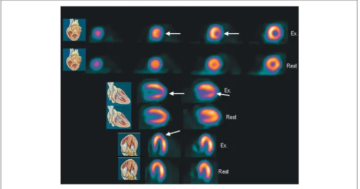
Figure 1 - Dipyridamole sestamibi scintigraphy showing transitory defects in the lateral and anterior walls. Ex: exercise.
Figure 2 - Severe stenosis of the left main coronary artery seen in the right anterior oblique view (A); successful ininnium stent implantation at the left main
coronary artery (B).
Case Report
Salemi et al
Coronary Lesion after Radiotherapy
(Arq Bras Cardiol 2011; 97(3) : e53-e55)
References
1. Hull MC, Price-Mendenhall N, Colgan ME. Subdiaphragmatic Hodgkin’s disease: the University of Florida experience. Int J Radiat Oncol Biol Phys. 2002;52(1):161-6.
2. Brennan S, Hann LE, Yahalom J, Oeffinger KC, Rademaker J. Imaging of late complications from mantle field radiation in lymphoma patients. Radiol Clin North Am. 2008;46(2):419-30.
3. Wethal T, Lund MB, Edvardsen T, Fosså SD, Pripp AH, Holte H, et al. Valvular dysfunction and left ventricular changes in Hodgkin’s lymphoma survivors: a longitudinal study. Br J Cancer. 2009;101(4):575-81.
4. Ng AK, Mauch PM. Late effects of Hodgkin’s disease and its treatment. Cancer J. 2009;15(2):164-8.
5. Yeh ET, Bickford CL. Cardiovascular complications of cancer therapy: incidence, pathogenesis, diagnosis, and management. J Am Coll Cardiol. 2009;53(24):2231-47.
cardiac complications but not secondary malignancies. Prevention also includes the use of computed-tomography-based treatment planning and the application of two fields. In addition, the association of RT and chemotherapy, known as combination-modality therapy with RT doses of 20 to 30Gy, and, of course, the treatment of cardiovascular risk factors can also be used to treat RT damage. Using statins to reduce inflammation and angiotensin-converting enzyme inhibitors may be useful in treating RIHD. Also, pentoxifylline and alpha-tocopherol have shown beneficial effects on RIHD when started before or 3 months after RT in rats10.
In conclusion, cardiotoxicity is one of the most important complications of cancer therapy. As more people survive cancer, teamwork composed by oncologists and
cardiologistsis importantin preventing cardiac problems related to cancer treatment.
Potential Conflict of Interest
No potential conflict of interest relevant to this article was reported.
Sources of Funding
There were no external funding sources for this study.
Study Association
This study is not associated with any post-graduation program.
6. Piovaccari G, Ferretti RM, Prati F, Traini AM, Gobbi M, Caravita L, et al. Cardiac disease after chest irradiation for Hodgkin’s disease: incidence in 108 patients with long follow-up. Int J Cardiol. 1995;49(1):39-43.
7. King V, Constine LS, Clark D, Schwartz RG, Muhs AG, Henzler M, et al. Symptomatic coronary artery disease after mantle irradiation for Hodgkin’s disease. Int J Radiat Oncol Biol Phys. 1996;36(4):881-9.
8. Reber D, Birnbaum DE, Tollenare P. Heart diseases following mediastinal irradiation: surgical management. Eur J Cardiothorac Surg. 1995;9(4):202-5.
9. Hancock SL, Donaldson SS, Hoppe RT. Cardiac disease following treatment of Hodgkin’s disease in children and adolescents. J Clin Oncol. 1993;11(7):1208-15.
10. Boerma M, Roberto KA, Hauer-Jensen M. Prevention and treatment of functional and structural radiation injury in the rat heart by pentoxifylline and alpha-tocopherol. Int J Radiat Oncol Biol Phys. 2008;72(1):170-7