669
Correspondence to: Sonja ŠALINGER-MARTINOVIĆ Durlan 23, 18000 Niš Serbia
Successful Retrieval of an Unexpanded Coronary
Stent from the Left Main Coronary Artery during
Primary Percutaneous Coronary Intervention
Sonja Šalinger-Martinović1, Siniša Stojković2, Milan Pavlović1, Zoran Perišić1,
Slobodan Obradović3, Svetlana Apostolović1, Milan Živković1, Nenad Božinović1 1Clinic for Cardiovascular Diseases, Clinical Centre, Niš, Serbia;
2Department of Cardiology, Clinical Centre of Serbia, Belgrade, Serbia; 3Cardiology Department, Military Medical Academy, Belgrade, Serbia
INTRODUCTION
Stent dislodgement or migration during coro-nary artery stenting is a rare complication and the incidence varies between 0.32-8.3% [1, 2]. Systemic and coronary embolizations are the consequences of stent dislodgement and can lead to serious complications such as the acute closure of the affected vessel, coronary throm-bosis, and myocardial infarction. In the past, manual crimping of stents was associated with a significantly increased risk of the stent disen-gagement and embolization. Although manu-ally crimped stents are seldom used, the prob-lem of stent dislodgement and potential embo-lization has not been completely eliminated [3]. We present a case of a successful retrieval of an unexpanded and slightly bended coronary stent from the left main coronary artery (LMCA) via the transfemoral approach.
CASE REPORT
A 70-year-old man was admitted to our hospital with an inferior wall acute myocardial infarc-tion with persistent ST segment elevainfarc-tion. Twelve hours after the onset of the symptoms, he was admitted to a local hospital and imme-diately transferred to our centre for primary
percutaneous coronary intervention (PCI). At the local hospital, the patient was treated with Aspirin 300 mg, Clopidogrel 300 mg, Enoxapa-rin 70 mg sc and analgesic agents. The electro-cardiogram revealed the ST segment elevation in inferior leads (D2, D3, aVF) with an abnor-mal R wave in V1-V3 leads (R/S ratio >1 in V1). The values of the cardiac markers of myocardial necrosis were elevated: TnI 12 ng/ml, CKMB mass 300 ng/ml. The emergency echocardio-graphic examination showed inferoposterior akinezia, with a preserved global systolic func-tion (EF 60%). The patient was sent to the cath-eterization laboratory and treated with addi-tional Clopidogrel dose (300 mg), Enoxaparin 0.3 mg/kg (20.0 mg iv) and agent for gastropro-tection (proton pump inhibitor).
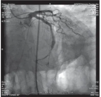
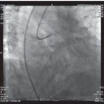
Using the right femoral approach (6 F arte-rial sheath), the coronary angiography revealed an occlusion in the mid-portion of the left circumflex artery (LCX) (Figure 1). After the implantation of a temporary pace-maker due to conduction disturbances, a 6 French (Fr) EBU 3.75 guiding catheter (Cordis Corporation, Miami Lakes, Florida) was used to engage the LMCA. The first attempt to cross the occluded segment of the LCX with a Zinger medium guidewire (Medtronic Cardiovascular Inc.) was unsuccessful. After this attempt, a Crosswire NT guidewire (TERUMO) was advanced across SUMMARY
Introduction Dislodgement and embolization of the new generation of coronary stents before their deployment are rare but could constitute a very serious complication.
Case Outline We report a case of a stent dislodgement into the left main coronary artery during the primary coronary intervention of infarct related left circumflex artery in a patient with acute myocardial infarction. The dislodged and unexpanded bare-metal stent FlexMaster 3.0x19 mm (Abbot Vascular) was stranded and bended in the left main coronary artery (LMCA), probably by the tip of the guiding catheter, but stayed over the guidewire. It was successfully retrieved using a low-profile Ryujin 1.25x15 balloon catheter (Terumo) that was passed through the stent, inflated and then pulled back into the guiding catheter. After that, the whole system was withdrawn through the 6 F arterial sheath via the transfemoral approach. After repeated cannulation via the 6F arterial sheath, additional BMW and ATW guidewires were introduced into the posterolateral and obtuse marginal branches and a bare-metal stent Driver (Medtronic Cardiovascular Inc) 3.0x18 mm was implanted in the target lesion.
Conclusion Stent dislodgement is a rare but potentially life-threatening complication of the percuta-neous coronary intervention. This incident occurring in the LMCA in particular during an acute myocar-dial infarction requires to be urgently resolved. The avoidance of rough manipulation with the guiding catheter and delivery system may help in preventing this kind of complications.
Keywords: STEMI; primary angioplasty; bare metal stent; dislodgement
670
doi: 10.2298/SARH1110669S
Šalinger-Martinović S. et al. Successful Retrieval of an Unexpanded Coronary Stent from the Left Main Coronary Artery
the lesion without difficulty into the distal LCX (Figure 2). Following this, a Balance Middle Weight (BMW) guide-wire (Guidant, Advanced Cardiovascular System Inc., CA, USA) was advanced in the LCX posterolateral branch, and an All Track Wire (ATW) (Cordis, USA) in the obtuse marginal branch (Figure 3). A Sprinter balloon catheters (Medtronic Cardiovascular) 1.5×20 mm and 20×20 mm were used to predilate the culprit lesion and both side branches at 16 atmospheres. After repeated intracoro-nary administration of nitro-glycerine (200 µg + 200 µg) a TIMI 3 flow was achieved (Figure 4). Than, the intention was to stent the lesion with a bare-metal stent FlexMaster 3.0×19 mm (Abbot Vascular), over the BMW guidewire in the posterolateral branch jailing the second guidewire in order to protect the obtuse marginal branch. While trying
to advance the stent across the guiding catheter, the guiding catheter got decannulated from the LMCA. In an attempt to recannulate the left coronary artery, the balloon cathe-ter and stent were pulled back to advance the guiding cath-eter. The attempt was unsuccessful, the guidewire twisted inward making a loop, and the unexpanded stent peeled off from the balloon and slightly bended in the LMCA (Figure 5). The balloon was removed and a Ryujin 1.25×15 balloon catheter (Terumo) was gently passed over the straightened guidewire through the stent (Figure 6). The balloon was inflated at 8 atm and carefully withdrawn inside the guid-ing catheter along with the dislodged stent. The whole system, including the guiding catheter, the balloon cathe-ter, the dislodged stent and the guidewires were withdrawn through the femoral sheath. After repeated cannulation,
Figure 1. Occlusion of the infarct related left circumflex artery
Figure 2. Advanced Crosswire NT guidewire across the lesion into the distal LCX
Figure 3. BMW guidewire advanced in the LCX posterolateral branch, and an ATW guidewire in the obtuse marginal branch
671
www.srp-arh.rs Srp Arh Celok Lek. 2011;139(9-10):669-672
additional BMW and ATW guidewires were introduced into the posterolateral and obtuse marginal branches. A bare-metal stent Driver (Medtronic Cardiovascular Inc.) 3.0×18 mm was implanted in the target lesion at 12 atm. After that a post-dilatation of the proximal part of the stent was performed using a Maverick balloon catheter (Boston Scientific) 3.25×9 mm at 18 atm. Following this, the jailed guidewire was removed, and the TIMI 3 flow remained in both side branches (Figure 7).
DISCUSSION
Dislodgement and embolization of new generation coro-nary stents before deployment is rare and may lead to chal-lenging complications of intracoronary stenting. The inci-dence of stent dislodgement is uncommon and is lower now than at the time when stents were manually crimped, with reports varying between 0.32% and 8%. Dislodge-ment of a stent can be secondary to severe coronary angu-lation, calcified coronary arteries, inadequate coronary artery predilatation and direct stenting [4].
Despite the infrequent incidence of stent dislodgement, the consequences may be severe, sometimes fatal, and oper-ators are generally not familiar with this kind of compli-cation [5]. Adequate delivery and placement of coronary stents are dependent not only on proficient handling, but also on various properties [1]. Flexibility, structure, shape, and radial strength affect the durability and deliverability of stents. The stent visibility is also important in delivery and retrieval procedures [6].
The dislodging may occur at the guide tip when travers-ing the coronary artery proximal to the lesion, or in the lesion itself. If the guide choice and stability are not proper, the guide will be displaced from the coronary ostium as the stent exits the guide and meets resistance in the coronary artery [7]. The stent cannot be pushed on because there is
Figure 5. Dislodged and slightly bended stent in left main coronary artery Figure 6. Ryujin 1.25×15 balloon catheter passed over the straightened guidewire through the stent
Figure 7. Final result: TIMI 3 flow after post dilatation of the bare-metal stent Driver 3.0×18 implanted in the target lesion
672
doi: 10.2298/SARH1110669S
Šalinger-Martinović S. et al. Successful Retrieval of an Unexpanded Coronary Stent from the Left Main Coronary Artery
A complication such as undeployed stent in the LMCA can be life-threatening [2]. Such a complication may lead to emergency surgery, or may be treated percutane-ously. The dislodged stent can be deployed locally, can be crushed to the arterial wall by a balloon or stent, or retrieved by a double guidewire technique. Other meth-ods include usage of devices such as a bioptome, a loop basket, a gooseneck snare, or an alligator forceps cath-eter [2, 3, 9]. The advent of IVUS imaging has played a significant role in enhancing visualization of stent deploy-ment. The use of IVUS can perhaps improve percutane-ous management of lost stents, further reducing the need for surgical retrieval [10].
In our case, the dislodged and unexpanded stent was stranded and bended in the LMCA, probably by the tip
of the guiding catheter, but stayed over the guidewire. It was removed from this location using a low profile deliv-ery balloon and advancing it through the stent, inflating the balloon on low pressure and than pulling back into the guiding catheter. The guiding catheter, balloon and stent were removed via the femoral sheath.
Stent dislodgement is a rare but potentially life-threat-ening complication of the percutaneous coronary inter-vention. This incident occurring in the LMCA in partic-ular during an acute myocardial infarction requires to be urgently resolved. The avoidance of rough manipulation with the guiding catheter and delivery system may help in preventing this kind of complications. This complication may, on occasion, be treated percutaneously preventing a more serious outcome.
REFERENCES
1. Colkesen Y, Baltali M, Acil T, Tekin G, Tekin A, Erol T, et al. Coronary and systemic stent embolization during percutaneous coronary interventions. A single center experience. Int Heart J. 2007; 48:129-36.
2. Patel MT, Shah SC, Gupta AK, Ranjan A. Successful retrieval of transradially delivered unexpanded coronary stent from the left main coronary artery. Indian Heart J. 2002; 54:715-6.
3. Goldberg A, Kemer A, Anne G, Gruberg L. Clinical images: one stent lost in two arteries. J Invas Cardiol. 2004; 16(3):163-4.
4. Ahmar W, Malaiapan Y, Meredith IT. Transradial retrieval of a dislodged stent from the left main coronary artery. J Invasive Cardiol. 2008; 20:545-7.
5. Brilakis ES, Best PJ, Elesber AA, Barsness GW, Lennon RJ, Holmes DR, et al. Incidence, retrieval methods, and outcomes of stent loss during percutaneous coronary intervention: a large single-center experience. Catheter Cardiovasc Interv. 2005; 66:333-40.
6. Lohavanichbutr K, Webb JG, Carere RG, Solankhi N, Jarochowski M,
D’yachkova Y, et al. Mechanisms, management, and outcome of failure of delivery of coronary stents. Am J Cardiol. 1999; 83:779-81. 7. Uyuan C, Gunduz H, Arinc H, Akademir R. Embolised stent into the
circumflex coronary artery during percutaneous coronary intervention. Int Heart J. 2006; 47:125-9.
8. Yimaz H, Belgi A, Demir I, Basarc I, Sancaktar O. Successful transcatheter retrieval of an embolized stent from the left ventricle. J Invas Cardiol. 2002; 14(8):466-468.
9. Meisel SR, DiLeo J, Rajakaruna M, Pace B, Frankel R, Shani J, et al. A technique to retrieve stents dislodged in the coronary artery followed by fixation in the iliac artery by means of balloon angioplasty and peripheral stent deployment. Catheter Cardiovasc Interv. 2000; 49:77-81.
10. George JC, Fiss D, Bashir R. Management of dislodged coronary stent using intravascular ultrasound. Cath Lab Digest Archive. 2009; 17(11):36-9.
КРАТАК САДРЖАЈ
Увод Скли зну ће са ба ло на пре уград ње и ми гра ци ја стен то-ва но ве ге не ра ци је ве о ма су рет ке, али по тен ци јал но опа сне, ком пли ка ци је пер ку та не ко ро нар не ан ги о пла сти ке. При каз бо ле сни ка При ка зу је мо слу чај скли зну ћа стен та у глав но ста бло ле ве ко ро нар не ар те ри је то ком при мар-не пер ку та мар-не ко ро нар мар-не ан ги о пла сти ке ле ве цир кум флек-сне ар те ри је код бо ле сни ка с акут ним ин фарк том ми о кар-да. Стент од нер ђа ју ћег че ли ка (Flex Ma ster 3.0×19 mm, Ab bot Vas cu lar) ма ни пу ла ци јом вр ха ка те те ра скли знуо је са ба ло-на у глав но ста бло ле ве ко ро ло-нар не ар те ри је, али је остао ло-на ко ро нар ној жи ци. Из ва ђен је по мо ћу ни ско про фил ног ба-лон-ка те те ра (Ryujin 1.25×15 mm, Te ru mo) ко ји је по ста вљен и ин су фли ран уну тар стен та, а за тим уву чен у ка те тер за јед но
са стен том. По сле то га, чи тав си стем, укљу чу ју ћи и ка те тер, из ву чен је кроз фе мо рал ни шит од 6 F. На кон по нов не ка ну-ла ци је пре ко ар те риј ског увод ни ка од 6 F ко ро нар не жи це
BMW и ATW су пла си ра не у по сте ро ла те рал ну и оп ту зну мар-ги нал ну гра ну, а стент од нер ђа ју ћег че ли ка (Dri ver 3.0×18 mm, Med tro nic Car di o va scu lar Inc.) по ста вљен је на ме сто сте но зе. За кљу чак Скли зну ће не у гра ђе ног стен та, прем да рет ка, мо же би ти по жи вот опа сна ком пли ка ци ја. Скли зну ће стен-та у глав но сстен-та бло ле ве ко ро нар не ар те ри је то ком при мар-не пер ку та мар-не ко ро нар мар-не ан ги о пла сти ке зах те ва хит но ре-ша ва ње. Не жни јом ма ни пу ла ци јом ка те те ром во ди чем и но са чем стен та мо же се спре чи ти ова ква вр ста про бле ма. Кључ не ре чи:STE MI; при мар на ан ги о пла сти ка; стент од нер-ђа ју ћег че ли ка; спа да ње стен та
У пе о
л ење
еек п д
о
ко о
о
те т
л
о
т л
ле е
ко о
е
те је
токо
п
е
пе кут е
ко о
е
те е
је
Соња Шалингер-Мартиновић1, Синиша Стојковић2, Милан Павловић1, Зоран Перишић1, Слободан Обрадовић3,
Светлана Апостоловић1, Милан Живковић1, Ненад Божиновић1
1Клиника за кардиоваскуларне болести, Клинички центар, Ниш, Србија; 2Клиника за кардиологију, Клинички центар Србије, Београд, Србија; 3Клиника за кардиологију, Војномедицинска академија, Београд, Србија