Comparison of Outcome between Chagas Cardiomyopathy and
Idiopathic Dilated Cardiomyopathy
Amanda P. Barbosa
1, Augusto Cardinalli-Neto
2, Ana Paula Otaviano
2, Bianca F. da Rocha
2, Reinaldo B. Bestetti
2 Faculdade de Medicina de São José do Rio Preto1; Divisão de Cardiologia do Hospital de Base de São José do Rio Preto2, São José do Rio Preto, SP, BrazilAbstract
Background: Little is known about the outcome of patients with Chagas cardiomyopathy in comparison to that of patients with Idiopathic Dilated Cardiomyopathy in the contemporary era.
Objective: To compare the outcome of chagasic patients with chronic systolic heart failure secondary to Chagas cardiomyopathy with that observed in patients with IDC in the contemporary era.
Methods: A total of 352 patients (246 with Chagas cardiomyopathy, 106 with Idiopathic Dilated Cardiomyopathy) prospectively followed at our Institution from January, 2000 to January, 2008 were included. All patients received standard contemporary medical therapy.
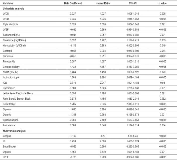
Results: In Cox proportional hazards model multivariate analysis, digoxin use (Hazard Ratio=3.17; 95% Confidence Interval 1.62 to 6.18; p=0.001), need of inotropic support (Hazard Ratio=2.08; 95% Confidence Interval 1.43 to 3.02; p<0.005), left ventricular ejection fraction (Hazard Ratio=0.97; 95% Confidence Interval 0.95 to 0.99; p<0.005), and Chagas cardiomyopathy etiology (Hazard Ratio=3.29; 95% Confidence Interval 1.89 to 5.73; p<0.005) were positively associated with mortality, whereas Beta-Blocker therapy (Hazard Ratio=0.39; 95% Confidence Interval 0.26 to 0.56; p<0.005) was negatively associated with mortality. Survival probability for patients with Chagas cardiomyopathy at 8, 24, and 49 months was 83%, 61%, and 41%, respectively, and for patients with Idiopathic Dilated cardiomyopathy 97%, 92%, and 82%, respectively (p<0.005).
Conclusion: In the current era of heart failure therapy, patients with Chagas cardiomyopathy have a poorer outcome in comparison to patients with Idiopathic Dilated Cardiomyopathy. (Arq Bras Cardiol 2011;97(6):517-525)
Keywords: Chagas disease; chagas cardiomyopathy; cardiomyopathy, dilated; trypanosomiasis.
Mailing Address: Amanda Pires Barbosa •
Av. Doutor Fontes, 791 - Bairro Xis - 14870-620 - Jaboticabal, SP, Brazil E-mail: [email protected]
Manuscript article March 24, 2011; revised manuscript received May 09, 2011; accepted May 13, 2011.
cardiac death11, and chronic systolic heart failure12. The
clinical picture of chronic systolic heart failure of patients with Chagas cardiomyopathy is quite similar to that found in patients with IDC, with a few exceptions related to higher frequency of pacemakers and amiodarone use13.
Outcome is poorer in Chagas cardiomyopathy patients with decompensated acute heart failure in comparison to non-Chagas cardiomyopathy patients over a median follow up of 25 months14,15. This is probably
secondary to increased activation of the renin-angiotensin system, increased cytokines levels, more severe cardiac impairment, and hemodynamic instability16. Nonetheless,
a direct comparison of outcome of Chagas cardiomyopathy with IDC patients with decompensated heart failure is still lacking.
The prognosis of patients with chronic Chagas cardiomyopathy also seems to be different from that seen in patients with IDC. Two studies have shown a poorer prognosis of patients with Chagas cardiomyopathy in comparison to patients with IDC among several etiologies of heart failure17,18. Recently, another study comparing
specifically outcome of Chagas cardiomyopathy with that of IDC patients has shown a better prognosis for patients with IDC19. Clearly, more data on a direct comparison of
Introduction
Idiopathic Dilated Cardiomyopathy (IDC) has an incidence of 17.9/100,000 inhabitants in the general population1. It is the third cause of chronic systolic heart
failure2, with an annual mortality of 95% in
population-based series and 69% in tertiary referral cohorts3.
Chagas disease, which is caused by the protozoan T.cruzi
and transmitted to humans through a sucking bug, affects about 11 million people, whereas other 90 million are at risk of acquiring the disease in South America4. The disease
has spread throughout the world due to international immigration, with approximately 750,000 affected persons living outside South America nowadays5.
The clinical manifestations of Chagas disease are thromboembolic phenomena6,7, chest pain8, atrioventricular
outcome of patients with chronic heart failure secondary to Chagas cardiomyopathy with patients with IDC are needed. Accordingly, the purpose of this study was to compare the outcome of patients with chronic systolic heart failure secondary to Chagas cardiomyopathy with that observed in patients with IDC in the contemporary era.
Methods
All patients routinely and prospectively followed at the Cardiomyopathy Outpatient Service of our Institution from January, 2000 to January, 2008 with the diagnosis of Chagas cardiomyopathy and IDC were considered for the study. The diagnosis of Chagas cardiomyopathy was made on the basis of a positive serology and a left ventricular ejection fraction <55% on echocardiography according to the Teicholz method, or a left ventricular ejection fraction <50% on Radionuclide Ventriculography. The diagnosis of IDC considered the same echocardiographic criteria, as described above, and the absence of concomitant obstructive coronary artery disease. The latter was ruled out by either coronary arteriogram or myocardial scintigraphy in those patients not amenable for coronariography.
The diagnostic work up consisted of history-taking, complete physical examination, standard laboratory tests, 12-lead ECG, and transthoracic Doppler echocardiography. Clinical status, heart rate, and systemic arterial pressure were noted on admission. Patients in the New York Heart Association Classes III/IV were given diuretics, angiotensin converting enzyme inhibitors (ACEI)/Angiotensin Receptor Blocker (ARB), and digoxin to alleviate symptoms. B-Blocker therapy was started immediately after clinical compensation. Patients in the New York Heart Association classes I/II were preferentially treated with Beta-Blockers and ACEI/ARB.
Patients were censored at the time of death, heart transplantation or spontaneous dropout.
Statistical Analysis
Unless otherwise indicated, continuous variables with normal distribution are presented as mean ± standard deviation, whereas continuous variables with non-normal distribution are given as median (25%, 75% percentile). Categorical variables are shown as numbers and proportions (%). Continuous variables were compared by the Mann-Whitney or the t Test for unpaired samples. Categorical variables were tested by the X2 test. The Spearman test was
used to establish correlations between continuous variables. A Cox proportional hazards model was used to discover independent predictors of mortality for the study population. Variables associated with mortality in the Univariate model at the p level <0.05 were entered into the multivariate model with a forward approach. When several continuous variables were correlated in the Univariate model, only that one with the greatest Wald coefficient was included in the Multivariate model.
A ROC curve was used to select the best cut-off point of a continuous variable to predict mortality. An area under the curve >0.50 was considered statistically significant.
Survival probability was estimated by the Kaplan-Meier method. Comparison of survival probability between groups was made with the log rank test. In all circumstances, a p value < 0.05 was considered to be of statistical significance.
Results
A total of 352 patients fulfilled inclusion criteria and were entered in the study. Two hundred forty six (70%) patients were diagnosed as having Chagas cardiomyopathy, and 106 (30%) with IDC. Table 1 shows baseline characteristics of Chagas and IDC patients. Twenty patients underwent heart transplantation, all of them with Chagas cardiomyopathy, and were censored at the time of cardiac procedure.
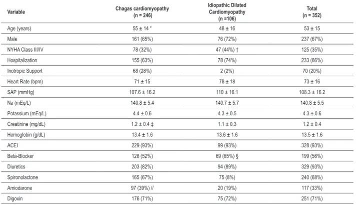
Patients with Chagas cardiomyopathy needed more inotropic support, had lower severity of functional status and lower heart rate than patients with IDC. By contrast, patients with IDC were found to have longer follow up period and higher proportion of Beta-Blocker therapy in comparison to Chagas cardiomyopathy patients.
Table 2 shows the electrocardiographic findings as well as the echocardiographic features. Patients with Chagas cardiomyopathy had higher proportion of right bundle branch block associated with left anterior fascicular block in the 12-lead electrocardiogram, higher proportion of pacemaker use, and lower proportion of left bundle branch block as compared with patients with IDC. At echocardiography, patients with Chagas cardiomyopathy had higher proportion of segmental wall motion abnormalities, whereas IDC patients were found to have larger left ventricular diastolic diameter. Mean left ventricular ejection fraction was higher in patients with Chagas cardiomyopathy than in patients with IDC.
Results of Univariate and Multivariate analysis by the Cox proportional hazards model are given in Table 3. Importantly, Chagas disease etiology was the powerful predictor of all-cause mortality for the study population.
Duration of follow up was 28 ± 27 months. Overall, 135 (38%) patients died during the study period. One-hundred-nine patients died in the Chagas cardiomyopathy patients group, whereas 16 (15%) patients died in the IDC patients group (p<0.005). Survival probability of patients with Chagas cardiomyopathy at 8, 24, and 49 months was 83%, 61%, and 41%, respectively, whereas survival probability for IDC patients at 8, 24, and 49 months was 98%, 92%, and 82%, respectively (p <0.005). Figure 1 illustrates these data.
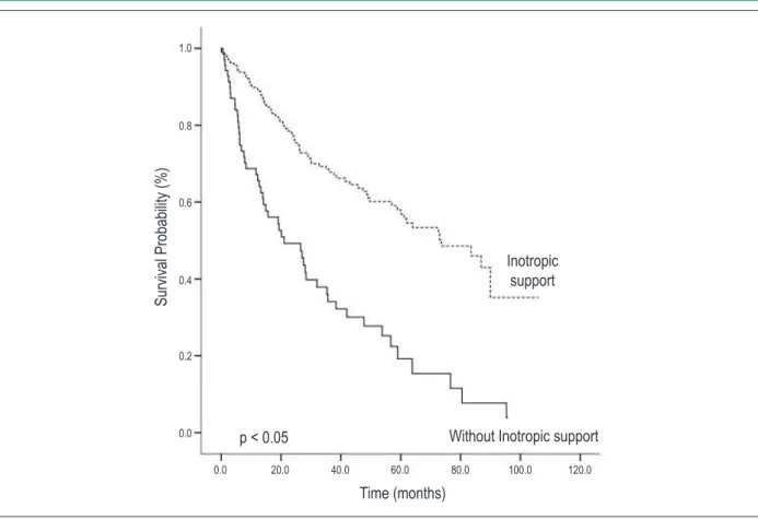
Survival probability for patients on inotropic support at 8, 26, and 47 months was 69%, 47%, and 28%, respectively, and survival probability of patients who did not need inotropic support at 8, 26, and 47 months was 93%, 75%, and 63%, respectively (Figure 2).
Patients receiving Beta-Blocker therapy had a survival probability at 8, 26, and 47 months of 95%, 85%, and 77%, respectively, whilst survival probability of patients not taking Beta-Blockers at 8, 26, and 47 months was 79%, 50%, and 30% (p<0.05), as seen in Figure 3.
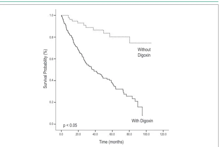
months was 98%, 91%, and 84%, respectively (p<0.05). Figure 4 shows these data.
A left ventricular ejection fraction < 32% was the best cutoff point to predict mortality. Probability of survival for patients with a left ventricular ejection fraction <32% at 8, 26, and 47 months was 85%, 59%, and 43%, respectively. Probability of survival for patients with a left ventricular ejection fraction equal to or greater than 32% at 8, 26, and 47 months was 90%, 77%, and 64%, respectively (p <0.01). Figure 5 shows these findings.
Discussion
This investigation clearly shows that Chagas etiology is an independent predictor of all-cause mortality in a population consisting of patients with IDC and Chagas Cardiomyopathy. Furthermore, survival probability was markedly decreased in Chagas Cardiomyopathy in comparison to IDC patients.
Comparison of outcome between Chagas cardiomyopathy and IDC patients has scarcely been reported. Freitas et al18
have studied 1,220 patients with severe heart failure (New York Heart Association Class III/IV), 454 of them (37%) with IDC and 242 (20%) with Chagas cardiomyopathy. They observed that Chagas etiology of heart failure was associated with the worst outcome in that population. However, in comparison with our work, the population studied by Freitas et al18 differs
as to patients’ heterogeneity, severity of syndrome and lack of Beta-Blocker therapy.
Very recently, Nunes et al19 have performed a specific
comparison of outcome between Chagas cardiomyopathy and IDC patients. They studied 287 patients (224 with Chagas cardiomyopathy, 63 with IDC), mean age of 49±13 years, followed for 39 months on average. They found that New York Heart Association Class, left ventricular ejection fraction, right ventricular function as determined by the Tei method, and left atrial volume were independent predictors of all-cause mortality. In addition, they observed a lower survival probability in patients with Chagas in comparison with patients with IDC. Compared with our investigation, the work by Nunes et al14 differs as to the smaller sample
size of patients enrolled, the low percentage of patients on Beta-Blocker therapy, the same proportion of total mortality in both groups, and the type of predictors of mortality.
The poor prognosis of Chagas cardiomyopathy in comparison to IDC patients, as well as the prognostic determinants observed in this study, can be related to the peculiarities of Chagas cardiomyopathy. The interplay of autoimmunity, microvascular spasm, and autonomic dysfunction is believed to play a pivotal role in the pathogenesis of Chagas cardiomyopathy20.
In fact, the association of mononuclear cell infiltrate throughout the myocardium with large areas of confluent fibrosis,21 similar to what is seen in catecholamine
cardiomyopathy22 and not seen in IDC patients23, can
lead to a more severe ventricular remodeling process and, ultimately, death. The presence of myocardial inflammatory
Table 1 - Clinical and standard laboratory tests characteristics at baseline (n=352)
Variable Chagas cardiomyopathy (n = 246)
Idiopathic Dilated Cardiomyopathy
(n =106)
Total (n = 352)
Age (years) 55 ± 14 * 48 ± 16 53 ± 15
Male 161 (65%) 76 (72%) 237 (67%)
NYHA Class III/IV 78 (32%) 47 (44%) † 125 (35%)
Hospitalization 155 (63%) 78 (74%) 233 (66%)
Inotropic Support 68 (28%) 2 (2%) 70 (20%)
Heart Rate (bpm) 71 ± 15 78 ± 18 73 ± 16
SAP (mmHg) 107.6 ± 16.2 110 ± 16.1 108.3 ± 16.2
Na (mEq/L) 140.8 ± 5.4 140.7 ± 5.7 140.8 ± 5.5
Potassium (mEq/L) 4.4 ± 0.6 4.3 ± 0.5 4.3 ± 0.6
Creatinine (mg/dL) 1.2 ± 0.4 ‡ 1.1 ± 0.3 1.2 ± 0.4
Hemoglobin (g/dL) 13.4 ± 1.6 13.6 ± 1.6 13.5 ± 1.6
ACEI 229 (93%) 99 (93%) 328 (93%)
Beta-Blocker 128 (52%) 69 (65%) § 199 (56%)
Diuretics 203 (82%) 94 (89%) 329 (93%)
Spironolactone 165 (67%) 75 (8%) 240 (68%)
Amiodarone 97 (39%) // 20 (19%) 117 (33%)
Digoxin 176 (71%) 75 (72%) 251 (71%)
Table 2 - Electrocardiographic and echocardiographic indings of the study population
Variable Chagas cardiomyopathy (n = 246)
Idiopathic Dilated Cardiomyopathy
(n = 106)
Total (n = 352)
Atrial Fibrillation 69 (28%) 31 (29%) 100 (28%)
ICD 3 (9%) 12 (11%) 35 (10%)
Pacemaker 124 (50%) * 15 (14%) 139 (39%)
Left Bundle Branch Block 41 (17%) 54 (51%) * 95 (27%)
Right Bundle Branch Block 99 (40%) * 13 (12%) 112 (32%)
Left Anterior Fascicular Block 99 (40.2%) 24 (22.6%) † 123 (34.9%)
Necrosis 12 (5%) 10 (9%) 22 (6%)
VPC 113 (46%) 75 (72%) 188 (53%)
LVDD (mm) 64.5 ± 8.9 67.7 ± 9.7 ‡ 65.5 ± 9.2
LVSD (mm) 53.5 ± 10.4 56.4 ± 10.8 54.4 ± 10.6
Right Ventricle (mm) 25 ± 7.4 24.2 ± 7.7 24.8 ± 7.5
SWMA 91 (37%) * 15 (14.2%) 106 (30.1%)
LVEF 35.2 ± 12.8 32.2 ± 10.5 34.3 ± 12.2
ICD - Implantable Cardioverter Deibrillator; VPC - Ventricular Premature Contractions; LVDD - Left Ventricular Diastolic Diameter; LVSD - Left Ventricular Systolic Diameter; SWMA - Segmental wall motion abnormalities; LVEF - Left Ventricular ejection fraction; (*) p < 0.0005; (†) p < 0.002; (‡) p = 0.02.
Figure 1 - Survival probability of patients with Chagas cardiomyopathy versus Idiopathic Dilated Cardiomyopathy (IDC) patients.
S
ur
vi
va
l P
ro
ba
bi
lity
(
%)
IDC
Time (months)
p < 0.005
1.0
0.8
0.6
0.4
0.2
0.0
Table 3 - Univariate and Multivariate analysis by Cox proportional hazards model
Variables Beta Coeficient Hazard Ratio 95% CI p value
Univariate analysis
LVDD 0.027 1.027 1.008-1.046 0.005
LVSD 0.035 1.035 1.018-1.053 <0.005
Right Ventricle 0.026 1.026 1.004-1.048 0.021
LVEF -0.032 0.969 0.954-0.983 <0.005
Sodium (mEq/L) -0.044 0.957 0.933-0.981 0.001
Creatinine (mg/100ml) 0.532 1.702 1.197-2.419 0.003
Hemoglobin (g/100ml) -0.113 0.893 0.802-0.995 0.040
Captopril -0.006 0.994 0.989-0.999 0.014
Carvedilol -0.050 0.951 0.927-0.976 <0.005
Furosemide 0.007 1.007 1.003-1.010 <0.005
Chagas etiology 1.432 4.187 2.483-7.059 <0.005
NYHA (III e IV) 0.404 1.498 1.058-2.122 0.023
Inotropic support 1.063 2.894 2.039-4.109 <0.005
ICD 0.716 2.047 1.001-4.186 0.05
Pacemaker 0.589 1.803 1.285-2.530 0.001
Left Anterior Fascicular Block 0.398 1.488 1.061-2.088 0.021
Right Bundle Branch Block 0.375 1.455 1.033-2.049 0.032
BetaBlocker 1.205 3.336 2.313-4.810 <0.005
Digoxin -1.695 0.184 0.099-0.341 <0.005
Diuretic -1.318 0.268 0.125-0.573 0.001
Spironolactone 0.904 2.469 1.583-3.853 <0.005
Amiodarone 0.500 1.648 1.174-2.314 0.004
Multivariate analysis
Chagas -1.193 3.29 1.89-5.73 <0.005
IS 0.733 2.080 1.431-3.024 <0.005
Beta-Blocker -0.952 0.386 0.263-0.565 <0.005
Digoxin 1.154 3.170 1.624-6.184 0.001
LVEF -0.32 0.969 0.952-0.986 <0.005
CI - Conidence Interval; LVDD - Left ventricular diastolic diameter LVSD - Left ventricular systolic diameter; LVEF - Left ventricular ejection fraction; NYHA - New York Heart Association class; ICD - Implantable Cardioverter-Deibrillator; IS - Inotropic Support.
cells are capable of producing TNF and interleukin 6, which are increased in serum of Chagas but not in IDC patients, and are prognostic markers of unfavorable outcome24. Furthermore, the sympathetic myocardial
overactivity seen only in Chagas cardiomyopathy patients may worsen the remodeling process25. The presence of
microvascular spasm can elicit myocardial ischemia and the presence of alterations in interstitial matrix26 can further
worsen myocardial ischemia, thus stimulating ventricular remodeling21 in Chagas cardiomyopathy patients, a
phenomenon not yet detected in patients with IDC. Collectively, these findings pose Chagas cardiomyopathy patients at greater risk of death than do IDC patients.
In this study, the use of digoxin was an independent predictor of all-cause mortality. In patients with Chagas cardiomyopathy, digoxin treatment has been shown to be an independent predictor of mortality27. Moreover, serum
digoxin levels have been found to be inadequate in almost half of patients with Chagas cardiomyopathy28. In the context
of patients with IDC, a multivariate analysis performed on 180 patients with IDC showed that digoxin use is not an independent predictor of all-cause mortality29. Our study,
Figure 2 -Survival probability of patients according to the need of inotropic support (IS).
S
ur
vi
va
l P
ro
ba
bi
lity
(%)
Time (months)
p < 0.05
0.0 20.0 40.0 60.0 80.0 100.0 120.0
1.0
0.8
0.6
0.4
0.2
0.0
Inotropic
support
Without Inotropic support
Figure 3 - Survival probability of patients dichotomized according to treatment with Beta-Blockers.
S
ur
vi
va
l P
ro
ba
bi
lity
(
%)
Time (months)
p < 0.05
0.0 20.0 40.0 60.0 80.0 100.0 120.0
1.0
0.8
0.6
0.4
0.2
0.0
With
Beta-Blockers
Figure 4 -Survival probability of patients dichotomized according to treatment with digoxin.
S
ur
vi
va
l P
ro
ba
bi
lity
(%)
Time (months)
p < 0.05
0.0 20.0 40.0 60.0 80.0 100.0 120.0
Without
Digoxin
With Digoxin
1.0
0.8
0.6
0.4
0.2
0.0
Figure 5 - Survival probability of patients dichotomized according to left ventricular ejection fraction (LVEF).
S
ur
vi
va
l P
ro
ba
bi
lity
(
%)
Time (months)
p < 0.01
0.0 20.0 40.0 60.0 80.0 100.0 120.0
LVEF <= 32%
1.0
0.8
0.6
0.4
0.2
0.0
References
1. Codd MB, Sugrue DD, Gersh BJ, Melton LJ 3rd. Epidemiology of idiopathic dilated and hypertrophic cardiomyopathy: a population-based study in Olmsted County, Minnesota, 1975-1984. Circulation. 1989;80(3):564-72.
2. Maron BJ, Towbin JA, Thiene G, Antzelevitch C, Corrado D, Arnett D, et al. Contemporary definitions and classification of the cardiomyopathies: an American Heart Association Scientific Statement from the Council on Clinical Cardiology, Heart Failure and Transplantation Committee; Quality of Care and Outcomes Research and Functional Genomics and Translational Biology Interdisciplinary Working Groups; and Council on Epidemiology and Prevention. Circulation. 2006;113(14):1807-16.
3. Sugrue DD, Rodeheffer RJ, Codd MB, Ballard DJ, Fuster V, Gersh BJ. The clinical course of idiopathic dilated cardiomyopathy: a population-based study. Ann Intern Med. 1992;117(2):117-23.
4. World Health Organization (WHO). A human rights - based approach to neglected tropical diseases. [Accessed on 2001 Jan 18]. Available from: http://who.int/neglected_diseases.
5. Schmunis GA. Epidemiology of Chagas disease in non-endemic countries: the role of international migration. Mem Inst Oswaldo Cruz. 2007;102(Suppl 1):75-85.
6. Carod-Artal FJ, Gascon J. Chagas disease and stroke. Lancet Neurol. 2010;9(5):533-42.
7. Nunes MC, Barbosa MM, Ribeiro AL, Barbosa FB, Rocha MO. Ischemic cerebrovascular events in patients with Chagas cardiomyopathy: a prospective follow-up study. J Neurol Sci. 2009;278(1-2):96-101.
8. Bestetti RB, Ariolli MT, do Carmo JL, Passos AD, Santos CR, Machado-Jr OB, et al. Clinical characteristics of acute myocardial infarction in patients with Chagas’ disease. Int J Cardiol. 1992;35(3):371-6.
9. Biolo A, Ribeiro AL, Clausell N. Chagas cardiomyopathy--where do we stand after a hundred years? Prog Cardiovasc Dis. 2010;52(4):300-16.
10. Bestetti RB, Santos CRF, Machado-Jr OB, Ariolli MT, Carmo JL, Costa NK, et al. Clinical profile of patients with Chagas’ disease before and during ventricular tachycardia. Int J Cardiol. 1990;29(1):39-46.
11. Bestetti RB, Cardinalli-Neto A. Sudden cardiac death in Chagas´ heart disease in the contemporary era. Int J Cardiol. 2008;131(1):9-17.
12. Bestetti RB, Theodoropoulos TAD, Cardinalli-Neto A, Cury PM. Treatment of chronic systolic heart failure secondary to Chagas’ heart disease in the current era of heart failure therapy. Am Heart J. 2008;156(3):422-30.
13. Braga JC, Reis F, Aras R, Costa ND, Bastos C, Silva R, et al. Clinical and therapeutics aspects of heart failure due to Chagas disease. Arq Bras Cardiol. 2006;86(4):297-302.
14. Cardoso J, Novaes M, Ochiai M, Regina K, Morgado P, Munhoz R, et al. Chagas cardiomyopathy: prognosis in clinical and hemodynamic profile C. Arq Bras Cardiol. 2010;95(4):518-23.
15. Oliveira MT Jr, Canesin MF, Munhoz RT, del Carlo CH, Scipioni A, Ramires JA, et al. Major clinical characteristics of patients surviving 24 months or more after hospitalization due to decompensated heart failure. Arq Bras Cardiol. 2005;84(2):161-6.
16. Silva CP, Del Carlo CH, Oliveira Junior MT, Scipioni A, Strunz-Cassaro C, Ramires JA, et al. Why do patients with chagasic cardiomyopathy have worse outcomes than those with non-chagasic cardiomyopathy? Arq Bras Cardiol. 2008;91(6):358-62.
17. Bestetti RB, Muccillo G. Clinical course of Chagas’ heart disease: a comparison with dilated cardiomyopathy. Int J Cardiol. 1997;60(2):187-93.
Another interesting finding of this investigation refers to the ability of the underuse of Beta-Blocker therapy to predict all-cause mortality. Lack of Beta-Blocker use has been found to be a predictor of all-cause mortality in patients with Chagas cardiomyopathy27. Furthermore, a randomized trial carried out
on a small population of patients30, and a retrospective study31,
have suggested a beneficial impact on mortality in Chagas cardiomyopathy patients. A specific study performed on patients with IDC showing a beneficial effect of Beta-Blockers on mortality is lacking. This study, therefore, can suggest a role for Beta-Blocking therapy for patients with both conditions.
Left ventricular ejection fraction and inotropic support are well known prognostic indicators of patients with Chagas cardiomyopathy27,31. However, they have not been identified
as prognostic markers in IDC patients in the contemporary era. Thus, this study can also suggest a role for such variables in the prognosis of patients with IDC as well.
The main limitation of this study is its observational nature. For this reason, the prognostic indicators identified — Beta-Blockers and digoxin use — should be received with caution due to the potential for risk overestimation and selection bias, as the sicker patients could be unable to tolerate Beta-Blocker therapy. Therefore, our study should be regarded as hypothesis generation, and the association of drugs with mortality should be tested in a randomized trial.
In order to try to minimize bias, however, we used a Cox proportional hazards model in which an explanatory
variable controls the other and vice-versa, thus minimizing the potential for bias. Furthermore, our data were prospectively collected from patients followed in an outpatient care unit specialized in heart failure treatment, and all patients received the same evidence-based treatment under the supervision of the same physician. Therefore, the recognition of predictors of all-cause mortality for patients with Chagas cardiomyopathy and IDC in this study can be of some value in clinical practice, inasmuch as prognostic determinants can influence the decision-making regarding therapeutic approaches.
In conclusion, patients with Chagas cardiomyopathy have a poorer prognosis in comparison to IDC patients in the current era of heart failure therapy.
Potential Conflict of Interest
No potential conflict of interest relevant to this article was reported.
Sources of Funding
This study was funded by CNPq.
Study Association
18. Freitas HFG, Chizzola PR, Paes AT, Lima AC, Mansur AJ. Risk stratification in a Brazilian hospital-based cohort of 1220 outpatients with heart failure: role of Chagas’ heart disease. Int J Cardiol. 2005;102(2):239-47.
19. Nunes MCP, Barbosa MM, Ribeiro AL, Fenelon LMA, Rocha MO. Predictors of mortality in patients with dilated cardiomyopathy: relevance of Chagas disease as an etiological factor. Rev Esp Cardiol. 2010;63(7):788-97.
20. Bestetti RB. Role of parasites in the pathogenesis of Chagas’ cardiomyopathy. Lancet. 1996;347(9005):913-4.
21. Rossi MA, Ramos SG, Bestetti RB. Chagas’ heart disease: clinical-pathological correlation. Front Biosci. 2003;8:e94-109.
22. Bestetti RB, Ramos CP, Figueredo-Silva J, Sales-Neto VN, Oliveira JSM. Ability of the electrocardiogram to detect myocardial lesions in isoproterenol-induced rat cardiomyopathy. Cardiovasc Res. 1987;21(12):916-21.
23. Nunes VL, Ramires FJA, Pimentel WS, Fernandes F, Ianni BM, Mady C. O papel do acúmulo de colágeno no interstício miocárdico na sobrevida dos pacientes com cardiomiopatia dilatada idiopática e chagásica. Arq Bras Cardiol. 2006;87(6):757-62.
24. Mocelin AO, Issa VS, Bacal F, Guimarães GV, Cunha E, Bocchi EA. The influence of aetiology on inflammatory and neurohumoral activation in patients with severe heart failure: A prospective study comparing Chagas’ heart disease and idiopathic dilated cardiomyopathy. Eur J Heart Fail. 2005;7(5):869-73.
25. Bestetti RB, Coutinho-Netto J, Staibano L, Pinto LZ, Muccillo G, Oliveira JSM. Peripheral and coronary sinus catecholamine levels in patients with severe congestive heart failure due to Chagas’ disease. Cardiology. 1995;86(3):202-6.
26. Higuchi ML, Fukasawa S, Brito TD, Parzianello LC, Bellotti G, Ramires JA. Different microcirculatory and interstitial matrix patterns in idiopathic dilated cardiomyopathy and Chagas’ disease: a three dimensional confocal microscopy study. Heart. 1999;82(3)
27. Dib JA, Bestetti RB, Freitas PF, Theodoropoulos TA, Cardinalli-Neto A, Cordeiro JA. Predictors of all-cause mortality in chronic Chagas’ heart disease in the current era of heart failure therapy. Int J Cardiol. 2009;136(2):162-4.
28. Ferrari SJ, Bestetti RB, Cardinalli-Neto A, Bortoluzzi TB. Digoxin serum levels in patients with Chagas’ cardiomyopathy with chronic heart failure. Rev Soc Bras Med Trop. 2010;43(5):496-9.
29. Fauchier L, Babuty D, Cosnay P, Fauchier JP. Digoxin and mortality in idiopathic dilated cardiomyopathy. Eur Heart J. 2000;21(10):858-9.
30. Issa VS, Amaral AF, Cruz FD, Ferreira SM, Guimarães GV, Chizolla PR, et al. Beta-blocker therapy and mortality of patients with Chagas’ cardiomyopathya subanalysis of the REMADHE prospective trial. Circ Heart Fail. 2010;3(1):82-8.