Coma scales
A historical review
Ana Luisa Bordini1, Thiago F. Luiz1, Maurício Fernandes1, Walter O. Arruda2, Hélio A.G. Teive3
ABSTRACT
Objective: To describe the most important coma scales developed in the last fifty years. Method: A review of the literature between 1969 and 2009 in the Medline and Scielo databases was carried out using the following keywords: coma scales, coma, disorders of consciousness, coma score and levels of coma. Results: Five main scales were found in chronological order: the Jouvet coma scale, the Moscow coma scale, the Glasgow coma scale (GCS), the Bozza-Marrubini scale and the FOUR score (Full Outline of UnResponsiveness), as well as other scales that have had less impact and are rarely used outside their country of origin. Discussion: Of the five main scales, the GCS is by far the most widely used. It is easy to apply and very suitable for cases of traumatic brain injury (TBI). However, it has shortcomings, such as the fact that the speech component in intubated patients cannot be tested. While the Jouvet scale is quite sensitive, particularly for levels of consciousness closer to normal levels, it is difficult to use. The Moscow scale has good predictive value but is little used by the medical community. The FOUR score is easy to apply and provides more neurological details than the Glasgow scale.
Key words: coma, scales, consciousness, review.
Escalas de coma: uma revisão histórica RESUMO
Objetivo: Apresentar as escalas de coma de maior relevância desenvolvidas nos últimos cinqüenta anos. Método: Foi realizado levantamento bibliográfico nos bancos de dados Medline e Scielo compreendendo o período de 1969 a 2009 de acordo com as palavras-chave: coma scales, coma, disorders of consciousness, coma score, levels of coma.
Resultados: Foram encontradas cinco escalas principais, em ordem cronológica: Escala de coma de Jouvet, Escala de coma de Moscou, Escala de coma de Glasgow (GCS), Escala de Bozza-Marrubini e Escala FOUR (Full Outline UnResponsiveness), além de outras com menor repercussão e raramente usadas fora do seu país de origem. Discussão: Das cinco escalas principais, a GCS é, de longe, a mais usada. É de fácil aplicabilidade e bastante adequada para situações de trauma crânio encefálico (TCE), porém, apresenta falhas, como a impossibilidade de se testar o componente verbal em pacientes intubados, entre outras. A escala de Jouvet é bastante sensível, especialmente para níveis de consciência mais próximos do normal, no entanto, é de difícil execução. A escala de Moscou apresenta um bom valor preditivo, porém, é pouco usada pela comunidade médica. A escala FOUR é de fácil aplicação e fornece mais detalhes neurológicos se comparada à GCS.
Palavras-chave: coma, escalas, consciência, revisão.
Correspondence
Hélio A.G. Teive
Rua General Carneiro 1103/102 80060-150 Curitiba PR - Brasil E-mail: [email protected]
Received 1 May 2010 Accepted 11 May 2010
1Medical Students, Hospital de Clínicas, Federal University of Paraná, Curitiba PR, Brazil; 2Associate Professor of Neurology,
Hospital de Clínicas, Federal University of Paraná, Curitiba PR, Brazil; 3Associate Professor of Neurology, Head of the
he state of consciousness is characterized by the abil-ity to get in contact with realabil-ity, to recognize objects that are part of it and to interact with it. Consciousness has two main components: wakefulness and content. he irst relates to the degree of consciousness, i.e., it represents a quantitative aspect. he second, on the other hand, is a qualitative aspect and is made up of functions mediated by the cortex; these include cognitive abilities such as at-tention, sensory perception, explicit memory, language, the execution of tasks, temporal and spatial orientation and reality judgment. here can be wakefulness without the content of consciousness, as occurs in the vegetative state. However, the content of consciousness can only ex-ist in the wakeful state1,2.
Although the neurological and anatomical aspects of consciousness have been exhaustively studied, many as-pects remain unexplained. Wakefulness is related to the reticular activating system, a structure that originates in the tegmentum of the pons and mesencephalon and has projections into the diencephalon and cortical areas. The content of consciousness, on the other hand, de-pends on various cortical structures and their subcorti-cal connections1,3.
he spectrum of alterations in the level of conscious-ness varies progressively from obtundation, through de-lirium, torpor and stupor to coma. he last of these is the complete absence of wakefulness and content of con-science, which manifests itself as a lack of response to any kind of external stimuli1. A comatose state usually occurs
in two circumstances: difuse or extensive involvement of both hemispheres of the brain and situations in which there is a lesion in the brainstem1,2. Unilateral focal
le-sions very rarely lead to coma1,4. Coma can be caused by
structural lesions (lesions of the central nervous system, such as ischemic and hemorrhagic lesions) or nonstruc-tural ones (such as exogenous intoxication and metabolic disorders)1,3. It is potentially fatal and must be
investigat-ed quickly and systematically using a standardizinvestigat-ed neuro-logical examination3. Certain clinical parameters can be
used to correlate the anatomical and physiological aspects of coma with its etiology, such as state of consciousness, respiratory rhythmicity, pupillary size, eye movements, motor response, cranial nerves responses, evidence of trauma in neck or head, and optic fundi abnormalities3-8.
Coma scales arose because of the need to standardize the language used and so make written and spoken commu-nication of information related to coma between diferent health professionals easier. A further aim of coma scales is to provide a consistent system for following the evo-lution of the patient’s level of consciousness. Lastly, they can also provide prognostic data, allowing treatment to be optimized and costs rationalized6-8.
he aim of this study is to carry out a historical
analy-sis of the most important and widely adopted coma scales developed in the last ifty years that have been of greatest importance and had the greatest impact.
METHOD
he literature in the Medline and Scielo databases and in journals between 1969 and 2009 was reviewed using the keywords coma scales, coma, disorders of conscious-ness, coma score and levels of coma, as well as speciic terms for each scale. Studies that described or validated the scales were chosen.
RESULTS
We describe below the most important coma scales in the order in which they were published.
Jouvet scale
he Jouvet coma scale9, which was published in 1969,
evaluates two parameters: perceptivity and reactivity. he parameter reactivity is divided into three categories: spe-ciic, non-speciic and autonomic. Perceptivity includes a set of acquired responses, which depend on the integrity of the cortical function as well as that of the thalamocor-tical system. It is assessed by means of the following tests: [1] asking the patient to obey a written order; [2] ask-ing the patient where they are and what the day, month and year are; [3] asking the patient to obey a verbal com-mand. he individual can be classiied in one of ive cate-gories: P1: No loss of consciousness, neurologically nor-mal as far as level of consciousness is concerned. P2: his represents obtundation. Patients in this category are dis-oriented in time or space or are unable to obey a written command but can obey a verbal one. P3: his represents torpor. his category includes individuals with poor un-derstanding of language. A verbal command needs to be repeated many times for it to be obeyed, and even then it is carried out slowly. Blinking relex is normal. P4: Pa-tients who only have the blinking relex. P5: A complete absence of perception, indicating an organic or function-al impairment of the corticfunction-al neurons9.
and positive waking reaction if eyes are closed. R2: Eye opening but loss of orientation reaction with eyes open. R3: Loss of eye opening response9.
Patient response to pain can be divided into four cate-gories: D1: Normal response. Characteristic facial mimic, possibly with crying and limb withdrawal. D2: Loss of facial and vocal response to pain. Waking reaction when stimu-lated during sleep still present. Limb withdrawal. D3: Only limb withdrawal. D4: Absence of any response to pain9.
Autonomic reactivity provides an assessment of the autonomous nervous system response to painful stimu-li. Response to pain causes a period of apnea followed by tachypnea. Heart rate may increase or decrease. here are frequent vasomotor changes, causing rubor and sweating. Mydriasis is also common. his indicator can be used to include patients in one of two groups: V1: Autonomic re-sponses to painful stimuli are present. V2: Absence of au-tonomic response to pain9.
Lastly, the classic (tendon, cutaneous and swallowing) relexes are tested. he inal score on this scale is obtained by adding the numbers after the letters for each item as-sessed. he overall score varies between 4 (P1R1D1V1) and 14 (P5R3D4V2)9.
Based on the above classiications, his own clinical ob-servations and other cases reported in the literature,
Jou-vet identiied four states related to deep coma. he irst of these is reactive apathic hypoperceptive syndrome, which covers individuals in whom perception is altered but not eliminated (P3-P4). Autonomic reactivity and autonomic functions are also normal. he response to a painful stim-ulus, however, is partially altered. he second state cor-responds to hyperpathic-hypertonic aperceptivity syn-drome, which is equivalent to decortication. here is no perception at all (P5), and reactivity is normal. he rigidi-ty and lexor posturing found in decortication are present. he third state, areactive apathic normotonic aperceptiv-ity syndrome, is characterized by deep coma, in which survival is limited to a few weeks. Perceptiveness is absent (P5) and non-speciic reactivity is altered (R2-R3), as is response to pain (D2-D3). However, autonomic respons-es are normal, and in most casrespons-es there is no hypertonici-ty. Finally, the last state, areactive apathic and atonic aper-ceptivity syndrome, corresponds to brain death (Coma Dépassé) and only exists because of resuscitation tech-niques9 (Tables 1 and 2).
Moscow scale
he Moscow coma scale was developed by the Insti-tute for Research into Neurosurgery at the USSR Acad-emy of Medical Sciences10. It consists of a quantitative
Table 1. Levels of perceptivity in Jouvet’s coma scale9.
Perceptivity written ordersExecution of time and spaceOrientation in spoken orderExecution of Blinking to threat
P1 + + + +
P2 – + + +
P3 – – +/– +
P4 – – – +
P5 – – – –
Table 2. Levels of reactivity in Jouvet’s coma scale9.
Unspeciic reactivity Orientation reaction Eye opening reaction – –
R1 + +
R2 – +
R3 – –
Reactivity to pain Facial mimic Eye opening Limb withdrawal –
D1 + + +
D2 – + +
D3 – – +
D4 – – –
Autonomic reactivity Respiratory variation Vasomotor changes Cardiac rhythm changes Pupil size changes
V1 + + + +
scale for the indings of the neurological examination and a scale for classifying disorders of consciousness, thus al-lowing the indings of the examination to be correlated with certain clinical conditions.
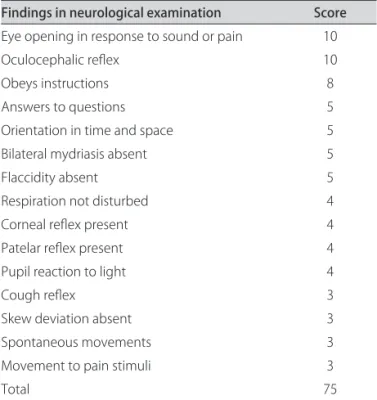
It was shown in a study that there is a critical value corresponding to 15 points as all the patients in the study whose scores after assessment were less than this value died10 (Tables 3 and 4).
Glasgow coma scale (GCS)
he Glasgow coma scale (GCS) is extensively used throughout the world by physicians and other health pro-fessionals. Since its introduction in 197411,12, it has proved
to be particularly suitable for characterizing the severity of changes in consciousness, especially in patients sufer-ing from traumatic brain injury. At the time the scale was published, the authors, Jennett and Teasdale, believed that the lack of guidelines for describing patients with altered consciousness caused diiculties with communication be-tween diferent centers and also made it diicult to com-pare groups of patients treated using diferent methods14.
Unlike Plum and Posner1, who concentrated on
explain-ing precisely and accurately the diagnosis of stupor and coma, Jennett and Teasdale limited themselves to devel-oping a practical method for obtaining an overall idea of the level of consciousness5.
he total score on the Glasgow scale is obtained by as-sessing the following three parameters: eye opening, best verbal response and best motor response. he score varies between 3 and 15 points, and values of 8 or less correspond to serious conditions requiring intubation11,12 (Table 5).
Bozza-Marrubini scale
In 1983, Bozza-Marrubini reviewed the existing sys-tems for classifying coma13. First, he separated them into
systems using scales and those using scores. In systems in the former category (those using scales), the clinical parameters are considered to be dependent and
continu-Table 3. Moscow coma scale: quantitative scale of alterations observed in neurological examination10.
Findings in neurological examination Score
Eye opening in response to sound or pain 10
Oculocephalic relex 10
Obeys instructions 8
Answers to questions 5
Orientation in time and space 5 Bilateral mydriasis absent 5
Flaccidity absent 5
Respiration not disturbed 4 Corneal relex present 4 Patelar relex present 4 Pupil reaction to light 4
Cough relex 3
Skew deviation absent 3 Spontaneous movements 3 Movement to pain stimuli 3
Total 75
Table 4. Moscow coma scale: classiications of consciousness levels10.
Consciousness level
Neurologics indings
VOR Open eyes to pain commandsFollows questionsAnswers Oriented bilateral mydriasisNon reactive Atonia
Total conscious + + + + + – –
Moderate torpor + + + + – – –
Deep torpor + + + – – – –
Vegetative state + + – – – – –
Moderate coma + – – – – – –
Deep coma – – – – – – +
Irreversible coma – – – – – + +
VOR: vestibulo-ocular relex.
ous; for example, a verbal response cannot be viewed as separate from a motor response, as this would theoret-ically result in diferent levels of consciousness. In sys-tems in the latter category (those using scores), this prem-ise is no longer valid, as diferent aspects of the patient are analyzed, a score is assigned for each of these and the scores are then added to give a number correspond-ing to the patient’s clinical condition. Accordcorrespond-ing to Boz-za-Marrubini13, the correct approach would be to use a
who have the greatest chance of beneitting from them23.
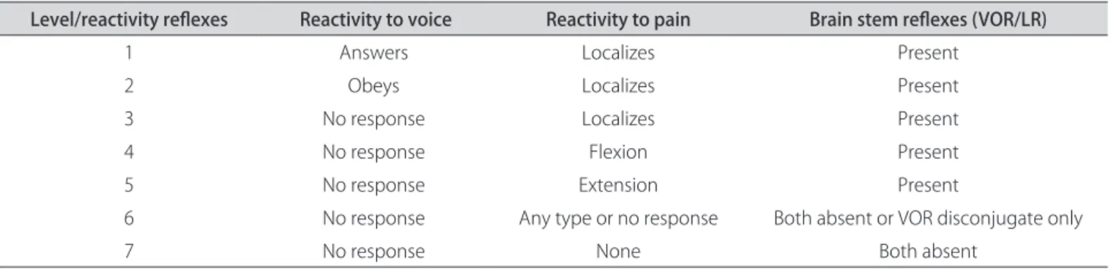
Based on this, he proposed a system of scales made up of 7 levels for classifying organic brain damage, which are described as: [1] he highest level, in which the patient is able to speak and obey commands, obviously not intu-bated; [2] Patient obeys commands. Eye opening is there-fore only one criterion and can be substituted by a similar one if, for example, the patient has an eyelid edema that makes eye opening impossible; [3] Patient is able to locate pain; [4] From this level on, the patient is no longer able to locate a painful stimulus but responds to it with abnor-mal lexion; [5] Limb extends abnorabnor-mally in response to pain; [6] From this level on, there is no brainstem relex or only disconjugate vestibulo-ocular relex, which does not happen in the levels above. In addition, there is no pat-tern of response to painful stimuli or no response. Stage preceding brain death; [7] Complete absence of response
to pain, and no brainstem relex. Stage corresponding to brain death13 (Table 6).
Full Outline UnResponsiveness - FOUR Score
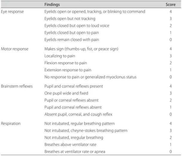
In 2005, Wijdicks et al. published a new coma scale, the FOUR score14. It involves assessment of the
follow-ing four components, each on a scale with a maximum of four: eye response, motor response, brainstem relex-es and rrelex-espiration. his scale is able to detect conditions such locked-in syndrome and the vegetative state, which are not detected by the GCS.
When assessing eye response, the best of three at-tempts is used. E4 indicates at least three voluntary move-ments in response to the examiner’s commands (for ex-ample, asking the patient to look up, look down and blink twice). If the patient’s eyes are closed, the exam-iner should open them and observe whether they track Table 5. Glasgow coma scale11.
Clinical parameter Points
Eyes Open Spontaneously 4
To verbal command 3
To pain 2
No response – 1
Best motor response To verbal command Obeys 6
To painful stimulus Localizes pain 5
Flexion withdrawal 4
Flexion abnormal (decorticate rigidity) 3 Extension (decerebrate rigidity) 2
No response 1
Best verbal response Oriented 5
Confused 4
Inappropriate speech 3
Incomprehensible speech 2
No response 1
Total (3-15 points)
Table 6. Bozza-Marrubini’s scale13.
Level/reactivity relexes Reactivity to voice Reactivity to pain Brain stem relexes (VOR/LR)
1 Answers Localizes Present
2 Obeys Localizes Present
3 No response Localizes Present
4 No response Flexion Present
5 No response Extension Present
6 No response Any type or no response Both absent or VOR disconjugate only
7 No response None Both absent
a moving object or the examiner’s index inger. If one of the eyes is afected by eyelid edema or trauma, the re-sponse of the healthy eye alone may be used. If there are no horizontal movements, check for vertical movements. E3 indicates the absence of any tracking movement with eyes open. E2 indicates eye opening in response to a loud sound, and E1 corresponds to eye opening in response to a painful stimulus. E0 indicates no eye opening even af-ter a painful stimulus14.
Motor response is assessed preferably at the upper extremities. A test is performed to determine if the pa-tient is able irst to abduct their thumb and simultaneous-ly lex their four ingers (thumbs up), lex their ingers and thumb together (ist) and then extend just their index and middle ingers (V sign). If they are able to do this, the pa-tient is classiied as M4. If the papa-tient’s only response is localization to pain, they are classiied as M3. Flexor re-sponse to pain is classiied as M2, extensor rere-sponse as M1 and a complete lack of response or generalized myo-clonus status is classiied as M014.
he brainstem relexes tested are the pupillary and corneal relexes. he corneal relex is tested by applying two or three drops of sterile saline solution from a dis-tance of 4 to 6 inches (to minimize corneal trauma as a
result of repeated examinations). Cotton swabs can also be used. When both (pupillary and corneal) relexes are absent, the cough relex is also tested. B4 indicates the presence of pupillary and corneal relexes. B3 indicates that one of the pupils is wide and ixed. B2 indicates the absence of one of the relexes. B1 corresponds to the ab-sence of both relexes. B0 indicates that all the relexes are absent, including the cough relex14.
For respiration, non-intubated patients with a nor-mal breathing pattern are classiied as R4, non-intubat-ed patients with a Cheyne-Stokes breathing pattern as R3 and non-intubated patients with an irregular breath-ing pattern as R2. Patients on mechanical ventilation are classiied in R1 if they are breathing above the ventilator rate (indicating that the respiratory center is still work-ing) and in R0 if they are breathing at the ventilator rate or have apnea14.
If the patient scores zero in all the categories, the ex-aminer should consider the possibility of a diagnosis of brain death14 (Table 7).
DISCUSSION
Coma scales have been developed throughout the world to standardize both the communication between Table 7. Full Outline of UnResponsiveness - FOUR Score14.
Findings Score
Eye response Eyelids open or opened, tracking, or blinking to command 4 Eyelids open but not tracking 3 Eyelids closed but open to loud voice 2 Eyelids closed but open to pain 1 Eyelids remain closed with pain 0
Motor response Makes sign (thumbs-up, ist, or peace sign) 4
Localizing to pain 3
Flexion response to pain 2
Extension response to pain 1 No response to pain or generalized myoclonus status 0
Brainstem relexes Pupil and corneal relexes present 4
One pupil wide and ixed 3
Pupil or corneal relexes absent 2 Pupil and corneal relexes absent 1 Absent pupil, corneal, and cough relex 0
members of health teams and the assessment of the clini-cal evolution of severely afected patients. By far the most commonly used scale is the Glasgow coma scale. Vari-ous other scales have been developed, some of which are seldom used outside their country of origin14. Examples
of these are the Innsbruck coma scale6 and the Japanese
scale15. hey all generally involve assessing the patient and
awarding a score that gives an overall idea of their level of consciousness.
he main advantage of the Jouvet scale is that it allows anatomo-clinical correlations to be established. However, the scale is complex, diicult to use and time-consuming and thus unsuitable for emergencies such as TBI. Com-pared with the Glasgow scale, it is more sensitive for lev-els of consciousness that are close to normal.
he Moscow scale is rarely used nowadays. Only one paper about this scale was found in our review of the lit-erature10. In the study described in the paper, 58
traumat-ic brain injury (TBI) vtraumat-ictims with Glasgow scores of three were also assessed with the Moscow scale. Of these 58 pa-tients, only 69% died, whereas all those who had scores of less than 15 on the Moscow scale died. his inding led to the deinition of a critical value of 15 points, below which the prognosis is brain death. he study concluded that the Moscow scale has good predictive value10.
he Glasgow scale was developed using simple pa-rameters for the speciic purpose of allowing less experi-enced doctors and other health professionals to produce an accurate report of a patient’s state of consciousness. Nevertheless, it has become the target of various criti-cisms in recent decades, and a number of studies have al-ready described its strengths and weaknesses5. Eye
open-ing, for example, is considered to indicate wakefulness, but it should be remembered that eye opening does not mean that the content of consciousness is intact (as in a persistent vegetative state). he fact is that the Glas-gow scale does not provide either a suicient number of or suitable tools to cover the whole spectrum of chang-es in consciousnchang-ess. Rather, it is limited to diagnosis of the state of coma and does not allow more precise dis-tinctions between the other states of consciousness to be made.5 Because of this its usefulness for inferring a
prog-nosis is limited, especially in patients with intermediate scores. As it lacks precision, the Glasgow scale is not suit-able for monitoring changes of certain magnitudes in the state of consciousness5,14,16,17.
In addition, Jennett and Teasdale speciied that the score should be calculated based on examination of the patient six hours after the traumatic brain injury18. Patients
with TBI are stabilized much sooner, and neuromuscular blocking drugs are often used to make it easier to trans-port and intubate agitated patients. All these circumstanc-es interfere in the validity of the initial score obtained19-21.
Another problem when applying the Glasgow scale is that the verbal component cannot be tested in intubat-ed patients. Some physicians use the lowest score possi-ble1, while others infer the verbal response based on
oth-er indings of the neurological examination. Furthoth-ermore, abnormal brainstem relexes, altered breathing patterns or the need for mechanical ventilation can indicate the severity of the coma, but the Glasgow scale does not cov-er these parametcov-ers14.
he Bozza-Marrubini scale was an attempt to com-bine the standardized language of the Glasgow scale with exact descriptions of each clinical level. It is worth high-lighting the eforts made by Bozza-Marrubini to ind al-ternative ways to assess the same item, as in the case of the response to a verbal command, where the commands can include the alternatives “close your eyes” and “stick your tongue out”, as seen in level 2 of the scale.13
Lastly, the FOUR score is easy to use and provides more neurological details than the Glasgow score, partly because it includes brainstem relexes. Another advantage is that it allows diferent stages of herniation and other disorders such as locked-in syndrome and the vegetative state to be identiied. It does not include verbal response and therefore has a higher predictive value for patients in intensive care14. A recent study showed that the scale can
be used successfully by diferent professionals from out-side the ield of neurosciences22.
Although scales are of tremendous importance in as-sessing disorders of consciousness, it should be stressed that instruments intended to assess something as com-plex as consciousness naturally have certain limitations. For some authors the items on a scale and the values as-signed to them are still not able to consistently specify and quantify in all possible clinical coma situations the extent to which the various cerebral cortical functions related to the level of consciousness have been afected1,5,7,14.
ACKNOWLEDGMENTS – We would like to thank Professor Elvira Kim (Languages Department of Federal University of Paraná) for translation of reference 10.
REFERENCES
1. Posner JB, Saper CB, Schif ND, Plum F. Plum F, Posner JB. Diagnosis of Stupor and Coma. 4th edition. Contemporary Neurology Series. Oxford
Uni-versity Press, New York, 2007.
2. Bateman D. Neurological assessment of coma. J. Neurol Neurosurg Psy-chiatry 2001;71(Suppl 1):S13-S17.
3. Stevens RD, Bhardwaj A. Approach to the comatose patient. Crit Care Med 2006;34:31-41.
4. Liao YJ, So YT. An approach to critically ill patients in coma. West J Med 2002;176:184-187.
5. Segatore M, Way C. The Glasgow coma scale: time for change. Heart Lung 1992;21: 548-557.
6. Benzer A, Mittrtschifthaler G, Marosi M, et al. Prediction of non-survival after trauma: Innsbruck Coma Scale. Lancet 1991;338: 977-978.
8. Adukauskiene D, Budryte B, Karpec D. Coma: etiology, diagnosis and treatment. Medicina (Kaunas) 2008;44:812-819.
9. Jouvet M. Coma and other disorders of consciousness. Handbook of Clinical Neurology. North-Holland Publishing Company. Amsterdam, 1969. 10. Shakhnovich AR, Mamadaliev AM, Abakumova LIa. The prognosis of the
outcomes of comatose states in the irst 24 hours following craniocerebral trauma. Zh Vopr Neirokhir Im N N Burdenko 1991;6:11-12.
11. Teasdale G, Jennett B. Assessment of coma and impaired consciousness: a practical scale. Lancet 1974;2:81-84.
12. Jennett B. The history of Glasgow coma scale: an interview with Professor Bryan Jennett. Interview by Carole Rush. Int J Trauma Nurs 1997;3: 114-118. 13. Bozza-Marrubini M. Classiications of coma. Intensive Care Med 1984;10:
217-226.
14. Wijidicks E, Bamlet WR, Maramatton BV, Manno EM, McClelland RL. Vali-dation of a new coma scale: The FOUR Score. Ann Neurol 2005;58: 585-593. 15. Sakai K, Iwahashi K, Terada K, Gohda Y, Sakurai M, Matsumoto Y. Outcome
after external decompression for massive cerebral infarction. Neurol Med Chir (Tokyo) 1998;38:131-135.
16. Starmark JE, Stalhammar D, Holmgren E, Rosander B. A comparison of the glasgow coma Scale (GCS) and the reaction level scale (RLS85). J Neu-rosurg 1988;69: 699-706.
17. Balestreri M, Czosnyka M, Chatield DA, et al. Predictive value of Glasgow coma scale after brain trauma: change in trend over the past ten years. J Neurol Neurosurg Psychiatry 2004;75:161-162.
18. Jennett B, Teasdale G, Galbraith S, et al. Severe head injuries in three coun-tries. J Neurol Neurosurg Psychiatry 1977;40:291-298.
19. Marion DW, Carlier PM. Problems with inicial Glasgow coma scale as-sessment caused by prehospital treatment of patients with head injuries: results of a national survey. J Trauma;1994;36:89-95.
20. Wiese MF. British hospitals and diferent versions of the Glasgow coma scale: telephone survey. BMJ 2003;327:782-783.
21. Stuke L, Dias-Arrastia R, Gentilello LM, Shai S. Efect of alcohol on Glasgow coma scale in head-injured patients. Ann Surg 2007;245:651-655. 22. Stead LG, Wijidicks E, Bhagra A, et al. Validation of a new coma scale,