83 Lundgren MSFS et al. Extracranial doses in radiosurgery for brain tumors
Radiol Bras. 2012 Mar/Abr;45(2):83–86
Extracranial doses in patients submitted to stereotactic
radiosurgery for brain tumors
*
Doses extracranianas em pacientes submetidos a radiocirurgia estereotáxica para tumores cerebrais
Maria da Salete Fonseca dos Santos Lundgren1, Helen Jamil Khoury2, Sérgio Azevedo3
Objective: To estimate extracranial doses on eyes, thyroid, chest and pelvis in patients submitted to radiosurgery with 6 MV linear accelerator. Materials and Methods: The present study evaluated 11 patients, 7 of them with primary, and 4 with secondary brain tumors. In the latter group, 2 patients had two lesions. Thermoluminescent dosimeters were utilized to estimate the extracranial dose. Radiosurgery cones ranges between 1.50 and 3.75 cm and doses between 1300 and 2000 cGy. Results: Mean patients’ age was 52 years, and 63.6% of them were women and 36.4%, men. Lesion locations were the following: right acoustic nerve (1), frontal (2), parietal (5), right occipital (1), cerebellum (2) and parasagittal (2). Mean received doses were the following: 5.1 cGy between the eyes; 4.8 cGy in the right eye; 6.5 cGy in the left eye; 4.2 cGy in the thyroid; 1.65 cGy in the chest; and 0.45 cGy in the pelvis. Conclusion: The results demonstrate that that although the eye doses do not exceed the tolerance limits for occurrence of lens opacity, it is important that the risks associated with radiation doses are taken into consideration by radiotherapists in the planning of cranial radiosurgery procedures.
Keywords: Brain tumor; Radiotherapy; Radiosurgery; Dosimetry.
Objetivo: Estimar a dose extracraniana nos olhos, tireoide, tórax e pelve em pacientes submetidos a radiocirurgia com acelerador linear de 6 MV. Materiais e Métodos: Foram avaliados 11 pacientes com tumores cerebrais primários (7 pacientes) e secundários (4 pacientes), sendo que dois destes apresentavam duas lesões. Para a estimativa da dose extracraniana, foram utilizados dosímetros termoluminescentes. Foram utilizados cones de 1,50 a 3,75 cm e as doses de radiação variaram de 1300 a 2000 cGy. Resultados: A idade média dos pacientes foi de 52 anos, sendo 63,6% do sexo feminino e 36,4% do sexo masculino. As localizações das lesões foram: nervo acústico direito (1), frontal (2), parietal (5), occipital direito (1), cerebelar (2) e parassagitais (2). Os valores médios das doses recebidas na região entre os olhos foram de 5,1 cGy; no olho direito, de 4,8 cGy; no olho esquerdo, de 6,5 cGy; na tireoide, de 4,2 cGy; no tórax, de 1,65 cGy; e na pelve, de 0,45 cGy. Conclusão: Estes resultados mostram que embora as doses não ultrapassem os limites de tolerância para ocorrência da opacidade do cristalino, é importante que os médicos radio-terapeutas considerem os riscos de dose de radiação nessas regiões durante o planejamento de procedimentos de radiocirurgia craniana.
Unitermos: Tumor cerebral; Radioterapia; Radiocirurgia; Dosimetria.
Abstract
Resumo
* Study developed at Instituto de Radioterapia Waldemir Mi-randa, Recife, PE, Brazil.
1. PhD, MD, Radiotherapist at Instituto de Radioterapia Wal-demir Miranda, Head of the Unit of Radiotherapy, Hospital Uni-versitário Oswaldo Cruz, Recife, PE, Brazil.
2. PhD, Full Professor, Department of Nuclear Energy, Univer-sidade Federal de Pernambuco (UFPE), Recife, PE, Brazil.
3. Medical Physicist, Instituto de Radioterapia Waldemir Mi-randa, Recife, PE, Brazil.
Mailing Address: Dra. Maria da Salete Fonseca dos Santos Lundgren. Instituto de Radioterapia Waldemir Miranda. Rua Pa-cífico dos Santos, 60, Derby. Recife, PE, Brazil, 52010-030. E -mail: [email protected]
Received June 7, 2011. Accepted after revision February 10, 2012.
Lundgren MSFS, Khoury HJ, Azevedo S. Extracranial doses in patients submitted to stereotactic radiosurgery for brain tumors. Radiol Bras. 2012 Mar/Abr;45(2):83–86.
0100-3984 © Colégio Brasileiro de Radiologia e Diagnóstico por Imagem
ORIGINAL ARTICLE
collimators scattering, as well as scattering in the patient(4,5). The estimation of the dose received by the patient in extracranial or-gans is relevant, particularly for those sub-mitted to radiosurgery and with long life expectancy. Secondary effects resulting from radiation may manifest specially in pediatric patients and patients with benign brain diseases.
It is important to highlight that dosim-etry in patients submitted to radiosurgery is of paramount relevance, considering the possibility of occurrence of biological ef-fects. The eyes deserve enhanced attention, considering the possibility of crystalline lens opacification, since the threshold for ing ration into a sterotactically defined
intracranial target. Such procedure is uti-lized in the treatment of small-sized lesions measuring < 4 cm and is performed with Cobalt-60 gamma beams or high-energy X-ray beams (linear accelerator (acelerador linear)(1,2). The advantage of this type of procedure is the application of high doses of radiation on the target to br treated with minimal radiation delivered to surrounding structures(3). However, parts of the patient’s body located out of the area to be treated may receive secondary radiation because of scattering originating from parts of the apparatus and room walls, radiation leaks through the shielding of the source head,
INTRODUCTION
ioniz-84
Lundgren MSFS et al. Extracranial doses in radiosurgery for brain tumors
Radiol Bras. 2012 Mar/Abr;45(2):83–86 cataract radioinduction is only 0.5 Gy, in
acute and fractionated exposures(6–9). The presente study was aimed at esti-mating the dose received in extracranial regions and organs such as eyes, thyroid, chest and pelvis.
MATERIALS AND METHODS
The present study evaluated 11 patients submitted to radiosurgery in Instituto de Radioterapia Waldemir Miranda – Centro de Radiocirurgia de Pernambuco, Recife, PE, Brazil, utilizing a Varian Clinac 600C 6 MV linear accelerator, with a Radionics radiosurgery system of circular collimators. The study sample included seven female and four male patients, whose minimum age was 26 years and maximum age was 83 years. The patients were treated for primary brain tumors (seven) and secondary brain tumors (four). Among the patients with secondary tumors, two presented two brain lesions.
For estimating the dose in extracranial regions, TLD-100 thermoluminescent do-simeters encapsulated in pairs in numeri-cally identified plastic envelopes. For each patient and for each treated lesion, enve-lopes with dosimeters were placed on the following regions: between the eyes, on the external corners of both eyes, on the region of the thyroid gland, on the chest (on the lower third of the sternum) and on the pel-vis (hypogastric region). After the dosim-eters positioning according to the tumor location and size, the treatment was per-formed with doses ranging from 1300 cGy to 2000 cGy. The cones diameter ranged from 1.50 cm and 3.75 cm. The number of fields (arches) ranged between three and six, and the angulation of the accelerator head, between 45° and 150°. Once the ra-diosurgery was completed, the TLDs were removed and taken to the Laboratory of Thermoluminescent Dosimetry of the Group of Dosimetry and Nuclear Instru-mentation, Department of Nuclear Energy, Universidade Federal de Pernambuco, for reading. As each envelope contained two dosimeters, two reading were obtained for each point. The mean value was deter-mined and a net value was obtained by subtracting the reading of the non-irradi-ated dosimeter (background reading). The
value of the net reading obtained at each point was converted into dose by utilizing the dosimeters calibration curve previously determined with dosimeters irradiation in the linear accelerator, with previously known doses. All the patients were given explanations on the present study and signed a term of free and informed consent.
RESULTS
Table 1 presents the characteristics of all the patients included in the present study, as well as the tumor sites. Based on such data, one observes that 63.6% of the pa-tients were women and 36.4% were men; and the mean patients’ age was 52 years.
Table 2 shows the location of the tu-mors, the size of the utilized cone, values of total treatment doeses, as well as values of the doses observed in the evaluated ex-tracranial regions, as follows: between the eyes, external corner of righ eye, external corner of lef eye, thyroid, chest (between the breasts) and pelvis (below the umbilicar scar). The locations of the lesions were the following: right acoustic nerve (1), frontal (2), parietal (5), right occipital (1), cerebel-lar (2) and parasagittal (2), which are re-gions whre brain tumors are typically found.
Figure 1 shows the distribution of val-ues of radiation doses received in extrac-ranial regions according to the Box & Whiskers chart. In such type of chart, the rectangle extremities represent the first and third quartiles of the data frequency distri-bution, and the bar crossing the rectangle
represents the median. The lower extrem-ity represents the minimum value and the upper, the maximum value. The external circles represent the data set outliers which, generally, are represented on the chart, but are not computed in the calculations of the mean and median.
Table 3 shows the mean values and stan-dard deviation for doses obtained at the different evaluated extracranial sites.
DISCUSSION
The mean patients’ age is similar to the one observed in the study developed by Yu et al.(5), who have studied the extracranial dose in 104 patients submitted to Gamma Knife radiosurgery. As their results are con-sidered, it is observed that the mean value of radiation doses received by the region between the eyes was 5.1 cGy, with a maxi-mum dose of 17.46 cGy. For the right eye region, the mean dose was 4.8 cGy and the maximum dose, 12.06 cGy. The left eye region received a mean dose of 6.5 cGy and maximum dose of 32.3 cGy. The dose in the eyes, as expected, depends on the position of the organ in relation to the primary ra-diation beam. The analysis of the data has shown that the mean dose value in the re-gions of the eyes ipsilateral to the lesion was 5.3 ± 2.4 (cGy) and in the region of the eyes contralateral to the lesion was 3.9 ± 1.1 (cGy). Such results are similar to the ones found by Ma et al.(7), corresponding to a mean dose value in the region of the eyes ipsilateral to the lesion of 7.6 ± 0.6 (cGy), and lower than the values found by
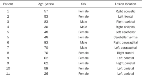
Table 1 Characteristics of patients submitted to radiosurgery and tumors location.
Patient
1 2 3 4 5 5 6 7 8 9 9 10 11
Age (years)
57 53 83 30 48 48 83 70 70 62 62 59 26
Sex
Female Female Male Male Female Female Male Male Female Female Female Female Female
Lesion location
Right acoustic Left frontal Right parietal Right occipital Left cerebellar Cerebellar vermis Right parasagittal Left parasagittal
Right frontal Left parietal Right parietal
85 Lundgren MSFS et al. Extracranial doses in radiosurgery for brain tumors
Radiol Bras. 2012 Mar/Abr;45(2):83–86
eye with the treated area, the total radiation dose applied and the utilized arches angu-lation. The values of doses observed in the present study are similar to data in the lit-erature and are below the threshold for occurence of opacity in the human crystal-line lens, which ranges between 0.5 and 2.0 Gy, for a single exposure(9). The study de-veloped by Yu et al.(5) presented a mean value of 24 cGy in the lateral region of the eyes for 104 patients submitted to Gamma Knife radiosurgery. A study developed by Novotný Jr et al.(10) also presents mean dose values of 22 cGy in the region of the eyes. In the thyroid, the mean dose value in the present study was 4.2 cGy, with the maximum value of 12.6 cGy. The analysis of data on Table 2 demonstrates that the dose in the thyroid varies as a function of the lesion location. Treatament of tumors in the cerebellar region result in higher doses in the region of the thyroid. Such values are similar to the ones reported by Novotný Jr et al.(10) and Yu et al.(11), and lower than the ones reported by Yu et al. in a study developed in 1997(5). The mean dose value in the region of the chest was 1.65 cGy and the maximum value was 3.71 cGy. For the region of the pelvis, the mean dose value was 0.45 cGy and the maximum value was 1.54 cGy.
CONCLUSIONS
In the present study, the values of radia-tion doses observed in extracranail regions Table 2 Tumor location, cones sizes, total treatment doses and values of doses in extracranial regions.
Lesion location
Right acoustic Left frontal Right parietal Right occipital Left cerebellar Cerebellar vermis Right parasagittal Left parasagittal
Right frontal Left parietal Right parietal
Left parietal Left parietal
Cone (cm)
1.5 1.75 3.75 2.75 3.5 1.5 3.25 3.25 3.25 1.5 1.25 1.75 2
Total
1300 2000 1800 1800 1500 2000 1500 1500 1500 1800 1900 1820 1800
Between the eyes
2 4 6.92 5.14 6.29 8.86 3.35 2.55 17.46
4.05 0.55 3.31 2.43
Right eye
6 3.8 6.92 6.16 3.09 3.66 3.8 4.82 12.06
2.84 3.21 3.34 2.8
Left eye
4 4.38 6.36 3.79 5.12 5.57 3.69 3.73 5.75 32.3 2.88 5.02 2.34
Thyroid
3 3.45 5.19 3.99 12.65
5.53 2.46 1.81 4.9 3.06 3.21 2.52 3.2
Chest
0.68 1.17 1.56 1.26 3.28 2.37 1.52 1.07 1.36 3.71 1.45 1.07 0.97
Pelvis
0.06 0.35 0.31 0.25 0.56 0.79 0.3 0.13 0.29 1.54 0.54 0.42 0.33 Dose (cGy)
Table 3 Mean values and standard deviation for extracranial doses.
Sites
Between the eyes Right eye
Left eye Thyroid Chest Pelvis
Mean dose (cGy)
5.1 4.8 6.5 4.2 1.6 0.4
Standard deviation
± 4.3 ± 2.6 ± 7.8 ± 2.8 ± 0.9 ± 0.3
Figure 1. Radiation doses distribution in extracranial regions. (OC, between the eyes; OD, right eye; OE, left eye; Tir, thyroid).
86
Lundgren MSFS et al. Extracranial doses in radiosurgery for brain tumors
Radiol Bras. 2012 Mar/Abr;45(2):83–86 are lower than the values reported by
stud-ies in the literature approaching treatment with cranial Gamma Knife radiosurgery. The dose values for extracranial regions of the patient reach levels as high as 30 cGy in the region of the eyes, and 12 cGy in the region of the thyroid. The dose in the thyrois varies as a function of the lesion location. Treatment of tumors in the cer-ebellar region result in higher doses in the thyroid. Suchs results demonstrate that, although the doses in the eyes do not ex-ceed the tolerance threshold for occurence of opacity of the crystalline lens, it is im-portant that radiotherapists take the risks of radiation doses in these regions into con-sideration during the planning of cranial radiosurgery.
REFERENCES
1. Cruz JC, Salvajoli JV, Weltman E, et al. Controle de qualidade em radiocirurgia. Radiol Bras. 1997; 30:163–70.
2. Cruz JC, Segreto RA, Segreto HRC. Estudo do-simétrico de campos pequenos de raios X utili-zados em radiocirurgia com um acelerador linear de 6 MV. Rev Imagem. 2003;25:257–67. 3. Souza CH, Monti CR. Dosimetria dos cones
ra-diocirúrgicos Radionics de diâmetros de 5 mm a 50 mm para um feixe de 6 MV de um acelerador linear Mevatron MD digital. Radiol Bras. 2001; 34:95–100.
4. Kase KR, Svensson GK, Wolbarst AB, et al. Mea-surements of dose from secondary radiation out-side a treatment field.Int J Radiat Oncol Biol Phys. 1983;9:1177–83.
5. Yu C, Luxton G, Apuzzo ML, et al. Extracranial radiation doses in patients undergoing gamma knife radiosurgery. Neurosurgery. 1997;41:553–9. 6. Liang CL, Ho MW, Lu K, et al. An investigation of eye lens dose of stereotactic radiosurgery for
trigeminal neuralgia using Leksell Gamma Knife model C. J Neurosurg. 2006;105 Suppl:112–6. 7. Ma L, Chin L, Sarfaraz M, et al. An investigation
of eye lens dose for gamma knife treatments of trigeminal neuralgia. J Appl Clin Med Phys. 2000;1:116–9.
8. International Commission on Radiological Pro-tection. The 2007 Recommendations of the Inter-national Commission on Radiological Protection. ICRP Publication 103. Ann ICRP. 2007;37:1– 332.
9. International Commission on Radiological Pro-tection. ICRP ref 4825-3093-1464. Statement on tissue reactions. Approved by the Commission on April 21, 2011.
10. Novotný J Jr, Novotný J, Hobzová L, et al. Trans-portation dose and doses to extracranial sites during stereotactic radiosurgery with the Leksell Gamma Knife. Stereotact Funct Neurosurg. 1996; 66:170–83.