3 8 0 Arq Bras Oftalmol. 2013;76(6):380-2
Relato de Caso |
Case RepoRtINTRODUCTION
Pigmented paravenous retinochoroidal atrophy (PPRCA) is a rare ocular disease that is characterized by paravenous retinal pigment epithelium (RPE) and choroidal atrophy often with overlying intrare-tinal bone spicule pigment deposition along reintrare-tinal veins(1). The cause
is unknown, and the course of the degeneration is unpredictable. Patients are usually asymptomatic, and the diagnosis is based on the characteristic fundus appearance. Fluorescein angiography, electro-physiological and visual ield tests may conirm the diagnosis(1).
Few cases have been reported. Most of them were sporadically and detected in male patients(2). While some reports have suggested
that PPRCA has a congenital origin(3), others suggest a primary retinal
degeneration(2). It has also been associated with mutations in the
human CRB1 gene, which is known to cause other retinal dystrophies, such as retinitis pigmentosa and Coats-like exudative vasculopathy(4).
An association with Norrie´s syndrome was also described. Finally, an inlammatory cause, such as syphilitic etiology, has been suggested by Hsing-Hsiang(5).
As a rare condition, there is scant information in the literature re garding anatomical indings and about how the retinal layers are involved in PPRCA. In fact, it seems to exist a considerable variability in the extent and degree to which the retina is afected(2). In the present
case report, we provide new indings and a detailed description of the anatomical involvement of the inner retina using high-resolution retinal imaging [Spectral-Domain Optical Coherence Tomography (SD-OCT)] to evaluate a patient with PPRCA.
Pattern of inner retinal layers involvement in pigmented paravenous retinochoroidal
atrophy as determined by SD-OCT: case report
Padrão de envolvimento das camadas retinianas internas na atroia retinocoroidiana pigmentada
paravenosa determinado pelo SD-OCT: relato de caso
Daniela laura Melo Junqueira1, Flavio Siqueira SantoS lopeS1,2, luíS GuStavo Biteli1,2, tiaGo SantoS prata1,2
Submitted for publication: August 02, 2013. Accepted for publication: October 07, 2013
Study carried out at Hospital Medicina dos Olhos, São Paulo, SP, Brazil.
1 Physician, Hospital Medicina dos Olhos, São Paulo (SP), Brazil;
2 Physician, Department of Ophthalmology, Universidade Federal de São Paulo, São Paulo (SP),
Brazil.
Funding: No specific financial support was available for this study.
Disclosure of potential conflicts of interest: D.L.M.Junqueira, None; F.S.S.Lopes, None; L.G.Biteli, None; T.S.Prata, None.
Correspondence address: Tiago Santos Prata. Department of Ophthalmology. Universi-dade Federal de São Paulo. Rua Botucatu, 821 - São Paulo (SP) - 04023-062 - Brazil E-mail: [email protected]
ABSTRACT
Pigmented paravenous retinochoroidal atrophy is an ocular disease characterized by outer retina and choroidal atrophy often with overlying intraretinal bone spicule pigment deposition along the retinal veins. As a rare condition, there is scant infor-mation in the literature regarding the pattern of inner retinal layers involvement. We present a case of a 41-year-old white man initially referred for a glaucoma evaluation. Fundoscopy revealed patches of retinochoroidal atrophy and light pigmentation extending from the optic nerve head along the inferior-temporal retinal veins in both eyes. Using different spectral-domain optical coherence tomography (SD-OCT ) protocols we identified a significant thinning of the inner retinal layers along the inferior-temporal veins, but with a lucid interval surroun-ding the optic nerve head. Standard automated perimetry revealed a superior absolute arcuate scotoma sparing the central fixation (good structure-functional correlation). This pattern of inner retinal layers involvement was not previously described. We believe SD-OCT added significantly to the anatomical description of this case. Physicians should consider these new anatomical findings and correlate them with functional status while assessing these patients.
Keywords: Atrophy; Retinal ganglion cells; Choroid/pathology; Retina/pathology; Retinal nerve fiber layer; Tomography, optical coherence; Visual field; Case report
RESUMO
Atrofia retinocoroidiana pigmentada paravenosa é uma doença ocular caracterizada por atrofia localizada da coroide e da retina externa associada a áreas de pigmentação em espícula óssea depositada ao longo das veias retinianas. Como é uma condição rara, há pouca informação na literatura sobre o padrão de envolvimento das camadas mais internas da retina. Relatamos o caso de um homem branco, de 41 anos, encaminhado incialmente para avaliação de glaucoma. Apresentava à fundoscopia áreas de atrofia retinocoroidiana com pigmentação leve sobrejacente, estendendo-se desde o disco óptico e seguindo ao longo da veia temporal inferior da retina em ambos os olhos. Por meio de diferentes protocolos da tomografia de coerência óptica de domínio espectral (SD-OCT ) identificamos um afinamento significante das camadas internas da retina ao longo da veia temporal inferior, mas com uma área de intervalo lúcido ao redor do disco óptico. A perimetria automatizada acromática revelou um escotoma arqueado superior absoluto, poupando a fixação central em ambos os olhos e correspondendo às áreas de atrofia ao longo das veias retinianas (boa correlação anátomo-funcional). Este padrão de envolvimento das camadas retinianas internasnão havia sido descrito anteriormente. Acreditamos que o SD-OCT contribuiu significativamente para a des-crição anatômica desse caso e que estes novos achados devam ser considerados e correlacionados com o estado funcional ao avaliar esses pacientes.
Junqueira DLM, et al.
3 8 1 Arq Bras Oftalmol. 2013;76(6):380-2 CASE REPORT
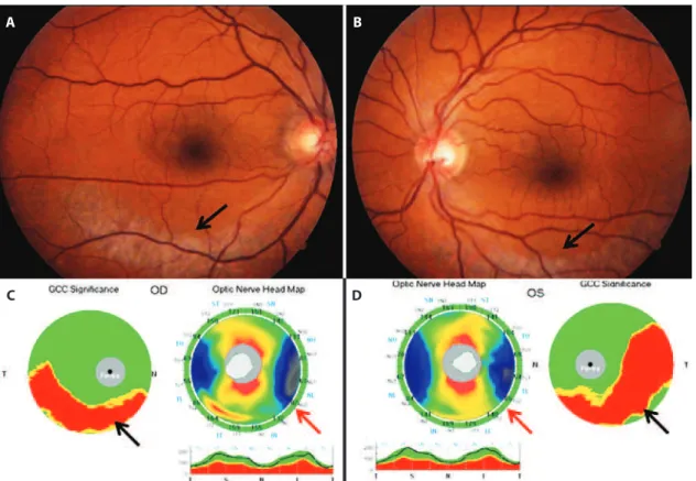
A 41-year-old white man was initially referred for a glaucoma evaluation, with no signiicant complaints. His past medical history revealed chemotherapy and radiotherapy treatment for a lymphoma two years earlier. Ophthalmological and familiar history was unre-markable. During ophthalmologic examination he presented with a best-corrected visual acuity of 20/20 in both eyes (dynamic refraction of -2.00 diopters OU). Slit-lamp biomicroscopy was unremarkable. Gonioscopy disclosed a normal open angle and the intraocular pres-sure was 14 mmHg OU [central corneal thickness of 540 µm (OD) and 538 µm (OS)]. Both fundi showed patches of retinochoroidal atrophy and light pigmentation along the inferior-temporal retinal veins (Figures 1A and B). The optic nerve head was well deined with 0.5 cup-to-disc-ratio and a healthy neuroretinal rim 360o OU.
Functional evaluation (visual ield test) by standard automated perimetry (Humphrey SITA-Standard 24-2, Carl Zeiss Meditec, Dublin, CA) revealed a superior absolute arcuate scotoma, sparing the central ixation in both eyes and corresponding to the areas of atrophy along the inferior-temporal retinal veins (Figures 2A and B). A detailed struc-tural analysis was undertaken using the RTVue-100 SD-OCT (Optovue Inc, Fremont, CA). With the advent of this technology, a signiicant improvement in imaging resolution was achieved (axial resolution of 5 μm in tissue and a scan speed of 26,000 A-scans/second), allowing segmentation and better identiication of each retinal layer(6). For
this speciic case, two scan protocols were chosen: the peripapillary retinal nerve iber layer (RNFL) scan and the macular ganglion cell complex (GCC) scan, which comprises three inner retinal layers: the RNFL, ganglion cell layer and inner plexiform layer(6). SD-OCT results
revealed a signiicant thinning of the inner retinal layers (as assessed by the GCC protocol) along the inferior-temporal veins OU (average
superior-inferior GCC thickness diference was 32.1 µm). However, the circum-peripapillary RNFL was preserved, forming a lucid interval sur-rounding the optic nerve head (Figures 1C and D). Typical outer retina indings (previously described in the literature) were also observed. There was a good structure-functional correlation between the in-ferior inner retinal layers involvement (Figures 2C) and the superior functional loss documented (Figures 2A and B).
A complete systemic work-up was undertaken, including com-plete blood cell counts, serum electrolytes, serum protein electro-phoresis and erythrocyte sedimentation rate (all within the normal range). Tuberculin skin test was negative. There was no serologic evidence of syphilis, toxoplasmosis, systemic lupus eritematosis and cytomegalovirus.
DISCUSSION
Pigmented paravenous retinochoroidal atrophy is an asympto-matic and slowly progressive disease with respect to the loss of peripheral retinal function, usually detected in a routine examination. The diagnosis of PPRCA is usually made by the characteristic fundus appearance and other causes including chorioretinal degeneration and inlammatory diseases must be ruled out(7). Classically, the main
sites of injury are the outer retinal layers and choroid, with RPE and choroidal atrophy(1). In this case report, we have demonstrated a
signiicant thinning of the inner retinal layers along the region of paravenous retinochoroidal atrophy (approximately 30% thinner compared to the unafected region) with a lucid interval surrounding the optic nerve head. In addition, these anatomical indings showed good correlation with patient’s functional loss. To the best of our kno-wledge, this is the irst report describing the pattern of inner retinal layers involvement in PPRCA with SD-OCT.
A
C
B
D
Pattern of inner retinal layers involvement in pigmented paravenous retinochoroidal atrophy as determined by SD-OCT: case report
3 8 2 Arq Bras Oftalmol. 2013;76(6):380-2
Looking carefully at the data published about the use of high-re-solution imaging in cases o PPRCA, we found only two case reports in the literature. Diferently form ours, the main focuses of these two publications were a detailed investigation of the outer retina(8) and
the fovea(9). Fleckenstein et al used the SD-OCT to assess lines of
increased fundus autoluorescence with a crescent-like distribution surrounding the area of RPE atrophy in a case of PPRCA(8). These lines
corresponded to the junction between a zone with preserved retinal layers on the SD-OCT scan and a zone where the presumed external limiting membrane appeared to rest directly on the RPE layer. More recently, Ghosh et al.(9) reported on the use of the SD-OCT in a case of
PPRCA, showing a lamellar macular hole in the involved eye. As none of these two cited papers reported on the GCC and peripapillary RNFL anatomy, our indings cannot be directly compared to them. However, we believe that by using high-resolution imaging, these papers as a group add signiicantly to the evolving knowledge about the retinal involvement in PPRCA.
In summary, using diferent SD-OCT protocols to investigate the pattern of inner retina involvement in PPRCA, we found a signiicant thinning of the inner layers, forming a lucid interval surrounding the optic nerve head. We believe SD-OCT added signiicantly to the anatomical description of this case, as this pattern of inner retina involvement was not previously described. From now on, we believe that these new indings should be considered and correlated with functional status while assessing patients with PPRCA.
REFERENCES
1. Choi JY, Sandberg MA, Berson EL. Natural course of ocular function in pigmented paravenous retinochoroidal atrophy. Am J Ophthalmol. 2006;141(4):763-5. 2. Kükner AS, Yilmaz T, Celebi S, Aydemir O, Ulaş F. Pigmented paravenous re tino
-choroidal atrophy. A literature review supported by seven cases. Ophthalmologica. 2003;217(6):436-40.
3. Noble KG. Hereditary pigmented paravenous chorioretinal atrophy. Am J Ophthal-mol. 1989;108(4):365-9.
4. McKay GJ, Clarke S, Davis JA, Simpson DA, Silvestri G. Pigmented paravenous chorio-retinal atrophy is associated with a mutation within the crumbs homolog 1 (CRB1) gene. Invest Ophthalmol Vis Sci. 2005;46(1):322-8.
5. Hsing-Hsiang C. Retino-choroiditis radiata. Am J Ophthalmol. 1948;41:1485-7. 6. Seong M, Sung KR, Choi EH, Kang SY, Cho JW, Um TW, et al. Macular and peripapillary
retinal nerve iber layer measurements by spectral domain optical coherence tomo-graphy in normal-tension glaucoma. Invest Ophthalmol Vis Sci. 2010;51(3):1446-552. 7. Haustrate FM, Oosterhius JA. Pigmented paravenous retinochoroidal atrophy. Doc
Ophthalmologica. 1986;63(3):209-37.
8. Fleckenstein M, Charbel Issa P, Helb HM, Schmitz-Valckenberg S, Scholl HP, Holz FG. Correlation of lines of increased autoluorescence in macular dystrophy and pig-mented paravenous retinochoroidal atrophy by optical coherence tomography. Arch Ophthalmol. 2008;126(10):1461-3.
9. Ghosh B, Goel N, Batta S, Raina UK. SD-OCT in pigmented paravenous retinochoroidal atrophy. Ophthalmic Surg Lasers Imaging. 2012;43(3):e41-3.
A B
C