Original Article
7 8 Arq Bras Oftalmol. 2016;79(2):78-81 http://dx.doi.org/10.5935/0004-2749.20160024
INTRODUCTION
Migraine is a chronic disease involving both neurological and vas-cular system abnormalities and is characterized by single-sided, epi-sodic attacks of headache. Gastrointestinal complaints and sensitivity to bright light and loud noises may accompany this headache(1), and migraine occurs most frequently between the ages of 35 and 45 years(2). Although the etiology of migraine is unknown, there are some theo-ries for its pathogenesis. At the end of the 1930s, Graham and Wolf identiied a dilatation in the temporal artery during migraine attacks as a cause for the headache. Wolf advocated that the aura preceding migraine symptoms is due to vasospasms that reduce the blood low to the brain(3).
Several studies have shown that vascular changes occur in the ocular system during migraine attacks. Cerebral blood low in the occi-pital hemisphere has been shown to diminish during migraine attacks, especially in migraine with aura. Hypoperfusion has been reported to
afect the retina and the optic nerve, as well as result in ganglion cell loss(4). The retinal nerve iber layer (RNFL) contains the axons of the retinal ganglion cells. Therefore, measurement of the mean peripa-pillary RNFL thickness is expected to provide beneits for monitoring the progressive loss of ganglion cells and axons in migraine patients.
Optical coherence tomography (OCT) can be used to obtain high-resolution images of the anterior and posterior segments of the eye. Today, modern technological OCT devices are employed for the in vivo quantitative measurement of the peripapillary RNFL, GCL, and choroid layer in various neuro-ophthalmological diseases. The reduction in RNFL thickness relects the loss of ganglion cells and axons in migraine patients(4-6).
We expected the retina and optic nerve to be afected in recurrent migraine attacks with aura due to hypoperfusion. We evaluated the thicknesses of the peripapillary RNFL, ganglion cell complex, subfo-veal choroid, and choroid layer at six distinct points using spectral domain (SD)-OCT.
Retinal nerve fiber layer, ganglion cell complex, and choroidal thicknesses in migraine
Espessuras da camada de ibras nervosas retinianas, complexo de células ganglionares e coroide na enxaqueca
Hatice Nur colak1, Feride ayliN kaNtarci1, MeHMet GurkaN tatar1, MeHMet eryilMaz2, HasiM uslu1, HasaN Goker1, aydiN yildiriM1, BuleNt Gurler1
Submitted for publication: May 14, 2015 Accepted for publication: November 14, 2015
1 Department of Ophthalmology, Fatih University Medical Faculty Hospital, Istanbul, Turkey. 2 Department of Neurology, Fatih University Medical Faculty Hospital, Istanbul, Turkey.
Funding: No specific financial support was available for this study.
Disclosure of potential conflicts of interest: None of the authors have any potential conflicts of interest to disclose.
Corresponding author: Hatice Nur Colak. Fatih University Medical Faculty Hospital - Istanbul 34844 Turkey - E-mail: [email protected]
ABSTRACT
Purpose: To evaluate the thicknesses of the peripapillary retinal nerve fiber layer (RNFL), ganglion cell complex (GCL), and choroid layer using spectral domain optical coherence tomography (SD-OCT) for investigating the effects of vascular changes on the eye and optic nerve in patients who have migraine with aura.
Methods: Forty-five patients who had migraine with aura (migraine group) and 45 healthy individuals (control group) were enrolled in the study. Age, gender, duration after migraine diagnosis, intraocular pressure, and axial length measure-ments were recorded in each case. RNFL, GCL, and choroid layer thicknesses were measured using SD-OCT in all participants.
Results: The mean age was 36.1 ± 6.7 (20-45) years in the migraine group and 35.7 ± 8.6 (19-45) years in the control group. There was no significant difference in the RNFL thicknesses of the temporal and nasal quadrants (p>0.05). The RNFL thicknesses of the superior and inferior quadrants were significantly lower in the migraine group compared with those in the control group (p=0.001, p<0.01, res-pectively). Measurements for the superior and inferior GCL were not significantly different between the groups (p>0.05). Subfoveal, temporal, and nasal choroidal thickness measurements at 500 µm, 1000 µm, and 1500 µm were significantly lower in the migraine group than in the control group (p=0.001; p<0.01, respectively).
Conclusions: Compared with the controls, the RNFL and choroid layer were determined to be thinner in patients who had chronic migraine with aura.
Keywords: Migraine with aura; Optic nerve; Nerve fibers/pathology; Choroid/pa-thology; Retinal ganglion cells; Eye/blood supply; Optical coherence tomography
RESUMO
Objetivo: Avaliar as espessuras de camada peripapilar de fibras nervosas retinianas (RNFL), complexo de células ganglionares (GCL) e da coroide utilizando a tomografia de coerência óptica de domínio espectral (SD-OCT ), a fim de investigar os efeitos das alterações vasculares no olho e nervo óptico em pacientes que apresentam enxaqueca com aura.
Métodos: Quarenta e cinco pacientes que apresentavam enxaqueca com aura (grupo enxaqueca) e 45 indivíduos saudáveis (grupo controle) foram incluídos no estudo. Idade, sexo, duração da enxaqueca, pressão intraocular e medidas de comprimento axial foram registrados em cada caso. Medidas da RNFL, GCL e espessuras da coroide foram obtidas com SD-OCT em todos os participantes.
Resultados: A média da idade foi de 36,1 ± 6,7 (20-45) anos no grupo enxaqueca e 35,7 ± 8,6 (19-45) anos no grupo controle. Não houve diferença significativa em espessuras RNFL nos quadrantes temporal e nasal (p>0,05). A espessura da RNFL nos quadrantes superiores e inferiores foram significativamente menores no grupo de enxaqueca em comparação ao grupo controle (p=0,001; p<0,01). Medidas da GCL superior e inferior não mostraram diferença significativa entre os grupos (p>0,05). Espessuras subfoveais, temporais e nasais da coroide (CT) a 500 µm, 1000 µm e 1500 µm foram significativamente menores no grupo de enxaqueca em relação ao grupo controle (p=0,001; p<0,01).
Conclusões: Comparados aos controles, as espessuras da RNFL e coroide foram mais finas em pacientes que apresentavam enxaqueca crônica com aura.
Colak HN, et al.
7 9 Arq Bras Oftalmol. 2016;79(2):78-81 METHODS
This cross-sectional study included patients who were diagnosed with migraine with aura at Fatih University Faculty of Medicine (study group), together with age-matched normal individuals (control group). Informed consent, which was obtained from all individuals, was pre-pared in accordance with the principles of the Declaration of Helsinki and was approved by the local ethics committee.
The study group was selected from patients who were diagnosed with migraine with aura (45 patients) according to the 2004 criteria of the International Headache Society (IHS)(2). A detailed anamnesis, including the history of headache, was obtained from each patient. Physical and neurological examinations were performed, and radio-logical examinations (computed tomography or magnetic resonance imaging) were performed as required. The control group in our study consisted of 45 age- and sex-matched healthy volunteers.
The inclusion criteria were as follows: age between 18 and 45 years, spherical or cylindrical refractory error less than +/–2 D, visual acuity of 20/20, intraocular pressure (IOP) <18 mmHg, cup-to-disc ratio <0.4, and no ophthalmological pathologies. The exclusion criteria were as follows: glaucoma, cataracts, previous eye surgery, systemic diseases such as diabetes, hypertension, history of prophylactic migraine treat ment, including calcium channel blocker, beta blockers, and anti-epileptics, to avoid their pharmacological efects on the retina.
Complete ophthalmological examination, including visual acuity, anterior segment, anterior chamber angle, and fundus examinations, as well as IOP measurement, central corneal thickness (CCT) measu-rement, and OCT measurements, were performed for each patient at our Ophthalmology Department. Best-corrected visual acuity was recorded using Snellen charts. Ocular axial lengths and CCT were measured using an ultrasonic biometry and pachymetry device (US-4000 Echoscan; NIDEK, Gamagori, Japan). Peripapillary RNFL, cen-tral macular thickness (CMT), GCL, and choroidal thickness (CT) were measured using an SD-OCT device (RS-3000; NIDEK). SD-OCT mea-surements were performed between 09:00-12:00 am by the same person 30 min after the dilation of the pupils with 0.5% tropicamide solution. Only the scans that had a signal strength of at least 6 or above with good reliability were included in the analysis.
The peripapillary RNFL thickness was measured by circular scanning around the optic nerve in an area with a diameter of 3.4 mm. We recor-ded the RNFL thicknesses at the superior, inferior, temporal, and nasal quadrants, and the mean RNFL thickness values. CMT was measured at the superior and inferior GCL regions. For CT analysis, measurements were performed vertically between the outer hyperrelective border of the retinal pigment epithelial layer and the choroid-sclera border, at the subfoveal area, and at six extrafoveal areas (500 µm, 1000 µm, 1500 µm nasal side of the fovea, and 500 µm, 1000 µm, and 1500 µm temporal to the fovea). Data obtained from the migraine patients were compared with those from the control group.
S
TATISTICALANALYSISStatistical analyses were performed on NCSS (Number Cruncher Statistical System) 2007 & PASS (Power Analysis and Sample Size) 2008 Statistical Software (Kaysville, UT, USA). Along with descriptive statis-tics (mean, standard deviation, median, frequency, ratio, minimum, and maximum), Student’s t-test was used for the comparison of normally distributed variables between the two groups. A repeated measures test (repeated measures analysis of variance) was used for the com-parison of normally distributed parameters within a group, and Bonfer-roni correction was used for the evaluation of dual comparisons. The comparison of qualitative data was performed with Yates’ continuity correction test (Yates’ chi-square test with correction). Signiicance was evaluated at the p<0.01 and p<0.05 levels.
RESULTS
The present study included 90 individuals; 73 (81.1%) of them were female and 17 (18.9%) were male. The mean age of the individuals
was 35.9 ± 7.6 (19-45) years. There were 37 (82.8%) females and 8 (17.8%) males in the migraine group, and 36 (80%) females and 9 (20%) males in the control group. The mean age in the migraine group was 36.1 ± 6.5 (20-45) years and in the control group was 35.7 ± 8.6 (19-45) years. There was no signiicant diference between the groups with regard to age and sex (p=0.159, p=0.124, respectively).
For the migraine patients, the mean duration after diagnosis was 8.8 ± 6.9 (1-28) years, and the mean IOP in this group was 16 ± 3 (10-22) mmHg. The mean CCT measurement was 553.4 ± 31.8 (490-645) µm. The demographic and clinical characteristics of the migraine and control groups are given in table 1.
The mean CMT was 226.3 ± 12.4 µm in the migraine group and 226.6 ± 15.6 µm in the control group. The measurements of the axial length and CMT were not signiicantly diferent between the groups (p=0.907, p=0.929, respectively; Table 2). As for the measurements of the RNFL thickness, the temporal and nasal quadrant thicknesses were not statistically signiicantly diferent between the groups (p=0.298, p=0.395, respectively), whereas the mean quadrant thickness values were signiicantly lower in the migraine group than in the control group (p=0.016). Similarly, the superior and inferior thickness measurements were signiicantly lower in the migraine group than in the control group (p=0.001, p=0.001, respectively; Table 2, Figure 1).
Table 1. Demographic and clinical characteristics of the migraine group
N=45 Mean ± SD (min-max)
Age (years) 036.1 ± 06.5 (20-45)
Years since diagnosis (years) 008.8 ± 06.9 (01-28)
IOP (mmHg) 016.0 ± 03.0 (10-22)
CCT (µm) 553.4 ± 31.8 (490-645)
SD= standard deviation; IOP (mmHg): intraocular pressure; CCT= central corneal thickness.
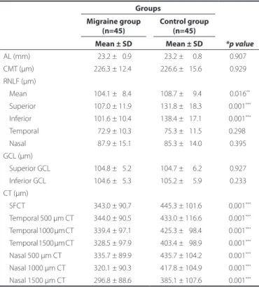
Table 2. Mean OCT analysis results of the migraine and control groups
Groups
*p value
Migraine group (n=45)
Control group (n=45) Mean ± SD Mean ± SD
AL (mm) 023.2 ± 00.9 23.2 ± 000.8 0.907**
CMT (µm) 226.3 ± 12.4 226.6 ± 015.6 0.929**
RNLF (µm)
Mean 104.1 ± 08.4 108.7 ± 009.4 0.016**
Superior 107.0 ± 11.9 131.8 ± 018.3 0.001***
Inferior 101.6 ± 10.4 138.4 ± 017.1 0.001***
Temporal 072.9 ± 10.3 075.3 ± 011.5 0.298***
Nasal 087.9 ± 15.1 085.3 ± 014.0 0.395**
GCL (µm)
Superior GCL 104.8 ± 05.2 104.7 ± 006.2 0.927**
Inferior GCL 104.6 ± 05.3 105.2 ± 005.9 0.233**
CT (µm)
SFCT 343.0 ± 90.7 445.3 ± 101.6 0.001***
Temporal 500 µm CT 344.0 ± 90.5 433.0 ± 116.6 0.001***
Temporal 1000 µm CT 339.4 ± 97.1 425.3 ± 098.4 0.001***
Temporal 1500 µm CT 328.5 ± 97.9 0403.4 ± 098.9 0.001***
Nasal 500 µm CT 335.7 ± 89.9 435.7 ± 104.2 0.001***
Nasal 1000 µm CT 320.1 ± 90.3 417.8 ± 104.9 0.001***
Nasal 1500 µm CT 296.8 ± 88.6 385.1 ± 107.6 0.001***
*= Student’s t-test; **=p<0.05; ***= p<0.01.
Retinal nerve fiber layer, ganglion cell complex, and choroidal thicknesses in migraine
8 0 Arq Bras Oftalmol. 2016;79(2):78-81
The superior and inferior GCL thickness measurements were not statistically signiicantly diferent between the two groups (p=0.907, p=0.233, respectively; Table 2; Figure 2).
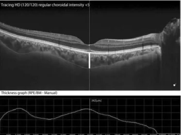
As for the CT measurements, the subfoveal, temporal, and nasal CT at 500 µm, 1000 µm, and 1500 µm were signiicantly thinner in the migraine group than in the control group (p=0.001; Table 2, Figures 3 and 4).
DISCUSSION
Migraine is a common neurological disease characterized by hea-daches together with transient focal symptoms called aura observed in some patients. Migraine patients with aura reportedly have increa-sed rates of ischemic stroke, cardiac disease, intracerebral hemorrhage, and mortality(4,7). Endothelial and vascular smooth muscle dysfunction as well as hypercoagulability have been reported as possible respon-sible factors for the pathogenesis of migraine(8). Due to the transient cerebral vasospasm that occurs in migraine, luctuations in perfusion during recurrent migraine attacks can lead to chronic retinal damage in the optic nerve head and the retina and eventually to ganglion cell death(4,9). Recurrent migraine attacks have been shown to induce chronic cerebral damage in several studies(10). Further, retinal infarcts due to the occlusion of the retinal artery have also been reported in migraine patients(11).
It has recently become possible to evaluate the thickness of the peripapillary RNFL and choroid layer using advanced OCT devices. This method has enabled the diagnosis and monitoring of various diseases. Today, RNFL analysis using OCT is commonly performed in patients with ocular hypertension and glaucoma(12,13). In our study, we compared the thicknesses of the RNFL, CMT, GCL, and choroid layer in migraine patients with aura to that in healthy individuals using SD-OCT.
In one study by Kara et al.in which Doppler ultrasound was used to demonstrate retinal vascular changes and perfusion in migraine patients, the resistance in the central retinal artery and posterior ciliary artery was found to be higher in migraine patients than in the control group(14).
In another study by Martinez et al., who compared 70 migraine patients and 53 normal individuals using OCT, the mean RNFL thickness was determined to be normal in migraine patients, but the RNFL thickness in the temporal quadrant was signiicantly thinner(4). Tan et al.evaluated RNFL thicknesses using laser polarimetry in their study, including 39 migraine patients, 15 with aura and 24 without aura, and 25 healthy individuals; they determined no reduction in the RNFL thickness, and they asserted that migraine had no efect on RNFL(15). Additionally, Sorkhabi et al. found that RNFL was thinner in the nasal quadrant in migraine patients(16).
In our study, while there was no statistically signiicant diference in the temporal and nasal quadrant RNFL thickness measurements between the groups (p>0.05), the mean RNFL thickness in migraine patients was found to be signiicantly lower compared with that in the control group (p<0.05). Similarly, the superior and inferior quadrant
Figure 4. Choroid layer thickness measurement of a patient in the migraine group. Figure 1. Measurements of the retinal nerve iber layer thickness in the migraine and
control groups.
Figure 2. Measurements of the ganglion cell complex thickness in the migraine and control groups.
Colak HN, et al.
8 1 Arq Bras Oftalmol. 2016;79(2):78-81 RNFL thicknesses were signiicantly lower in the migraine group than
in the control group (p<0.05).
Various visual complications have been reported in migraine, such as visual ield defects similar to that observed in glaucoma as well as retinal vasculopathy(17). Ischemic damage causes thinning in RNFL in glaucoma, and RNFL thickness measurements are used to eva luate the glaucomatous changes. Studies have shown that the peripapillary RNFL is consistently thinner in patients with glaucoma. Kook et al. observed thinning in RNFL in patients with normal-tension glaucoma (NTG) in the superior and inferior quadrants, consistent with our observation(18). Drance et al.determined that the progression of visual ield loss was faster in NTG patients who also had migraines compared with that in NTG patients who did not have migraines(19). Moreover, the reduction of RNFL thickness has also been reported in multiple sclerosis (MS), Alzheimer’s disease, and Parkinson’s di-sease(20-22). The GCL thickness could provide a better structural indica-tor of axonal loss compared with RNFL in certain optic neuropathies such as MS, non-arteritic anterior ischemic optic neuropathy, and compressive optic neuropathy. GCL analysis may provide a method for diagnosing and monitoring optic nerve disease(23).
In their study comparing female migraine patients to healthy women, Gipponi et al.did not ind any diference in the foveal thickness and ma-cular volume; however, they determined that superior RNFL and GCL thicknesses were signiicantly reduced only in migraine patients with aura but not in those without aura(24). In our study, we did not observe a signiicant diference in the CMT and GCL measurements of migraine patients with aura and healthy individuals (p>0.05).
The choroid layer is the most important vascular layer of the eye. Choroidal vascular insuiciency and reduction in CT result in the dysfunction of the retinal pigment epithelium and photoreceptor layers(25,26). In migraine, which is regarded as a neurovascular disease, the choroid layer can become thinner due to reduced blood low in the central retinal and posterior ciliary arteries(27). Diseases that afect the retina lead to reduced thickness of the choroid layer.
In their study evaluating CT in diabetic retinopathy, Regatieri et al. determined that CT can vary depending on the severity of retino-pathy caused by hypoxia in the retinal tissue, and they found that CT signiicantly decreased with increasing severity of retinopathy caused by hypoxia, especially in the presence of diabetic macular edema(28). Bourke et al.reported a correlation between untreated systemic hypertension and choroidopathy(29). The reduction of CT has been reported in a study with smokers, which was thought to be due to increased resistance in the retinal vasculature(30). Similarly, we observed that the subfoveal, temporal, and nasal CT measurements at 500 µm, 1000 µm, and 1500 µm were signiicantly lower in migraine patients compared with those in controls (p<0.01).
We found that the thicknesses of the RNFL and choroid layer were reduced in migraine patients compared with those in controls. Considering that patients with other diseases that could cause any retinal abnormalities, such as glaucoma (especially NTG) and systemic diseases, were not included in the study, this reduction is likely to be related to the migraine itself.
The limitations of our study are the exclusion of migraine patients without aura and the small number of cases in the study.
In conclusion, the RNFL and choroid layer thicknesses were determined to be thinner in migraine patients with aura compared with those in age-matched healthy subjects, and this is thought to be related to a progressive loss of ganglion cells and axons.
REFERENCES
1. Ozge A, Aydinlar E, Tasdelen B. Grey zones in the diagnosis of adult migraine without aura based on the International Classiication of Headache Disorders-III beta: exploring the covariates of possible migraine without aura. Pain Res Manag. 2015;20(1):1-7. 2. Smitherman TA, Burch R, Sheikh H, Loder E. The prevalence, impact, and treatment of
migraine and severe headaches in the United States: a review of statistics from national surveillance studies. Headache. 2013;53(3):427-36.
3. Graham JR, Wolf HG. Mechanism of migraine headache and action of ergotamine tartrate. Arch NeurPsych. 1938;39(4):737-63.
4. Martinez A, Proupim N, Sanchez M. Retinal nerve ibre layer thickness measurements using optical coherence tomography in migraine patients. Br J Ophthalmol. 2008;92(8): 1069-75.
5. Gipponi S, Scaroni N, Venturelli E, Forbice E, Rao R, Liberini P, et al. Reduction in retinal nerve iber layer thickness in migraine patients. Neurol Sci. 2013;34(6):841-5. 6. Shin HJ, Cho BJ. Comparison of retinal nerve iber layer thickness between Stratus
and Spectralis OCT. Korean J Ophthalmol. 2011;25(3):166-73.
7. Sacco S, Ricci S, Carolei A. Migraine and vascular diseases: a review of the evidence and potential implications for management. Cephalalgia. 2012;32(10):785-95. 8. Larrosa-Campo D, Ramón-Carbajo C, Para-Prieto M, Calleja-Puerta S, Cernuda-Morollón
E, Pascual J. Migraine as a vascular risk factor. Rev Neurol. 2012;55(6):349-58. 9. Cohen AS, Goadsby PJ. Functional neuroimaging of primary headache disorders. Curr
Pain Headache Rep. 2005;9(2):141-6.
10. Schwedt TJ, Chiang CC, Chong CD, Dodick DW. Functional MRI of migraine. Lancet Neurol. 2015;14(1):81-91.
11. Agostoni E, Rigamonti A. Migraine and small vessel diseases. Neurol Sci. 2012;33(1):51-4. 12. Yang Z, Tatham AJ, Zangwill LM, Weinreb RN, Zhang C, Medeiros FA. Diagnostic ability of retinal nerve iber layer imaging by swept-source optical coherence tomography in glaucoma. Am J Ophthalmol. 2015;159(1):193-201.
13. Shin HY, Park HY, Jung Y, Choi JA, Park CK. Glaucoma diagnostic accuracy of optical coherence tomography parameters in early glaucoma with diferent types of optic disc damage. Ophthalmology. 2014;121(10):1990-7.
14. Kara SA, Erdemoglu AK, Karadeniz MY, Altinok D. Color Doppler sonography of orbital and vertebral arteries in migraineurs without aura. J Clin Ultrasound. 2003;31(6):308-14. 15. Tan FU, Akarsu C, Güllü R. Retinal nerve iber layer thickness is unafected in migraine
patients. Acta Neurol Scand. 2005;112(1):19-23.
16. Sorkhabi R, Mostafaei S, Ahoor M, Talebi M. Evaluation of retinal nerve iber layer thickness in migraine. Ir J neurol. 2013;12(2):51-5.
17. McKendrick AM, Vingrys AJ, Badcock DR, Heywood JT. Visual ield losses in subjects with migraine headaches. Invest Ophthalmol Vis Sci. 2000;41(5):1239-47.
18. Kook MS, Cho JW, Sung KR, Hong JT, Um TW, Kang SY, et al. Macular and peripapillary retinal nerve iber layer measurements by spectral domain optical coherence tomo-graphy in normal tension glaucoma. Invest Ophthalmol Vis Sci. 2010;51(3):1446-52. 19. Drance S, Anderson DR, Schulzer M. Risk factors for progression of visual ield
abnor-malities in normal-tension glaucoma. Am J Ophthalmol. 2001;131(6):699-708. 20. Galetta KM, Calabresi PA, Frohman EM, Balcer LJ. Optical coherence tomography (OCT):
imaging the visual pathway as a model for neurodegeneration. Neurotherapeutics. 2011;8(1):117-32.
21. Kirbas S, Turkyilmaz K, Anlar O, Tufekci A, Durmus M. Retinal nerve iber layer thickness in Alzheimer disease. J Neuroophthalmol. 2013;33(1):58-61.
22. Monterio ML, Fermandes DB, Apóstolos-Pereira SL, Callegaro D. Quantiication of retinal neural loss in patients with neuromyelitis optica and multiple sclerosis with or withoutoptic neuritis using Fourier-domain optical coherence tomography. Invest Ophthalmol Vis Sci. 2012;53(7):3959-66.
23. Kardon RH. Role of the macular optical coherence tomography scan in neuro-ophthal-mology. J Neuroophthalmol. 2011;31(4):353-61.
24. Gipponi S, Scaroni N, Venturelli E, Forbice E, Rao R, Liberini P, et al. Reduction in retinal nerve iber layer thickness in migraine patients. Neurol Sci. 2013;34(6):841-5. 25. Osmanbasoglu OA, Alkin Z, Ozkaya A, Ozpınar Y, Yazici AT, Demirok A. Diurnal choroidal
thickness changes in normal eyes of Turkish people measured by spectral domain optical coherence tomography. J Ophthalmol. 2013;2013:687165. doi: 10.1155/2013/687165. 26. Lee SW, Yu SY, Seo KH, Kim ES, Kwak HW. Diurnal variation in choroidal thickness in
relation to sex, axial length, and baseline choroidal thickness in healthy korean subjects. Retina. 2014;34(2):385-93.
27. Linsenmeier RA, Braun RD. Oxygen distribution and consumption in the cat retina during normoxia and hypoxia. J Gen Physiol. 1992;99(2):177-97.
28. Regatieri CV, Branchini L, Carmody J, Fujimoto JG, Duker JS. Choroidal thickness in patients with diabetic retinopathy analyzed by spectraldomain optical coherence to-mography. Retina. 2012;32(3):563-8.
29. Bourke K, Patel MR, Prisant LM, Marcus DM. Hypertensive choroidopathy. J Clin Hypertens (Greenwich). 2004;6(8):471-2.