Case Report
Key words
Robotics; cardiac surgical procedures/methods; surgical procedures, minimally invasive; mammary arteries.
The use of robotic systems in cardiac surgeries aims at decreasing the surgical trauma. The totally endoscopic myocardial revascularization, assisted by the DaVinci robot (Intuitive Surgical, Sunnyvale, California) is feasible and the learning process must be carried out in steps. The first step is the preparation of the left internal thoracic artery, through the totally endoscopic approach.
The case presented here proposes the dissection of the right internal thoracic artery through complete sternotomy. It proposes a new step directed at the totally endoscopic procedure, aiming at decreasing the injuries caused by the learning curve.
Robotic Dissection of the Right Internal Thoracic Artery through
Median Sternotomy
Fabio Biscegli Jatene, Paulo Manuel Pêgo-Fernandes, Ramez Anbar, Fábio Antonio Gaiotto, Marcello S. Barduco,
Roberto Kalil Filho
Instituto de Ensino e Pesquisa do Hospital Sírio-Libanês, São Paulo, SP - Brazil
Mailing address: Fábio Biscegli Jatene •
Av. Dr. Enéas de Carvalho Aguiar, 44 - 5º andar - bloco 2 - sala 7 - Cerqueira César - 05403-000 - São Paulo, SP - Brazil
E-mail: [email protected], [email protected]
Manuscript received February 27, 2009; revised manuscript received April 07, 2009; accepted August 21, 2009.
There are several cardiovascular procedures that can be performed with robotic assistance. The atrial communication and mitral valve repair, the myocardial revascularization are the procedures with the highest number of publications4-6.
The learning curve for the use of robotic assistance consists of steps7-10. The exhausting training in experimental
models is crucial and establishes the beginning of the team’s experience9,10. The dissection of the left internal thoracic
artery is the first step to perform the robot-assisted totally endoscopic revascularization7,8. Usually, one starts directly with
the endoscopic dissection; however, difficulties in handling the equipment can happen, resulting in trauma and injury to the main graft used for the myocardial revascularization.
The present study proposes a former step when preparing the totally endoscopic left internal thoracic artery. We report the complete dissection of the right internal thoracic artery using the median sternotomy approach.
Case Report
In December 2008, a 54-year-old male patient with chronic coronary insufficiency, stable angina and candidate to myocardial revascularization was assessed. He had a history of arterial hypertension and dyslipidemia. The coronary angiography disclosed severe lesions in the anterior interventricular branch, diagonal branch and left marginal branch. The left ventricular function was normal.
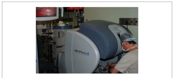
The proposed surgery was the revascularization of the anterior interventricular branch to the left internal thoracic artery; of the diagonal branch to the right internal thoracic artery and an aortic bypass surgery to the left marginal branch, in retro-aortic position. The dissection of the right internal thoracic artery was totally robot-assisted. The anesthesia preparation did not differ from the one routinely used for on-pump myocardial revascularization surgery. Mechanical ventilation was established without the need for selective orotracheal intubation. At the operating room, the robot was positioned to the patient’s right, aiming at establishing the access of the arms and the video to the right internal thoracic artery (Figure 2).
The median sternotomy was the chosen approach. The preparation of the right magna saphenous vein, removed from the right thigh, was carried out through mini-incisions. After the opening of the pericardial sac and exploration of the cavity, the left internal thoracic artery was prepared, through a skeletonized approach, without pleural opening. After the retractor was positioned, now to the right internal thoracic
Introduction
The development of new equipment and technologies has contributed to the decrease in surgical trauma in the area of cardiovascular surgery1. The decreased hospitalization time,
less frequent use of blood derivatives and better esthetic results are examples of important benefits resulting from minimally invasive procedures.
The thoracoscopic procedures in cardiovascular surgery started to be published in the beginning of the 1990s. Initially, they were video-assisted without robotic help2,3. Progressively,
the computed technology started to be incorporated by surgeons and robotic help became a reality. From a single mechanical arm controlled by voice and that supported only video, the telesurgery became available, completely assisted by a robot equipped with a three-dimensional console and four mechanical arms (DaVinci Intuitive Surgical, Sunnyvale, California) (Figure 1).
Case Report
Jatene et al Robotic dissection of the right mammary artery
Arq Bras Cardiol 2010;94(6) : e76-e78
Figure 1 -DaVinci Intuitive Surgical robot operation console, Sunnyvale, California.
Figure 2 -Robot set in position for the right internal thoracic artery preparation through median sternotomy. The robot is positioned to the patient’s right and three arms are working: video, forceps and electrocauterizer.
artery, the robot was brought closer. The operating table was placed in a 30-degree right lateral decubitus position.
Three mechanical arms were employed, as follows: the video, the electrocauterizer (EndoWrist - Intuitive Surgical) and forceps (EndoWrist deBakey - Intuitive Surgical) (Figure 2). The fourth arm remained inactive. The dissection was carried out under the same principles employed in the preparation of the left internal thoracic artery: skeletonized approach and with no opening of the pleural cavity. The electrocauterizer (Valleylab Force II) was kept at very low voltage throughout the entire procedure.
The robot-assisted right internal thoracic artery preparation time through the sternotomy was 50 minutes. The artery was dissected from its subclavian artery emergence to its bifurcation. The smaller branches were cauterized and the larger ones were clipped under direct vision. At the end of the preparation, no hematomas or lesions caused by traction or pinching were observed. The complete heparinization was carried out and the artery was cut. The flow was considered to be excellent.
Subsequently, the extracorporeal circulation circuit was installed. The surgery was uneventful and the proposed
Case Report
Jatene et al
Robotic dissection of the right mammary artery
Arq Bras Cardiol 2010;94(6) : e76-e78
revascularization was carried out. The total surgical time was 5 hours. The hospital discharge occurred seven days after the surgery, under excellent clinical conditions.
Discussion
The equipment used in the present case (DaVinci Intuitive Surgical, Sunnyvale, California) (Figure 1) presents high image resolution, satisfactory three-dimensional visualization, control for vibration decrease and allows free maneuvers with a broad range of movements. These are advantages that encourage and stimulate its use in minimally invasive procedures.
There are several reports on the preparation of internal thoracic arteries for totally endoscopic myocardial revascularization. However, the learning curve can be long and injuries can eventually occur7,8.
Several experimental models have been proposed for the training in robotic surgery9,10. Simulations with
non-living models present the robotic technology to the surgeon. Experimental animal models allow a closer approach to reality and are crucial before surgical procedures are performed in humans. The robot-assisted dissection of the right internal thoracic artery through median sternotomy can be the first step. When the median sternotomy is employed, the robotic arm access to the artery is facilitated, compared to the closed thorax. The possibility of immediate intervention in case of bleeding provides the surgeon with reassurance and tranquility, in adaptation to the technique. An assistant surgeon remains opposite the robot arms and has prompt access to the surgical field. The surgeon’s activity is identical to that performed when the totally endoscopic left internal thoracic artery preparation is carried out. The visual field is the same and the arms’ movements are also the same. The progress to the totally endoscopic preparation becomes natural.
tranquilidade The anesthesia control of the patient did not differ from the routine anesthesia used for myocardial
revascularization. There was no need for selective orotracheal intubation or decrease in ventilatory volume. Bleeding was negligible.
The time of preparation was satisfactory, considering the learning curve. Definitely, it would have been longer if the chosen preparation had been the totally endoscopic procedure. No injuries caused by the robot-assisted dissection were observed. The incidence of lesions during the learning phase is estimated at 6% and 2% is considered acceptable after long training7,8. The learning process using the conventional
sternotomy can decrease these figures, especially at the initial phase.
The learning curve must be considered in all surgical areas, mainly when the technological innovations are incorporated. We believe that the preparation of the right internal thoracic artery through median sternotomy can be an important initial step. It is a safe way to start the robot-assisted totally endoscopic cardiac surgery.
Acknowledgements
To Dr. Paulo Chap-Chap, Dr. Ricardo Abdalla and Dr. Marcelo Cerdan Torres, for their support of this procedure.
Potential Conflict of Interest
No potential conflict of interest relevant to this article was reported.
Sources of Funding
There were no external funding sources for this study.
Study Association
This study is not associated with any post-graduation program.
References
1. Jatene F, Gaiotto FA, Monteiro R. Cirurgia cardíaca minimamente invasiva. In: Tratado de Cardiologia da SOCESP. 2a ed. São Paulo: Manole; 2008. p. 2554-60. 2. Jatene FB, Fernandes PM, Stolf NA, Kalil R, Hayata AL, Assad R, et al. Cirurgia
de revascularização do miocárdio minimamente invasiva com utilização da videotoracoscopia. Arq Bras Cardiol. 1997; 68: 107-11.
3. Jatene FB, Pêgo-Fernandes PM, Hayata AL, Arbulu HE, Stolf NA, Oliveira SA, et al. VATS for complete dissection of LIMA in minimally invasive coronary artery bypass grafting. Ann Thorac Surg. 1997; 63: 110-3.
4. Ak K, Aybek T, Wimmer-Greinecker G, Ozaslan F, Bakhtiary F, Moritz A, et al. Evolution of surgical techniques for atrial septal defect repair in adults: a 10-year single-institution experience. J Thorac Cardiovasc Surg. 2007; 134: 757-64. 5. Chitwood WR Jr, Rodriguez E, Chu MW, Hassan A, Ferguson TB, Vos PW, et al.
Robotic mitral valve repairs in 300 patients: a single-center experience. J Thorac Cardiovasc Surg. 2008; 136: 436-41.
6. Schachner T, Feuchtner GM, Bonatti J, Bonaros N, Oehlinger A, Gassner E, et al. Evaluation of robotic coronary surgery with intraoperative graft angiography and postoperative multislice computed tomography. Ann Thorac Surg. 2007; 83: 1361-7.
7. Oehlinger A, Bonaros N, Schachner T, Ruetzler E, Friedrich G, Laufer G, et al. Robotic endoscopic left internal mammary artery harvesting: what have we learned after 100 cases? Ann Thorac Surg. 2007; 83: 1030-4.
8. Bonatti J, Schachner T, Bernecker O, Chevtchik O, Bonaros N, Ott H, et al. Robotic totally endoscopic coronary artery bypass: program development and learning curve issues. J Thorac Cardiovasc Surg. 2004; 127: 504-10.
9. Ishikawa N, Watanabe G, Hirano Y, Inaki N, Kawachi K, Oda M. Origami using da Vinci surgical system. Surg Endosc. 2007; 21: 1252-3.
10. Ishikawa N, Sun Y, Nifong W, Watanabe G, Chitwood WR. Thoracoscopic lobectomy with the da Vinci Surgical System. Innovations. 2006; 1: 169-70.