RBCCV 44205-1534 DOI: 10.5935/1678-9741.20140061
Analysis of transit time low of the right internal
thoracic artery anastomosed to the left anterior
descending artery compared to the left internal
thoracic artery
Análise da medida de luxo por tempo de trânsito da artéria torácica interna direita anastomosada para
a artéria interventricular anterior comparada com a artéria torácica interna esquerda
Rodrigo Milani
1. MD. MsC. PhD; Daniela de Moraes
1. MD; Aline Sanches
1. MD; Rodrigo Jardim
1.
MD; Thais Lumikoski
1. MD; Gabriela Miotto
1. MD; Vitor Hugo Santana
1. MD; Paulo Roberto
Brofman
1. MsC. PhD
1 Pontifícia Universidade Católica do Paraná (PUCPR). Curitiba,PR, Brazil.
Correspondence address: Rodrigo Milani
Rua Cezar Correia de Souza Jr. 54 – Curitiba. PR. Brazil – Zip code: 82015-220 E-mail: [email protected]
This study was carried out at Santa Casa de Misericórdia de Curitiba. Cu
-ritiba. PR. Brazil; Pontifícia Universidade Católica do Paraná (PUCPR), Curitiba, PR, Brazil.
No inancial support.
Article received on December 19th, 2013 Article accepted on March 14th, 2014 Abstract
Introduction: We evaluated with transit time low the perfor -mance of the right and left thoracic arteries when used as a graft for the left anterior descending artery.
Methods: Fifty patients undergoing surgery for myocardial revascularization without cardiopulmonary bypass were divided into two groups. In group A patients received graft of right inter -nal mammary artery to the anterior interventricular branch. In group B patients received graft of left internal mammary artery to the same branch. At the end of the operation the low was assessed by measuring transit time.
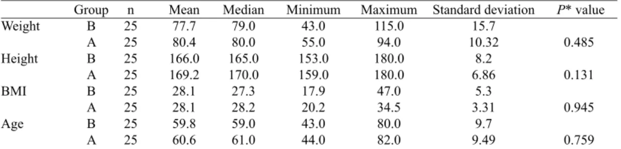
Results: In group A. mean age was 60.6±9.49 years. The average height and weight of the group was 80.4±10.32 kg and 169.2±6.86 cm. The average number of grafts per patient in this group was 3.28±1.49. The mean low and distal resistance obtained in right internal thoracic artery was 42.1±23.4 ml/min and 2.8±0.9 respectively. In group B. the mean age was 59.8±9.7 years. The average height and weight of this group was 77.7±14.22 kg and 166.0±8.2 cm. The average number of grafts per patient in this group was 3.08 ±0.82. The mean low and distal resistance
observed in this group was 34.2±19.1 ml/min and 2.0±0.7. There were no deaths in this series.
Conclusion: Right internal mammary artery presented a similar behavior to left internal mammary artery when anasto -mosed to the anterior interventricular branch of the left coronary artery. There was no statistical difference between the measured low obtained between both arteries.
Descriptors: Mammary Arteries. Coronary Artery Bypass. off-Pump. Internal Mammary-Coronary Artery Anastomosis.
Resumo
Introdução: Avaliamos por meio da medida de luxo por tempo de trânsito o desempenho das artérias torácicas direita e esquerda quando utilizadas como enxerto para revascularização da artéria interventricular anterior.
-Most authors agree that the use of both thoracic arteries is beneicial. They also believe that the right internal thoracic artery should be used mainly in the left coronary system. es -pecially in the circumlex artery and its branches. by making a retro-aortic[14] passage.
The use of the right internal thoracic artery to the ante -rior interventricular branch is no consensus. In the last joint guideline from the American Heart Association (AHA) and the Society for Thoracic Surgery (STS)[15]. when the best
distribution of grafts according to the coronary vessels was assessed. the use of the left internal thoracic artery to the an -terior interventricular branch received recommendation IB by keeping as the main graft for this branch. while the use of the right internal thoracic artery to the same branch was IIA class. In the present study we assessed ifty patients divided into two groups of twenty-ive comparing them by measuring the low for trafic time and the behavior of both thoracic arteries grafted to the anterior interventricular branch of the left coronary. Our main aim was to determine whether the low obtained at the anastomosis of the right internal thoracic artery to the anterior interventricular branch is similar to the data obtained with the “gold standard” which is the anasto -mosis of the left internal thoracic artery to the same branch of the left coronary artery.
METHODS
In this retrospective study. we assessed 50 patients under -going coronary artery bypass grafting without cardiopulmo -INTRODUCTION
The irst reports of experimental work for a possible myo -cardial revascularization are from the mid 50’s. A group of researchers from the Soviet Union made the anastomosis of the left internal thoracic artery to the anterior interventricular branch in dogs[1]. This work served as the basis for the re
-ports of the irst cases in humans[2]. In 1967. when this study
was presented at a cardiology symposium in Leningrad. the majority of those present agreed to a resolution that said cor -onary surgery was impossible and had no future. After more than 40 years after these initial reports. the surgeries for myo -cardial revascularization have become one of the most per -formed surgeries in the world and certainly the most studied. Several studies[3-6] show that even after so much time
passed and the steady advance of medicine in the last de -cades it remains the treatment of choice for patients with se -vere coronary disease. Obtaining consistent long-term results after performing this type of procedure depends mainly on the grafts to be used. The superiority of arterial grafts over saphenous vein grafts. especially the internal thoracic artery is well documented[7-9]. Studies comparing these two types of
grafts show a higher patency rate of 90% in 10 years for the internal thoracic artery versus 50% of saphenous vein grafts. being relected this difference in increased patient survival. Furthermore. several studies from different groups were pre -sented showing that the use of both internal thoracic arteries has a signiicant survival when a period of 20 years after sur -gery is evaluated[10-13].
Abbreviations. acronyms & symbols
AHA American Heart Association NYHA New York Heart Association
PI Pulsatility Index
STS Society for Thoracic Surgery
beram enxerto de artéria torácica interna direita para o ramo interventricular anterior. No grupo B. os pacientes receberam enxerto de artéria torácica interna esquerda para o mesmo ramo. Ao término da operação. o luxo foi avaliado por meio da medida de luxo por tempo de trânsito.
Resultados: No grupo A. idade média foi de 60.6±9.49 anos. A média de peso e altura do grupo foi de 80.4±10.32 Kg e 169.2±6.86 cm. A média de pontes por paciente neste grupo foi de 3.28±1.49.
O luxo médio e a resistência distal obtidos na artéria torácica interna direita foi de 42.1±23.4 ml/min e 2.8±0.9 respectivamente. No grupo B. a idade média foi de 59.8±9.7 anos. A média de peso e altura deste grupo foi de 77.7±14.2215.7 Kg e 166.0±8.2 cm. A média de pontes por paciente neste grupo foi de 3.08±0.82. O luxo médio e a resistência distal observados neste grupo foi de 34.2±19.1ml⁄min e 2.0±0.7. Não houve óbitos nesta série.
Conclusão: A artéria torácica interna direita apresentou um comportamento similar ao da artéria torácica interna esquerda quando anastomosada ao ramo interventricular anterior da coronária esquerda. Não houve diferença estatís -tica entre a medida de luxo obtida entre ambas as artérias.
nary bypass. divided into two groups of 25 patients each. In group A. there are patients who received graft of right inter -nal thoracic artery to the anterior interventricular branch. In group B. patients who received left internal thoracic artery to the same branch. In this study. we excluded all patients who required a procedure associated with revascularization. as well as patients who possibly undergoing cardiopulmonary bypass. In none of the ifty patients conversion to cardiopul -monary bypass was required.
In both groups. we used general anesthesia. and the approach was median sternotomy. The manner on which the anesthesia was performed and how the grafts were obtained have been subject to previous publication[16]. In
both groups. the internal thoracic artery was obtained us -ing skeletonization.
The strategy of revascularization differs somewhat be -tween groups. In both cases. we used a dose of 2 mg/kg of heparin. The initial approach was the same. with the pericar -dium opened widely. pulled in its anterior edge on both sides. With the help of a polyester tape 90 cm long and 3 cm wide. a section by Lima[17] was performed. This was applied between
the inferior vena cava and the left inferior pulmonary vein to better exposure of the heart without signiicant changes of hemodynamics. In all ifty patients a suction tissue sta -bilizer (Octopuss II ®. Medtronic) to perform anastomosis was used.
Moreover. in most cases. it was performed with an intra -luminal shunt. The strategy of revascularization. on which graft would be directed to coronary branch was decided at the time of operation. Rather. we chose the retroaortic right inter -nal thoracic artery to the circumlex artery and its branches and the left internal thoracic artery to the anterior interven -tricular branch. The anastomoses are also preferably per -formed in the middle third of the coronary artery and. if the graft did not reach the target artery satisfactorily we reversed the distribution.
In group A. the operation started by anastomosis of the right internal thoracic artery to the anterior interventricular branch. followed. in cases where it was necessary. through the sidewall and inally through the anterior interventricular branch. In group B. the operation started by the anastomoses of the inferior wall. followed by sidewall and inally the an -terior interventricular branch. The approach taken in group B. leaving the anterior interventricular artery last is due to a fear of injury by traction. This could occur while performing an anastomosis in the sidewall of an increased ventricle with the anastomosis of the left internal thoracic artery already performed to the anterior interventricular branch.
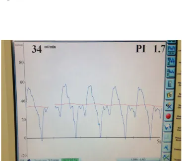
At the end of the anastomoses after heparin reversal. sys -tolic blood pressure was maintained around 110 mmHg and a heart rate average ranging between 80 and 100 beats per min -ute. No patient in the series needed use of vasoactive drugs to obtain the desired pressure. Moreover. with “3mm probe”
(Figure 1) connected to the lowmeter Medistim brand. mod -el Butterly Flowmeter. low measures by transit time and re -view of the pulsatility index. PI (Figure 2) were performed. To obtain these values . the patients were kept under apnea for a period of around 10 seconds. and the “probe” applied to the selected graft. According to the device manufacturer. lows higher than 10 ml/min. and a pulsatility index which measures the distal strength below 5 means an anastomosis of good quality.
After evaluation of the anastomoses. the revision of he -mostasis and closure of the chest were performed. Next. still under general anesthesia. patients were referred to the inten -sive care unit.
This study received a favorable opinion from the Re -search Ethics Committee of our institution under number 20827713.2.0000.0020.
Fig. 1 - 3 mm Probe
Statistical Analysis
To evaluate the association between two dichotomous qualitative variables we considered the Fisher exact test. The correlation between quantitative variables was evaluated by estimating the Pearson correlation coeficient. To compare the two groups with respect to quantitative variables we used the Student’s t test for independent samples. These compar -isons. considering more than two groups were performed using a model of analysis of variance factor (ANOVA). For multivariate analysis a model of Multiple Linear Regression was adjusted. P values <0.05 were considered statistically
signiicant. Data were analyzed using the SPSS v.20.0 soft -ware.
RESULTS
We assessed 50 patients undergoing coronary artery by -pass grafting without cardiopulmonary by-pass were divided into two groups of 25 each. In group A we put patients who received internal thoracic artery to the anterior interventric -ular branch. In group B we put patients who received a graft of the left internal thoracic artery graft to the same coronary branch.
In group A. the mean age was 60.6 ± 9.49 ranging be -tween 44 and 82 years and in group B 59.8 ± 9.7 be-tween 43 and 80 years. Patients in group A had a mean weight of 80.4 ± 10.32 kg. The mean weight from B group was 77.7 ± 15.7 kg. The average height of the group A was 169.2 ± 6.86 cm and in group B 166.0 ± 8.2 cm. Table 1 shows the values of age. weight. height and body mass index for both groups:
In assessing risk factors for coronary disease. in group A 21 (84%) patients were hypertensive. compared with 18 (72%) in group B (P=0.496). 10 (40%) patients in group A
were diabetics against ive (20%) in group B (P=0.217) and 6 (24%) patients in group A were smokers against seven (28%) in group B (P=1).
In group A. seven (28%) had undergone previous coro -nary angioplasty and eight (32%) had presented myocardial infarction against four (16%) who had undergone angioplasty in group B and four (16%) with history of myocardial infarc -tion. There was no statistical difference between the groups regarding previous coronary angioplasty or old myocardial infarction. Regarding the left ventricular function. if normal. moderate or poor compromise. Table 2 presents the data from both groups.
There was no statistical difference between the groups when evaluating left ventricular function (P: 0.461).
In the assessment of functional class. no patient in group A (0%) were in NYHA class I. 15 (60%) in class II. seven (28%) in class III. and three (12%) in class IV . In group B. three (12%) were in class I. 14 (56%) in class II. seven (28%) in class III and one (4%) in class IV. There was no statistical difference (P=0.258) between the two groups.
Table 3 presents the distribution of the number of individ -ual grafts per patient in each group.
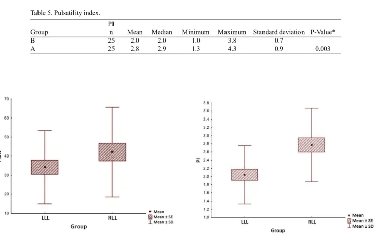
The central point of this study is the evaluation of coro -nary low. The evaluation of the measured low through the transit time showed an average low of 42.2 ± 23.4 ml/min in Group A and 34.2 ± 19.1 ml/min in group B. featuring a sta -tistically signiicant difference in favor of group A. We tested the null hypothesis that the mean FLOW is the same in both groups (RLL and LLL). versus the alternative hypothesis of means that are not all the same. Table 4 and Figure 3 shows the results obtained.
Finally. we assessed the pulsatility index. PI. that deter -mines the resistance after the anastomosis. The device man -ufacturer provides measurement values for low trafic time of less than 5 as indicative of a good quality anastomosis. In group A. the mean PI was 2.8±0.9 and in group B 2.0±0.7. Table 5 shows the results obtained from both groups.
Figure 4 shows a representation of the behavior of pulsa -tility index in both groups.
Table 1. Quantitative variables.
Weight Height BMI Age Group B A B A B A B A n 25 25 25 25 25 25 25 25 Mean 77.7 80.4 166.0 169.2 28.1 28.1 59.8 60.6 Median 79.0 80.0 165.0 170.0 27.3 28.2 59.0 61.0 Minimum 43.0 55.0 153.0 159.0 17.9 20.2 43.0 44.0 Maximum 115.0 94.0 180.0 180.0 47.0 34.5 80.0 82.0 Standard deviation 15.7 10.32 8.2 6.86 5.3 3.31 9.7 9.49
P* value
0.485
0.131
0.945
0.759
Table 2. Left ventricular function.
LV
Normal
Moderate
Poor
Total
B
12
48.00%
8
32.00%
5
20.00%
25
A
13
52.00%
10
40.00%
2
8.00%
25
Group
Table 3. Individual grafts. Number of grafts
1
2
3
4
6
Total
B 3
12.00%
6
24.00%
12
48.00%
4
16.00%
0
0.00%
25
A 0
0.00%
8
32.00%
13
52.00%
3
12.00%
1
4.00%
25
Group
Table 5. Pulsatility index.
Group
B A
n 25 25
Mean 2.0 2.8 PI
Median 2.0 2.9
Minimum 1.0 1.3
Maximum
3.8 4.3
Standard deviation 0.7 0.9
P-Value*
0.003
Table 4. Flow measurement.
Group
B A
n 25 25
Mean 34.2 42.2
Fluow
Median 31.0 34.0
Minimum 13.0 16.0
Maximum
82.0 94.0
Standard deviation 19.1 23.4
P-Value*
0.197
Fig. 3 - Flow Performance
DISCUSSION
During the past 20 years. numerous studies have been published related to the prevention. diagnosis and surgical treatment of coronary artery disease[18.19]. In these studies. the
advantage on the use of the thoracic artery is clearly demon -strated due to its superior patency. showing consistent long-term results and thus leading to an increased survival. This mainly occurs in groups where getting better results over the years is less frequent as in women and diabetics.
Currently. the most widely used strategy in coronary artery bypass operations is the use of an internal thoracic artery accompanied the other saphenous vein grafts[20.21].
However. atherosclerosis of saphenous vein is responsi -ble for the failure of CABG during the study. A growing number of diabetics has been referred for surgery due to poor results with coronary angioplasty. There was a reduc -tion in mortality. 5.8% vs. 20.6%. with respect to results obtained in patients who received at least one internal tho -racic artery[22].
The left internal thoracic artery presents patency greater than 90% after 10 years of its implantation. This is because less than 1% of them develops atherosclerosis with severe impairment[23].
As already emphasized above. the use of the mammary artery improves survival of patients. Studies show that when using both internal thoracic arteries. the survival increases signiicantly in 20 years[11-13]. Study presented by Ribeiro et
al.[24] shows very little difference from the histological point
of view between left and right internal thoracic arteries. not -ing only that the distal segment of the elastic layer is more prominent.
With these results clearly established. the central focus revolves around what is the best distribution for thoracic ar -teries. which vessel should receive the second thoracic ar -tery. and especially if it is appropriate to use the right internal thoracic artery to the anterior interventricular branch of left coronary. As noted earlier. our main distribution is the same advocated by Gerola et al.[14] with the right internal thorac
-ic artery directed to the circumlex artery and its branches through a retroaortic course.
The low measurement by transit time became available for clinical use in the late 90s[25,26]. It is a quick. simple and
objective way to verify the quality of the anastomosis in cor -onary surgeries. Flows below 10 ml/min associated with a pulsatility index greater than 5 indicate problems. The pul -satility index. PI. is a number obtained by dividing the dif -ference between the maximum low and minimum low by mean low[26].
One of the irst studies on the low measurement by transit time was published in 1998 by Walpoth et al.[25].
In this study. the authors assessed 46 anastomoses of the left internal thoracic artery to the anterior interventricu
-lar branch. Of these. 43 had normal PI low and. in the other three after anastomosis is performed again the PI low has been normalized. Based on these data. the au -thors concluded that the measured low by transit time is simple. reproducible and easy to perform. Moreover. they concluded that a low low in the graft with a high PI in -dicates need for revision of the anastomosis and after that the values tend to normalize. They also conirmed that this type of low measurement is cost-effective and probably prevents hemodynamic instability in the immediate post -operative period.
Leong et al.[27] assessed 116 patients with a total of 322
grafts. with 125 arterial and 197 venous. They performed the measurement of low by transit time. The average low for the internal thoracic artery to the anterior interventricu -lar branch was 37.4 ± 23.5 ml/min and 21.2 to 36 ml/min in the other grafts. In six patients the anastomosis error was detected and that being performed again. The authors found no statistical difference between PI low or arterial and ve -nous grafts. They concluded stating that this type of coronary lowmetry provides important and reliable information about the patency of each graft individually. It is able to accurately detect folds. twists and signiicant stenoses in grafts. allow -ing their immediate correction.
In our country. Cerqueira Neto et al.[28] published a study
showing the measured low of the left internal thoracic artery to the anterior interventricular branch in patients who un -derwent surgery with and without cardiopulmonary bypass. concluding that there is no difference in low between these two situations.
In our series. we assessed two groups of 25 patients each. comparing the performance of the left internal tho -racic artery to the right internal tho-racic artery. when grafted to the anterior interventricular branch. Many sur -geons still have some fear of putting the right internal thoracic artery to the anterior interventricular branch. The two groups were similar with respect to weight. height and age. In the group in which the right internal thoracic artery was used there were more diabetic patients. but without statistical difference.
When evaluating low. the group of left internal thoracic artery had an average low of 34.2±19.1 versus 42.2±23.4 ml/ min in the right thoracic artery group. Despite the increase. there was no signiicant difference between the two groups. In assessing the pulsatility index. PI. the irst group had a mean PI of 2.0±0.7 vs. 2.8±0.9 in the second group. Despite
the difference between the groups is signiicant (P=0.003). the values obtained in the group in which the thoracic artery was used showed values within the normal range. or that is. a PI below 5.
Authors’ roles & responsibilities
RM Analysis and interpretation of data. inal approval of the manuscript; conception and design of the study; implementation of operations and/or experiments; writing of the manuscript or revising it critically for its content DM Analysis and interpretation of data; conception and design
of the study; writing of the manuscript or revising it critically for its content
AS Analysis and interpretation of data; conception and design of the study; writing of the manuscript or revising it critically for its content
RJ Help in operations and data collection; performing operations and experiments
TL Help in operations and data collection; performing operations and experiments
GM Help in operations and data collection; performing operations and experiments
VHS Help in operations and data collection; performing operations and experiments
PRB Writing and revision of the article; analysis and interpretation of data
REFERENCES
1. Kolesov VI. Potashov LV. Surgery of coronary arteries. Eksp Khir Anesteziol. 1965;10(2):3-8.
2. Kolesov VI. Mammary artery-coronary artery anastomosis as
method of treatment for angina pectoris. J Thorac Cardiovasc Surg. 1967;54(4):535-44.
3. Grondin CM. Campeau L. Lespérance J. Enjalbert M. Bourassa MG. Comparison of late changes in internal mammary artery and saphenous vein grafts in two consecutive series of patients 10 years after operation. Circulation. 1984;70(3 Pt 2):I208-12.
4. Loop FD. Lytle BW. Cosgrove DM. Stewart RW. Goormastic M. Williams GW. et al. Inluence of the internal-mammary artery graft on 10-year survival and other cardiac events. N Engl J Med. 1986;314(1):1-6.
CONCLUSION
The results obtained in our study suggest that the right internal thoracic artery when anastomosed to the anterior interventricular branch presents an initial behavior very sim -ilar to that of the left internal thoracic artery. In cases where the left internal thoracic artery needs to be anastomosed to another coronary branch or that it is not available. the right internal thoracic artery should be considered as the substitute of choice. The initial low obtained when comparing the right and left arteries showed no signiicant difference.
5. Bidstrup BP. Underwood SR. Sapsford RN. Streets EM. Effect of aprotinin (Trasylol) on aorta-coronary bypass graft patency. J Thorac Cardiovasc Surg. 1993;105(1):147-52.
6. Barner HB. Mudd JG. Mark AL. Ahmad N. Dickens JF. Patency of internal mammary-coronary grafts. Circulation. 1992;54(6
Suppl):III70-3.
7. Sabik JF 3rd. Lytle BW. Blackstone EH. Houghtaling PL. Cosgrove DM. Comparison of saphenous vein and internal thoracic artery graft patency by coronary system. Ann Thorac Surg. 2005;79(2):544-51.
8. Benzon E. Choplain JN. Maguid YA. Aziz AA. Barra JA. Failure of internal thoracic artery grafts: conclusions from coronary angiography mid-term follow-up. Ann Thorac Surg. 2003;76(3):754-9.
9. Caes FL. Van Nooten GJ. Use of internal mammary artery for emergency grafting after failed coronary angioplasty. Ann Thorac Surg. 1994;57(5):1295-9.
10. Lytle BW. Blackstone EH. Loop FD. Houghtaling PL. Arnold JH. Akhrass R. et al. Two internal thoracic artery grafts are better than one. J Thorac Cardiovasc Surg. 1999;117(5):855-72.
11. Calafiore AM. Di Mauro M. Di Giammarco G. Teodori G. Iacò AL. Mazzei V. et al. Single versus bilateral
internal mammary artery for isolated first myocardial revascularization in multivessel disease: long-term clinical
results in medically treated diabetic patients. Ann Thorac Surg. 2005;80(3):888-95.
12. Martins SK. Santos MA. Tirado FHP. Martins Jr FCE. Malat HF.
Jatene AD. et al. Revascularização do miocárdio com emprego
de ambas artérias mamárias internas em pacientes com diabetes mellitus. Rev Bras Cir Cardiovasc. 2007;22(3):291-6.
13. Milani R. Brofman PR. Guimarães M. Barboza L. Tchaick RM. Meister Filho H. et al. Dupla artéria torácica esqueletizada versus
convencional na revascularização do miocárdio sem CEC em
diabéticos. Rev Bras Cir Cardiovasc. 2008;23(3):351-7.
14. Gerola LR. Puig LB. Moreira LFP. Gemha GP. Cividanes GVI. Santos RCM. et al. Dez anos de experiência com a artéria torácica interna direita através do seio transverso na revascularização da artéria circunlexa e seus ramos. Rev Bras Cir Cardiovasc. 1993;8(4):259-65.
15. Hills LD. Smith PK. Anderson JL. Bittl JA. Bridges CR. Byrne JG. et al. 2011 ACCF/AHA Guideline for Coronary Artery Bypass Graft Surgery: a report of the American College of Cardiology Foundation/American Heart Association Task Force on Practice Guidelines. Circulation. 2011;124(23):e652-735.
16. Milani RM. Análise dos resultados imediatos da operação para revascularização do miocárdio sem pinçamento total da aorta
17. Lima RC. Padronização técnica de revascularização da artéria circunlexa e seus ramos sem circulação extracorpórea [Tese de doutorado]. São Paulo: Escola Paulista de Medicina; 1999.
18. Edwards FH. Carey JS. Grover FL. Bero JW. Hartz RS. Impact of gender on coronary bypass operative mortality. Ann Thorac Surg. 1998;66(1):125-31.
19. Carey JS. Cukingnan RA. Singer LK. Health status after myocardial revascularization: inferior results in women. Ann Thorac Surg. 1995;59(1):112-7.
20. Calaiore AM. Di Mauro M. Di Giammarco G. Contini M. Vitolla G. Iacò AL. et al. Effect of diabetes on early and late survival after isolated irst coronary bypass surgery in multivessel disease. J Thorac Cardiovasc Surg. 2003;125(1):144-54.
21. Lev-Ran O. Braunstein R. Nesher N. Ben-Gal Y. Bolotin G. Uretzky G. Bilateral versus single internal thoracic artery grafting in oral-treated diabetic subsets: comparative seven-year outcome analysis. Ann Thorac Surg. 2004;77(6):2039-45.
22. The BARI Investigators. Influence of diabetes on 5-year mortality and morbidity in a randomized trial comparing CABG and PTCA in patients with multivessel disease: the Bypass
Angioplasty Revascularization Investigation (BARI). Circulation.
1997;96(6):1761-9.
23. Gurné O. Chenu P. Buche M. Louagie Y. Eucher P. Marchandise B. et al. Adaptive mechanisms of arterial and venous coronary bypass grafts to an increase in low demand. Heart. 1999;82(3):336-42.
24. Ribeiro MF. Kneubil MC. Aquino MS. Gomes GN. Mazzili P. Buffolo E. et al. Histomorphometric differences between the left and right internal thoracic arteries in humans. Rev Bras Cir Cardiovasc. 2008;23(1):1-6.
25. Walpoth BH. Bosshard A. Genyk I. Kipfer B. Berdat PA. Hess OM. et al. Transit-time low measurement for detection of early graft failure during myocardial revascularization. Ann Thorac Surg. 1998;66(3):1097-100.
26. D’Ancona G. Karamanoukian HL. Bergsland J. Is intraoperative measurement of coronary blood low: a good predictor of graft patency? Eur J Cardiothorac Surg. 2001;20(5):1075-7.
27. Leong DK. Ashok V. Nishkantha A. Shan YH. Sim EK. Transit-time low measurement is essential in coronary artery bypass grafting. Ann Thorac Surg. 2005;79(3):854-7.