Daniela Patrícia da Cunha Machado
Acute cyanide intoxication in industrialised countries
2011/2012Daniela Patrícia da Cunha Machado
Acute cyanide intoxication in industrialised countries
Mestrado Integrado em Medicina
Área: Toxicologia Médica
Trabalho efetuado sob a Orientação de: Professor Doutor Daniel Filipe de Lima Moura
E sob a Coorientação de: Dr. Luís Alexandre de Castilho Silva Coentrão
Trabalho organizado de acordo com as normas da revista: Clinical Toxicology
1 Title: Acute cyanide intoxication in industrialised countries
Authors:
• Daniela Patrícia da Cunha Machado
− Faculty of Medicine, University of Porto, Porto, Portugal • Daniel Filipe de Lima Moura
− Institute of Pharmacology and Therapeutics, University of Porto and Hospital de S. João EPE, Porto, Portugal
• Luís Alexandre de Castilho Silva Coentrão
− Institute of Pharmacology and Therapeutics, University of Porto and Hospital de S. João EPE, Porto, Portugal
− Nephrology Research and Development Unit, Faculty of Medicine, University of Porto and Hospital de S. João EPE, Porto, Portugal
Corresponding author:
• Daniela Patrícia da Cunha Machado
Institute of Pharmacology and Therapeutics, Faculty of Medicine, University of Porto Al. Prof. Hernâni Monteiro
4202 Porto, Portugal Phone: +351 220426700 Email: [email protected]
Keywords:
Poisoning; Jeweller; Hydroxocobalamin; Antidote
2
Index
Abstract ……… 3 Resumo ………. 5 Introduction ………. 7 Objectives ………. 7 Methods ……… 7Pathophysiology of cyanide poisoning ………... 8
Detoxification of cyanide ………. 9
Routes of cyanide toxicity • Inhalation ………. 10
• Ingestion ……….. 11
• Dermal contact and parenteral administration ……….... 15
Clinical manifestations and laboratory findings ……… 16
Diagnosis ……… 17 Treatment ……….. 17 Follow-up ………... 21 Conclusion ………. 22 Acknowledgments ………. 24 Declaration of interest ……….. 25 References ……….. 26 Table ………... 32
3 Abstract
Introduction. Cyanide is a rapidly acting poison existing in various forms. It is a toxicant in fire smoke and a substance used by some professional groups. Objectives. This article reviews the multiple routes and possible sources of acute cyanide poisoning and their association with certain occupations. Pathophysiology. Cyanide can kill within seconds or minutes to hours of exposure, through the inhibition of the mitochondrial cytochrome oxidase a3, causing intracellular hypoxia. Detoxification. Cyanide is mainly detoxified in the liver by the enzyme rhodanese to the less toxic thiocyanate. Other route is reaction with hydroxocobalamin to form cyanocobalamin excreted in urine. Inhalation. Modern structural fires can generate toxic levels of hydrogen cyanide, making the inhalation of smoke in a closed-space fire the most common cause of cyanide poisoning in industrialised countries. Inhalation of this toxic is also possible in occupational or industrial accidents. Ingestion. Suicide attempts are the most common reason for cyanide ingestion. There is a strong association between suicide by ingestion of cyanide and certain professional groups, namely jewellers, chemists or miners. Ingestion of this poison can also occur as an accident in some occupations. Nitriles are cyanogenic compounds with a delayed onset of cyanide toxicity, causing poisoning most often due to suicide attempts or accidental ingestion by young children. There are cyanogenic glycosides that can cause toxicity, normally occurring as an accidental poisoning. Dermal contact and parenteral administration. Dermal exposure and subcutaneous injection of cyanide are rare causes of poisoning. Sodium nitroprusside is an inorganic hypotensive agent that can be an iatrogenic source of cyanide. Clinical manifestations and laboratory findings. Early symptoms of poisoning represent the reflexive attempts of the organism systems to overcome hypoxia and late symptoms reflect these systems depression. Patients with acute cyanide intoxication have markedly elevated plasma lactate concentration, high anion gap metabolic acidosis and reduced arteriovenous oxygen
4 saturation difference. Diagnosis. The diagnosis is generally based on index of suspicion, clinical presentation and laboratory findings, since there is no blood cyanide test available on an emergent basis. Treatment. Management of patients with acute cyanide poisoning requires decontamination, supportive care and the early administration of an antidote. From the antidotes available, hydroxocobalamin seems to have the best risk-benefit profile and has been recommended as an antidote of first choice. Follow-up. Most patients who survive cyanide intoxication will be free of complications. Nonetheless, some may develop parkinsonism or other symptoms. Conclusion. Acute cyanide poisoning can occur through several routes. Smoke inhalation is the most common in industrialised countries. Many professional groups have prompt access to this toxic, so the possibility of poisoning should be strongly considered in those workers when they present a clinical picture and laboratory findings suggestive of cyanide. An antidote, preferably hydroxocobalamin, should be administered.
Keywords Poisoning; Jeweller; Hydroxocobalamin; Antidote
Word count:
5 Resumo
Introdução. O cianeto é um veneno de rápida ação existente em várias formas. Encontra-se presente no fumo resultante de incêndios, assim como é uma substância usada em algumas profissões. Objetivos. Este artigo faz uma revisão das múltiplas vias e possíveis fontes da intoxicação aguda com cianeto, assim como a sua associação com certas ocupações. Patofisiologia. O cianeto é letal em poucos segundos ou minutos a horas após a exposição, através da inibição da enzima citocromo oxidase a3 mitocondrial, causando hipóxia intracelular. Destoxificação. O cianeto é sobretudo destoxificado no fígado pela ação da enzima rodanase, com a sua conversão no composto menos tóxico tiocianato. Outra via existente é a reação com a hidroxocobalamina para formar cianocobalamina, excretada na urina. Inalação. Os incêndios em estruturas modernas podem produzir níveis tóxicos de ácido cianídrico, tornando a inalação de fumo num incêndio num espaço fechado a causa mais comum de intoxicação por cianeto nos países industrializados. A inalação deste tóxico também pode ocorrer em acidentes ocupacionais ou industriais. Ingestão. As tentativas de suicídio são o motivo mais comum para a ingestão de cianeto. Existe uma forte associação entre o suicídio pela ingestão deste tóxico e certos grupos profissionais, nomeadamente joalheiros, químicos ou mineiros. A ingestão de cianeto também pode ocorrer por acidente em algumas ocupações. Os nitrilos são compostos cianogênicos que provocam toxicidade pelo cianeto de início tardio, causando envenenamento normalmente devido a tentativas de suicídio ou ingestão acidental por crianças pequenas. Existem glicosídeos cianogênicos que podem causar toxicidade, normalmente ocorrendo de forma acidental. Contacto dérmico e administração parentérica. A exposição dérmica e injeção subcutânea de cianeto são causas raras de envenenamento. O nitroprussiato de sódio é um composto inorgânico hipotensor que pode ser uma fonte iatrogénica de cianeto. Manifestações clínicas e achados laboratoriais. Os sintomas precoces representam a tentativa inicial do organismo em superar a hipóxia e os
6 sintomas tardios refletem a depressão sistémica que acaba por ocorrer. Os pacientes com intoxicação aguda apresentam uma concentração plasmática de lactato acentuadamente elevada, uma acidose metabólica com aumento do “gap” aniónico e uma diferença artério-venosa de oxigénio reduzida. Diagnóstico. O diagnóstico geralmente baseia-se no índice de suspeição, apresentação clínica e achados laboratoriais, dado que não existe nenhuma análise sanguínea que detete o cianeto atempadamente num contexto de emergência. Tratamento. O tratamento da intoxicação aguda com cianeto passa pela descontaminação, cuidados de suporte e a administração precoce de um antídoto. Dos antídotos disponíveis, a hidroxocobalamina parece apresentar o melhor perfil risco-benefício e tem sido recomendada como o antídoto de primeira escolha. Seguimento. A maioria dos doentes que sobrevive ao envenenamento com cianeto não fica com complicações da intoxicação. No entanto, alguns podem desenvolver parkinsonismo ou outros sintomas. Conclusão. A intoxicação aguda por cianeto pode ocorrer através de diversas vias. A inalação de fumo é a mais comum nos países industrializados. Várias profissões têm acesso facilitado a esta substância, de tal forma que a possibilidade de envenenamento deve ser fortemente considerada nesses trabalhadores quando apresentarem um quadro clínico e achados laboratoriais sugestivos de cianeto. Um antídoto, preferencialmente a hidroxocobalamina, deve ser administrado.
Palavras-chave Envenenamento; Joalheiro; Hidroxocobalamina; Antídoto
7 Introduction
Cyanide is considered one of the most rapidly acting and deadly poisons (1). It can kill within seconds or minutes to hours of exposure, depending on the route and length of exposure, the form of cyanide, as well as the amount received (1, 2). Cyanide exists in gas, liquid and solid forms and has many natural, industrial, environmental and even iatrogenic sources (1, 3). Therefore, it is not surprising that it can cause human toxicity via multiples routes including inhalation, ingestion, dermal or conjunctival contact and parenteral administration (1). Although acute poisoning is infrequently, the increased understanding that cyanide can contribute to death of victims of smoke inhalation in enclosed space fires, the existence of multiple sources of cyanide, the frequent association with certain professional groups, the difficult in the diagnosis without an history of exposure and the availability of an antidote that can be used as empiric therapy in the prehospital and hospital settings make cyanide an agent of continued clinical interest.
Objectives
This article reviews acute cyanide poisoning with particular focus on the multiples routes of cyanide toxicity as well as the possible sources of cyanide and their association with some professional groups.
Methods
A search of published studies on cyanide poisoning was performed with PubMed (last accessed 27 December 2011). Search terms were “cyanide poisoning”, “cyanide exposure”, “cyanide intoxication”, “cyanide ingestion” and “cyanide inhalation”. The limits were articles
8 in English or in Portuguese and in humans. Studies regarding chronic intoxication were excluded. The search originated 1335 results. After reading the title and/or the abstract, were selected 230 relevant publications for further search of the full text, 106 of which were found. Another search using the terms “community” and “cyanide” was performed with Web of Knowledge, and from the 80 results only one article was selected and the abstract obtained. From the references of the obtained articles, two more were selected by their relevance. The full text was obtained for one and the abstract for the other.
From the total of 109 articles obtained, 63 were used as references for this review.
Pathophysiology of cyanide poisoning
Cyanide poisoning can rapidly culminate in incapacitation and death because it diffuses into tissues and binds to target organs within seconds after exposure. Inhaled and intravenous exposures produce more rapid onset of symptoms than does oral ingestion. Inhalation of hydrogen cyanide and other cyanide gases can be rapidly lethal because of fast diffusion of cyanide across alveolar membranes and direct distribution to organs via the bloodstream. In the same way intravenous administration can be lethal within seconds because of fast and direct exposure of target organs to this toxic. Dermal or oral ingestion may produce a delay in symptoms as concentrations rise in the bloodstream (1, 4).
The toxic action of cyanide is attributed to the cessation of aerobic cell metabolism. It inhibits the mitochondrial cytochrome oxidase a3,an enzyme necessary for the reduction of oxygen to water in the fourth complex of oxidative phosphorylation. This oxidative metabolism via the electron transport chain is responsible for creating large amounts of adenosine triphosphate (ATP) that is the primary source of cellular energy. Cyanide, which has a chemical structure
9 similar to that of oxygen, reversibly binds to the ferric iron portion of cytochrome oxidase a3, inhibiting the terminal enzyme in the respiratory chain and cease electron transport and oxidative phosphorylation. This results in a decrease of ATP production. As supplies of ATP become depleted and since the mitochondria is unable to extract or use the oxygen they are exposed to, the metabolism shifts to glycolysis through anaerobic metabolism, an inefficient mechanism for energy needs, that only produces a tiny amount of ATP. It also produces lactate. The decreased availability of ATP leads to cellular dysfunction and death (1, 4).
Detoxification of cyanide
There are natural sources of cyanide in the environment, such as cyanogenic foods. So it is not surprising the presence of endogenous mechanisms of cyanide detoxification in humans (1). About 80% of absorbed cyanide is detoxified within the liver, by the mitochondrial enzyme rhodanese. Rhodanese catalyzes the transfer of sulphur from a sulphate donor to cyanide, converting it to less toxic thiocyanate, which is then excreted in the urine. This pathway is rapidly and easily overwhelmed by high doses of cyanide in acute poisoning or in patients with impaired renal function. Other detoxification mechanisms include reaction with hydroxocobalamin (vitamin B12a) to form cyanocobalamin (vitamin B12), which is excreted in urine; and oxidation of cyanide to thiocyanate and other stable nontoxic compounds through enzymatic and nonenzymatic mechanisms. Small amounts of cyanide are eliminated as carbon dioxide by expiration, along with small amounts of hydrogen cyanide (1, 5). Unmetabolized cyanide has a bitter almond-like odor that is sometimes detected in cyanide poisoned patients. However, the ability to smell this odor seems to be genetically determined and about 50% of the population lacks the relevant gene (1, 2).
10 Routes of cyanide toxicity
• Inhalation
The most common cause of cyanide poisoning in western countries is inhalation of smoke from structural fires (2). The thermal breakdown of nitrogen-containing materials can produce toxic levels of hydrogen cyanide (6). Combustion of synthetics such as polyacrylonitrile, polyurethane, polyester, polyvinyl chloride, neoprene, modacrylic, fiberglass insulation, rubber, rayon, nylon, adhesive resins and melamine, as well as natural materials such as wool, silk, cotton, wood and paper, can generate high concentrations of hydrogen cyanide, particularly in modern structural fires, because of the abundance of these products (2-4, 6-8). Although carbon monoxide is the most commonly reported cause of smoke-inhalation mortality, Baud et al. suggest that cyanide poisoning may prevail over carbon monoxide poisoning as the cause of death in some fire victims (6). Thereby, acute cyanide poisoning should be suspected, at least as a concomitant intoxication, whenever fire victims have smoke inhalation in a closed-space fire, particularly if two or more of the following criteria are fulfilled:
1.
Soot in the mouth, nose or expectoration2.
Signs such as altered mental status, unconsciousness, convulsions and hemodynamic instability3.
Arterial blood sampling reveal metabolic acidosis with a lactate above 8 mmol/L, as the concentration of lactate increases proportional with the amount of cyanide poisoning (2, 9-11).Besides inhalation of fire smoke, cyanide inhalation is possible in occupational or industrial accidents (12-14). Global consumption of cyanide reaches 1.5 million tonnes annually. Both gaseous and solid forms are used in industries such as metallurgy, electroplating, metal
11 cleaning, leather tanning and photoengraving. It is also used in the gold and silver extraction, in the recovery of silver from photographic materials, in chemical manufacture, in the production of plastics, pigments and dyes, for the manufacture of methionine or other amino acids in the animal feed industry and as a pesticide/insecticide. However, by far the bulk of consumption of cyanide goes into methyl-methacrylate and into adiponitrile (a precursor of nylon) manufacture (3, 15, 16). In these industries and occupations, hydrogen cyanide can be produced by the contact of cyanide salts (sodium or potassium cyanide) with acids or acid salts, by the hydrolysis of other cyanide gases such as cyanogen chloride and can result from the combustion in fires at industrial sites, where cyanide is use (15, 17).
Although rare, cyanide inhalation has already been used to commit suicide (18).
• Ingestion
Cyanide ingestion can occur as a suicide and homicide attempts and as an accidental ingestion (3). Suicide attempt has been described as the most common reason for cyanide ingestion (19).
Although ingestion of cyanide is a quick and efficient method of suicide, it is not one of the leading suicidal causes of death (20). In a 10-year review of Kentucky Medical Examiner cases, among the 2864 cases of suicide analysed, only six resulted from cyanide poisoning (21). People who choose cyanide to commit suicide often have prompt access to the poison through their occupations. These include people involved in the industries stated above along with chemists, jewellers, laboratory scientists and technicians, among others (3, 5, 20, 22-26). Availability of a poison is one factor that determines the choice of a particular method of suicide. So it is not surprising that these occupational groups choose cyanide instead of others
12 poisons more commonly used (20). Throughout a 55-year period of study of the causes of death among female members of the American Chemical Society, among chemists who committed suicide, cyanide was the leading choice (39% of cases) (27). This study highlights the idea that the accessibility of cyanide in certain occupations may be a risk factor for this particular poisoning.
As regards the jewellers, there are also some reports describing cases of cyanide poisoning among these workers. The Southeast Asian Hmong community has a tradition in jewellery, and some cases of suicidal and accidental ingestion of metal polish cleaner solutions containing cyanide salts, used to clean jewels and for shining coins, has been described in this community (28-30). There are also a few articles reporting the use of potassium cyanide containing solutions by Portuguese goldsmiths in suicidal attempts (25, 31). A review of the records of all patients admitted during a 20-year period at the emergency department of one hospital in the north of Portugal displayed six cases of acute cyanide poisoning involving these workers. These cases resulted from intentional ingestion of potassium cyanide salt, which was used in a jewellery polish cleaner solution. A case of accidental ingestion by a two year old infant was also reported (25). Cyanide salts are used in jewellery industry in certain regions of Portugal in order to perform a “gold stripping process”. It involves the use of a mixture of water and potassium cyanide (70%), which is boiled in a recipient, preferably of alloy enamel. The jewels are immersed in this mixture and then is add to it a small amount of hydrogen peroxide (90%), which reacts allowing the pickling of gold. This cleaning bath is reserved in order to recover residuum’s of gold lost in the process, since about 1% of gold is normally lost in this method (verbal communication from a local goldsmith). This procedure makes gold brighter in appearance and is used by almost every company in the gold manufacturing industry, especially in smaller and medium-sized facilities (25). Cyanide salts
13 are easily available to Portuguese goldsmiths in retail markets. Cyanide salts are normally sold as powder, in a plastic container closed with a lid.
Historically, several murderers have employed cyanide to commit homicide and avoided detection for years, due in part to the fact that initial diagnosis is often missed in surreptitious cases by health care professionals (26). Owing to cyanide restricted availability to certain occupations, it is not surprising that some murderers belong to those professional groups (5, 32).
Cyanide ingestion can occur as an accident in some occupations, where it is use, or if someone, by mistake, ingest a product containing cyanide, thinking that is another substance (33, 34). Young children are a particular group of risk for accidental ingestion of substances containing poisons, placing these products in their mouths and/or ingest them as a means of exploration (35). In a retrospective review of cases of cyanide ingestion over seven years in the USA, 43 cases of children less than six years were reported, representing 10% of all paediatric cases. All of these 43 cases were unintentional (19).
Nitriles are cyanogenic compounds which generate cyanide when burned or when metabolized in the body. They can release hydrogen cyanide during combustion, or after absorption through the skin or gastrointestinal tract, they are metabolized by the liver and subsequently release cyanide into the body. The onset of toxicity from metabolized nitrile compounds may be delayed from two to eight hours after initial exposure, so the failure to observe symptoms of poisoning in the initial minutes and hours after nitrile exposure should not be interpreted as the absence of toxicity. The most toxic of the nitrile compounds are acetonitrile, acrylonitrile, isobutyronitrile and methyl acrylonitrile and the most commonly encountered are acetonitrile and propionitrile. They are used as solvents, glue and nail polish removers, in the manufacturing of plastics and biotechnology processes (3, 4, 15, 35, 36).
14 Although acetonitrile is a common industrial solvent, it is a rare cause of poisoning, most often found in suicide attempts or due to accidental ingestion by young children (35-37).
Laetrile is amygdalin, a cyanogenic glycoside that occurs in low concentrations in the seeds and fruit pits of Prunus species (e.g. cherry pits, bitter almonds and apricot pits) (4). It has been advocated as a treatment for various diseases, especially malignancies, but there has never been any scientific evidence of efficacy of amygdalin (38, 39). It is not approved for sale in the European Union, USA, Canada or Australia (38). Intravenous administration of amygdalin rarely causes toxicity as the substance is almost completely excreted unchanged in the urine. However, when ingested, it can release hydrogen cyanide by hydrolysis in the gut and then cause cyanide poisoning. This process occurs in the presence of heat, mineral acids, high doses of ascorbic acid and β-glucosidase (a component of the enzyme complex emulsin present in almonds and apricot kernels and produced by some gut bacteria) (39-41). The clinical effects of poisoning may become manifest in minutes to hours, because amygdalin does not undergo rapid conversion to cyanide (1, 40). Cyanide toxicity in this setting can occur as an accidental poisoning following ingestion of amygdalin with therapeutic intent, or as unintentional accident following laetrile ingestion, for example, by a child (38-40).
Natural sources of cyanide include amygdalin, as stated previously, and similar cyanogenic compounds found in a wide variety of plants, like apple or pear seeds, peach or apricot kernels, lima beans, bitter almonds, cherry or plum kernels (42, 43). As described, cyanide is present in various alimentary sources. Ingestion of cyanogenic plants is relatively common but acute poisoning is rare since in small quantities these foods may not cause adverse effects. However, they can be lethal if ingested in large quantities, especially if the fruit kernels are grinding or chewing, which increases toxicity by releasing emulsin from lysosomes (44, 45). Apricot kernels, bitter almonds and choke cherries have been reported to
15 cause cyanide poisoning (43-48). Normally, signs and symptoms of cyanide toxicity are delayed after ingestion (43).
• Dermal contact and parenteral administration
Dermal exposure is rare and most reported cases of toxicity from dermal contact involved total body immersion in tanks of cyanide solution. However, there are reported cases where firemen experienced toxic symptoms high likely due to absorption of highly concentrated hydrogen cyanide gas through their intact skin (14, 49).
Although a rarity, subcutaneous injection of cyanide has already been used in suicide and homicide attempts (50, 51).
Sodium nitroprusside is an inorganic hypotensive agent used to lower blood pressure in hypertensive crisis and congestive heart failure and for producing controlled hypotension in order to reduce bleeding during surgery. It is 44% cyanide by molar weight and can be an iatrogenic source of this toxic. Sodium nitroprusside is rapidly broken down nonenzymatically to cyanogens after intravenous administration (4, 52, 53). Under normal conditions cyanide is detoxified by enzymatic conversion to thiocyanate through the action of rhodanese in the liver, which is then normally excreted by the kidney. Cyanide toxicity may be due to a defect in cyanide metabolism, impaired renal function which may not lead to excretion of thiocyanate at a sufficient rate, or to an accumulation of thiocyanate as a consequence of prolonged sodium nitroprusside infusion or administration at excessive doses (4, 54).
16 Clinical manifestations and laboratory findings
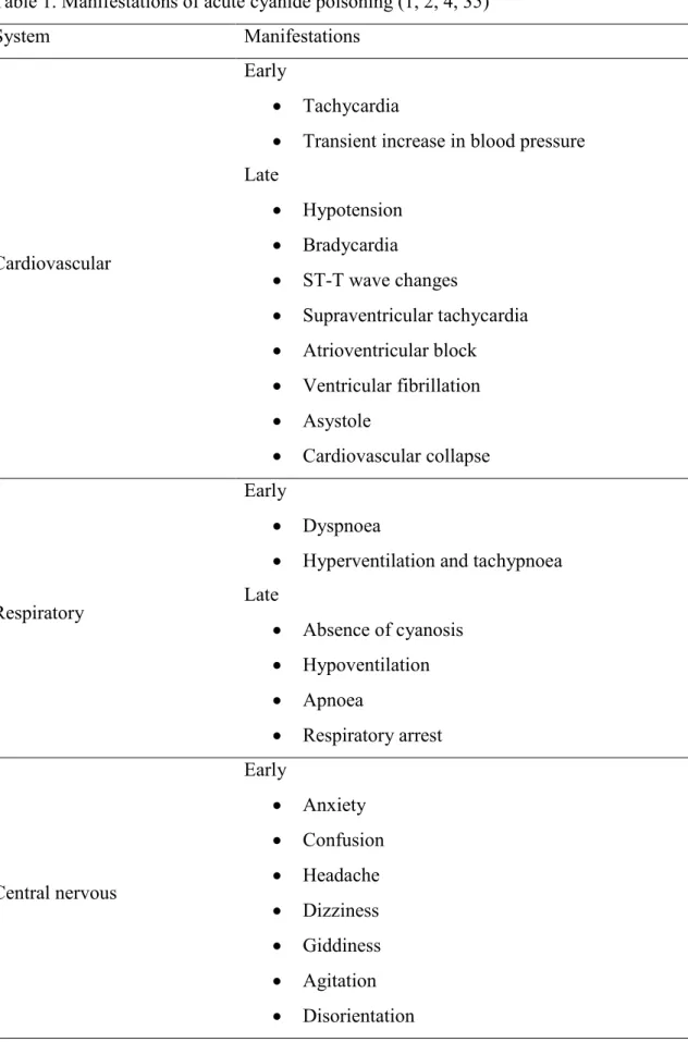
All body systems are affected by cyanide poisoning since cells of all tissues rely on oxygen and ATP. Therefore, clinical manifestations of acute cyanide toxicity are a reflection of intracellular hypoxia. The heart and brain are particularly susceptible to this poisoning because of their requirement for a continuous and large supply of oxygen and ATP for normal function. Early symptoms reflect reflexive attempts of the cardiovascular, respiratory and neurologic systems to overcome tissue hypoxia. Late symptoms reflect these systems depression as tissues fail to compensate for their failure to use oxygen (1). Table 1 lists common symptoms of acute cyanide intoxication.
Patients with acute cyanide poisoning have markedly elevated plasma lactate concentration, due to anaerobic metabolism. The normal range of plasma lactate concentration is 1 to 2 mmol/L, but in cyanide poisoning lactate concentration often exceeds 8 mmol/L. This value was shown to be highly sensitive (94%) and moderately specific (70%) for a toxic blood cyanide concentration of ≥1 mg/L, in a sample of 11 patients exposed solely to cyanide (11). The process that produces this rise in plasma lactate concentration leads also to a high anion gap metabolic acidosis, defined by an arterial blood pH of less than 7.35 with a plasma bicarbonate concentration of less than 22 mmol/L. Since cyanide poisoned cells are unable to extract and use oxygen from arterial blood, venous blood is often nearly as oxygenated as arterial blood, this leading to the characteristic laboratory finding on blood gas analysis of elevated venous oxygen with reduced arteriovenous oxygen saturation difference (<10 mmHg) in patients with acute cyanide poisoning (1, 4). Blood cyanide concentrations should be determined to confirm exposure to cyanide but the results of this test are generally not available in time to be useful in the initial diagnosis of poisoning. Moreover, blood cyanide concentrations can be misleading because of the rapid metabolism of cyanide, its instability in blood samples and the susceptibility of cyanide blood tests to interference (2). Blood
17 concentrations of cyanide greater than 0.5 mg/L are normally associated with cyanide toxicity, with levels approaching 3 mg/L or higher being potentially fatal if untreated (55).
Diagnosis
Since there is no blood cyanide test that can rapidly confirm poisoning, acute cyanide poisoning is presumptively diagnosed based on index of suspicion and clinical presentation. Rapid loss of consciousness and hemodynamic instability, in the presence of high anion gap metabolic acidosis with elevated plasma lactate (>8 mmol/L), particularly in someone in an occupation with potential exposure to cyanide, strongly suggest the possibility of acute cyanide poisoning (2).
Treatment
The first step in the initial management of acute cyanide poisoning is decontamination. Patients with suspected exposure to cyanide gas or fire smoke should be evacuated from the contaminated environment to an area with fresh air. Removal of contaminated clothing and washing of the skin with copious amounts of water should be done in cases of dermal exposure. In patients with suspected ingestion, emesis should not be induced. Activated charcoal may be administered if the victim is alert and the time is within one hour of the suspected ingestion, unless contraindicated. Early gastric lavage can also be done in the case of ingestion, if there is no contraindication (4, 56).
Supportive care should occur simultaneously with decontamination. The principles of basic life support triad (airway, breathing and circulation) apply to treatment of acute cyanide poisoning. The administration of 100% oxygen by nonrebreather mask or endotracheal tube
18 is indicated. Although this will not correct the problem, since cyanide poisoning involves deficient oxygen utilization instead of deficient availability, 100% oxygen may enhance the effectiveness of antidotal therapy and is also essential to effective treatment of concurrent carbon monoxide exposure. Critically ill patients should be given advanced life support, including endotracheal intubation for comatose patients, vasopressors for cardiovascular collapse, sodium bicarbonate for metabolic acidosis and anticonvulsants for controlling seizures (2, 4, 56).
After these measures are initiated, the key to treatment of acute cyanide poisoning is early administration of an antidote (4). The Cyanide Antidote Kit (CAK), 4-dimethylaminophenol (4-DMAP), dicobalt edetate and hydroxocobalamin are cyanide antidotes available in one or more countries around the world (57).
The CAK consists of amyl nitrite, sodium nitrite and sodium thiosulfate, which are given together to enhance the onset and duration of action relative to those achievable with administration of any of the components alone (57). Amyl nitrite is given via inhalation over 15 to 30 seconds while intravenous access is established. Then, sodium nitrite is administered intravenously over three to five minutes, followed by intravenous sodium thiosulfate administration during 30 minutes (4). Nitrite administration oxidizes ferrous (2+) iron to ferric (3+) iron in haemoglobin, causing the formation of methaemoglobin, which binds cyanide with greater affinity than cytochrome oxidase, releasing this enzyme for aerobic cellular respiration. Methaemoglobin serves as a temporary binding site for cyanide and thereby transiently reduces free (active) cyanide in the bloodstream (52). Sodium thiosulfate acts as a sulfhydryl donor that increases the rate of rhodanese-catalyzed transformation of cyanide to thiocyanate, which is renally excreted. The nitrites in the CAK may cause toxicity and pose potential risk to the patient. They can cause profound vasodilatation leading to hypotension, tachycardia and syncope. In addition, nitrite-induced methaemoglobinaemia
19 reduces the level of functional circulating haemoglobin, impairing oxygen transport to the cells. Therefore, it is desirable to monitor methaemoglobin levels, which should not exceed 20%. Methaemoglobinaemia can be especially dangerous for victims of cyanide poisoning from smoke inhalation, who already have a reduction in the oxygen-carrying capacity of the blood, secondary to concomitant carbon monoxide poisoning. Therefore, the CAK is generally considered unsuitable for use in the prehospital setting in those suspected of having cyanide poisoning from inhalation of fire smoke (4, 56, 57).
4-DMAP was first introduced in Germany as a cyanide antidote. It produces a profound methaemoglobinaemia, observed within a few minutes of its administration. Therefore, monitoring of methaemoglobin levels and availability of methylene blue for reversal of methaemoglobinaemia are recommended when 4-DMAP is used. It has the advantage that it may be administered intramuscularly and thus can be given by paramedical personnel. 4-DMAP has been used in combination with other antidotes and as sole antidotal therapy. It is associated with severe side effects, including reticulocytosis, nephrotoxicity and haemolysis. Necrosis at the site of intramuscular injection has also been seen and there is concern that absorption will be poor in the acute stage of intoxication/collapse, where there is peripheral shutdown. Given its safety profile, 4-DMAP is also not well suited for prehospital empiric treatment of acute cyanide poisoning, especially in fire smoke-inhalation victims (16, 55, 57).
Dicobalt edetate is currently used as a cyanide antidote in Europe. It is administered by fast (one minute) intravenous infusion and is very rapid in its effects, with the hypothesized mechanism of action being the chelation of cyanide to form cobalticyanide, which is much less toxic. But in the absence of cyanide to bind the free cobalt ions present in the solution containing the antidote, serious cobalt toxicity (cardiac and respiratory distress, abdominal pain and vomiting) can develop. Therefore, before giving dicobalt edetate one has to be certain that the victim is actually suffering from cyanide poisoning. Also, because it has been
20 suggested that glucose protects against cobalt toxicity, it is recommended that each injection of dicobalt edetate should be followed immediately by intravenous 50 mL of 50% glucose, even in patients with confirmed cyanide poisoning. Since this antidote is associated with significant toxicities, including vomiting, tachypnoea, chest pain, hypotension, ventricular arrhythmias, seizures, urticaria, facial, laryngeal or neck oedema and anaphylactic shock, it is not well suited for prehospital empiric treatment of acute cyanide poisoning and is recommended as a second-line antidote (15, 16, 55, 57).
Hydroxocobalamin combines with cyanide to form the renally excreted, nontoxic cyanocobalamin. In the blood, cyanocobalamin releases cyanide at a rate slow enough to allow the enzyme rhodanese to detoxify the cyanide in the liver. Cyanide has a greater affinity for hydroxocobalamin than for the cytochrome oxidase, releasing the mitochondria for aerobic cellular respiration (4). A dose of 5 g hydroxocobalamin is administered by rapid intravenous infusion when cyanide poisoning is suspected and a second dose has to be repeated according to seriousness (55). This antidote has a favourable tolerability and safety profiles. It has the advantage of not interfering with tissue oxygenation, unlike other antidotes (57). A transient and apparent harmless reddish-brown discoloration of the skin, mucous membranes and urine, are the most common drug-related side effects of treatment with this antidote. It occurs due to the red colour of the medication and appears to resolve within two to three days of administration of the drug (4, 9). Because hydroxocobalamin has maximum absorption at wavelengths used in many colorimetric assays it can interfere with certain laboratory blood tests, such as those for bilirubin, creatinine, creatine kinase, glucose, phosphorus, magnesium, serum iron, serum aspartate aminotransferase, carboxyhaemoglobin, methaemoglobin and oxyhaemoglobin. Whether the values are falsely high or low is specific to the auto-analyzer and none appear to be of major clinical significance. Also, the impact on colorimetric laboratory values is transient (4, 9, 57, 58). Hydroxocobalamin has also been
21 associated with headache, erythema, exceptional allergic reactions, mild and transient hypertension accompanied by reflex bradycardia (7, 9, 59). This transient elevation in blood pressure appears to not pose significant risks in individuals without cyanide toxicity and may improve the hemodynamic status in patients with acute cyanide poisoning who are hypotensive (59). The safety and tolerability profiles of hydroxocobalamin, its rapid onset of action, the fact that it is not harmful when administered to non-poisoned patients and is safe in smoke-inhalation victims are reasons that explain why hydroxocobalamin has been recommended as an antidote of first choice. It is used empirically in the prehospital and hospital settings for cases in which acute cyanide poisoning from ingestion or inhalation is suspected but cannot be confirmed within the short time available for initiating effective intervention (55, 57-59).
Sodium thiosulfate is synergistic with hydroxocobalamin and they are sometimes used together, especially in the critically ill patient. However, they should not be administered in the same intravenous line. It is believed that concurrent administration of sodium thiosulfate “recycles” the hydroxocobalamin binding and may reduce the amount of hydroxocobalamin required (9, 52, 58). Regarding cyanide toxicity induced by nitroprusside use, it may be prevented by sodium thiosulfate (infusion of 1 g to each 100 mg of sodium nitroprusside) or by hydroxocobalamin, while treatment of nitroprusside-induced acute cyanide poisoning with this later antidote is efficient (7, 55).
Follow-up
Discharge of the cyanide poisoned patient from hospital depends on the stabilization of symptoms and the resolution of acidosis and other metabolic abnormalities. Although most patients who survive cyanide toxicity will be free of complications, after discharge they
22 should be screening at regular intervals for delayed central nervous system effects (17). Patients may develop parkinsonism and other symptoms like dystonia, bradykinesia, slowed speech, personality changes, disordered memory and mathematical abilities (42, 56). These symptoms usually develop days to months after exposure to cyanide and are attributable either to the cerebral hypoxia from the acute intoxication process or to the direct toxic action of cyanide (56, 60, 61). Brain-imaging studies of patients with acute cyanide poisoning who manifested delayed central nervous systems effects show abnormalities in the basal ganglia, nigrostriatal pathways and the cerebral and cerebellar cortices (61, 62). Levodopa and dopamine agonists have been used in some of these cases, with apparent beneficial effects (61, 63).
Conclusion
The main causes of acute cyanide poisoning are inhalation of fire smoke, occupational or industrial accidents, suicide or homicide attempts, accidental ingestion and ingestion of cyanogenic substances, such as nitriles or cyanogenic plants. From these, smoke inhalation in a closed-space fire is the most common source in industrialised countries, and the possibility of cyanide intoxication in a fire victim should not be underestimated, especially if there is altered mental status or hemodynamic instability.
Many occupations have access to cyanide, so the possibility of attempted suicide, accidental ingestion or occupational accident with this poison should be considered by emergency physicians in an individual from those professional groups that presents with rapid loss of consciousness and cardiovascular instability, in the presence of elevated plasma lactate (> 8 mmol/L).
23 Hydroxocobalamin can be recommended as the first line antidote, in association with supportive treatment, in patients with suspected cyanide poisoning in the prehospital and hospital settings.
24 Acknowledgments
I am truly thankful to my co-authors, Daniel Moura MD, PhD and Luís Coentrão MD, for all the help, time, contribution and dedication.
25 Declaration of interest
26 References
1. Nelson L. Acute cyanide toxicity: mechanisms and manifestations. J Emerg Nurs 2006; 32:S8-11.
2. Borron SW. Recognition and treatment of acute cyanide poisoning. J Emerg Nurs 2006; 32:S12-8.
3. Schnepp R. Cyanide: sources, perceptions, and risks. J Emerg Nurs 2006; 32:S3-7. 4. Hamel J. A review of acute cyanide poisoning with a treatment update. Crit Care Nurse 2011; 31:72-81.
5. Musshoff F, Schmidt P, Daldrup T, Madea B. Cyanide fatalities: case studies of four suicides and one homicide. Am J Forensic Med Pathol 2002; 23:315-20.
6. Baud FJ, Barriot P, Toffis V, Riou B, Vicaut E, Lecarpentier Y, et al. Elevated blood cyanide concentrations in victims of smoke inhalation. N Engl J Med 1991; 325:1761-6. 7. Barillo DJ. Diagnosis and treatment of cyanide toxicity. J Burn Care Res 2009; 30:148-52.
8. Fortin JL, Giocanti JP, Ruttimann M, Kowalski JJ. Prehospital administration of hydroxocobalamin for smoke inhalation-associated cyanide poisoning: 8 years of experience in the Paris Fire Brigade. Clin Toxicol (Phila) 2006; 44 Suppl 1:37-44.
9. Hall AH, Dart R, Bogdan G. Sodium thiosulfate or hydroxocobalamin for the empiric treatment of cyanide poisoning? Ann Emerg Med 2007; 49:806-13.
10. Lawson-Smith P, Jansen EC, Hyldegaard O. Cyanide intoxication as part of smoke inhalation--a review on diagnosis and treatment from the emergency perspective. Scand J Trauma Resusc Emerg Med 2011; 19:14.
11. Baud FJ, Borron SW, Megarbane B, Trout H, Lapostolle F, Vicaut E, et al. Value of lactic acidosis in the assessment of the severity of acute cyanide poisoning. Crit Care Med 2002; 30:2044-50.
27 12. Seidl S, Schwarze B, Betz P. Lethal cyanide inhalation with post-mortem trans-cutaneous cyanide diffusion. Leg Med (Tokyo) 2003; 5:238-41.
13. Cherian MA, Richmond I. Fatal methane and cyanide poisoning as a result of handling industrial fish: a case report and review of the literature. J Clin Pathol 2000; 53:794-5.
14. Lam KK, Lau FL. An incident of hydrogen cyanide poisoning. Am J Emerg Med 2000; 18:172-5.
15. Guidotti T. Acute cyanide poisoning in prehospital care: new challenges, new tools for intervention. Prehosp Disaster Med 2006; 21:s40-8.
16. Cummings TF. The treatment of cyanide poisoning. Occup Med (Lond) 2004; 54:82-5.
17. Koschel MJ. Where there's smoke, there may be cyanide. Am J Nurs 2002; 102:39-42. 18. Musshoff F, Kirschbaum KM, Madea B. An uncommon case of a suicide with inhalation of hydrogen cyanide. Forensic Sci Int 2011; 204:e4-7.
19. Bebarta VS, Pitotti RL, Borys DJ, Morgan DL. Seven years of cyanide ingestions in the USA: critically ill patients are common, but antidote use is not. Emerg Med J 2011; 28:155-8.
20. Gill JR, Marker E, Stajic M. Suicide by cyanide: 17 deaths. J Forensic Sci 2004; 49:826-8.
21. Shields LB, Hunsaker DM, Hunsaker JC, 3rd, Ward MK. Toxicologic findings in suicide: a 10-year retrospective review of Kentucky medical examiner cases. Am J Forensic Med Pathol 2006; 27:106-12.
22. Kampe S, Iffland R, Korenkov M, Diefenbach C. Survival from a lethal blood concentration of cyanide with associated alcohol intoxication. Anaesthesia 2000; 55:1189-91.
28 23. De Busk RF, Seidl LG. Attempted suicide by cyanide. A report of two cases. Calif Med 1969; 110:394-6.
24. Fortin JL, Waroux S, Giocanti JP, Capellier G, Ruttimann M, Kowalski JJ. Hydroxocobalamin for poisoning caused by ingestion of potassium cyanide: a case study. J Emerg Med 2010; 39:320-4.
25. Coentrão L, Moura D. Acute cyanide poisoning among jewelry and textile industry workers. Am J Emerg Med 2011; 29:78-81.
26. Holstege CP, Forrester JD, Borek HA, Lawrence DT. A case of cyanide poisoning and the use of arterial blood gas analysis to direct therapy. Hosp Pract (Minneap) 2010; 38:69-74.
27. Walrath J, Li FP, Hoar SK, Mead MW, Fraumeni JF, Jr. Causes of death among female chemists. Am J Public Health 1985; 75:883-5.
28. Geller RJ, Alsop JA. Suicidal ingestions of metal polish cleaner in the Hmong community [abstract]. Clinical Toxicology 2005; 43:744-5.
29. Krieg A, Saxena K. Cyanide poisoning from metal cleaning solutions. Ann Emerg Med 1987; 16:582-4.
30. Garlich FM, Roberts DJ, Anderson DL, Kalugdan T. Suicidal cyanide deaths in the Hmong community [abstract]. Clinical Toxicology 2008; 46:617-.
31. Coentrão L, Neves A, Moura D. Hydroxocobalamin treatment of acute cyanide poisoning with a jewellery-cleaning solution. BMJ Case Reports 2010; doi:10.1136/bcr.01.2010.603.
32. Chin RG, Calderon Y. Acute cyanide poisoning: a case report. J Emerg Med 2000; 18:441-5.
29 34. Prajapati NC, Puri RK, Sarangi MP, Yadav S, Khalil A. Potassium cyanide poisoning. Indian Pediatr 1992; 29:903-5.
35. Geller RJ, Barthold C, Saiers JA, Hall AH. Pediatric cyanide poisoning: causes, manifestations, management, and unmet needs. Pediatrics 2006; 118:2146-58.
36. Mueller M, Borland C. Delayed cyanide poisoning following acetonitrile ingestion. Postgrad Med J 1997; 73:299-300.
37. Boggild MD, Peck RW, Tomson CR. Acetonitrile ingestion: delayed onset of cyanide poisoning due to concurrent ingestion of acetone. Postgrad Med J 1990; 66:40-1.
38. O'Brien B, Quigg C, Leong T. Severe cyanide toxicity from 'vitamin supplements'. Eur J Emerg Med 2005; 12:257-8.
39. Bromley J, Hughes BG, Leong DC, Buckley NA. Life-threatening interaction between complementary medicines: cyanide toxicity following ingestion of amygdalin and vitamin C. Ann Pharmacother 2005; 39:1566-9.
40. Hall AH, Linden CH, Kulig KW, Rumack BH. Cyanide poisoning from laetrile ingestion: role of nitrite therapy. Pediatrics 1986; 78:269-72.
41. Kalyanaraman UP, Kalyanaraman K, Cullinan SA, McLean JM. Neuromyopathy of cyanide intoxication due to "laetrile" (amygdalin). A clinicopathologic study. Cancer 1983; 51:2126-33.
42. Hall AH, Rumack BH. Clinical toxicology of cyanide. Ann Emerg Med 1986; 15:1067-74.
43. Sanchez-Verlaan P, Geeraerts T, Buys S, Riu-Poulenc B, Cabot C, Fourcade O, et al. An unusual cause of severe lactic acidosis: cyanide poisoning after bitter almond ingestion. Intensive Care Med 2011; 37:168-9.
30 44. Cigolini D, Ricci G, Zannoni M, Codogni R, De Luca M, Perfetti P, et al. Hydroxocobalamin treatment of acute cyanide poisoning from apricot kernels. Emerg Med J 2011; 28:804-5.
45. Suchard JR, Wallace KL, Gerkin RD. Acute cyanide toxicity caused by apricot kernel ingestion. Ann Emerg Med 1998; 32:742-4.
46. Akyildiz BN, Kurtoglu S, Kondolot M, Tunc A. Cyanide poisoning caused by ingestion of apricot seeds. Ann Trop Paediatr 2010; 30:39-43.
47. Shragg TA, Albertson TE, Fisher CJ, Jr. Cyanide poisoning after bitter almond ingestion. West J Med 1982; 136:65-9.
48. Pentore R, Venneri A, Nichelli P. Accidental choke-cherry poisoning: early symptoms and neurological sequelae of an unusual case of cyanide intoxication. Ital J Neurol Sci 1996; 17:233-5.
49. Dodds C, McKnight C. Cyanide toxicity after immersion and the hazards of dicobalt edetate. Br Med J (Clin Res Ed) 1985; 291:785-6.
50. Prieto I, Pujol I, Santiuste C, Poyo-Guerrero R, Diego A. Acute cyanide poisoning by subcutaneous injection. Emerg Med J 2005; 22:389-90.
51. Abeyasinghe NL, Perera HJM, Weerasinghe DSK. Case report - death by subcutaneous injection of cyanide in Sri Lanka. J Forensic Leg Med 2011; 18:182-3.
52. DesLauriers CA, Burda AM, Wahl M. Hydroxocobalamin as a cyanide antidote. Am J Ther 2006; 13:161-5.
53. Ehrlich E, Riesselmann B, Tsokos M. A series of hospital homicides. Leg Med (Tokyo) 2009; 11 Suppl 1:S100-2.
54. Cheung AT, Cruz-Shiavone GE, Meng QC, Pochettino A, Augoustides JA, Bavaria JE, et al. Cardiopulmonary bypass, hemolysis, and nitroprusside-induced cyanide production. Anesth Analg 2007; 105:29-33.
31 55. Megarbane B, Delahaye A, Goldgran-Toledano D, Baud FJ. Antidotal treatment of cyanide poisoning. J Chin Med Assoc 2003; 66:193-203.
56. Koschel MJ. Management of the cyanide-poisoned patient. J Emerg Nurs 2006; 32:S19-26.
57. Hall AH, Saiers J, Baud F. Which cyanide antidote? Crit Rev Toxicol 2009; 39:541-52.
58. Borron SW, Baud FJ, Megarbane B, Bismuth C. Hydroxocobalamin for severe acute cyanide poisoning by ingestion or inhalation. Am J Emerg Med 2007; 25:551-8.
59. Borron SW, Baud FJ, Barriot P, Imbert M, Bismuth C. Prospective study of hydroxocobalamin for acute cyanide poisoning in smoke inhalation. Ann Emerg Med 2007; 49:794-801.
60. Zaknun JJ, Stieglbauer K, Trenkler J, Aichner F. Cyanide-induced akinetic rigid syndrome: clinical, MRI, FDG-PET, beta-CIT and HMPAO SPECT findings. Parkinsonism Relat Disord 2005; 11:125-9.
61. Rachinger J, Fellner FA, Stieglbauer K, Trenkler J. MR changes after acute cyanide intoxication. AJNR Am J Neuroradiol 2002; 23:1398-401.
62. Kasamo K, Okuhata Y, Satoh R, Ikeda M, Takahashi S, Kamata R, et al. Chronological changes of MRI findings on striatal damage after acute cyanide intoxication: pathogenesis of the damage and its selectivity, and prevention for neurological sequelae: a case report. Eur Arch Psychiatry Clin Neurosci 1993; 243:71-4.
63. Valenzuela R, Court J, Godoy J. Delayed cyanide induced dystonia. J Neurol Neurosurg Psychiatry 1992; 55:198-9.
32 Table
Table 1. Manifestations of acute cyanide poisoning (1, 2, 4, 35)
System Manifestations
Cardiovascular
Early
• Tachycardia
• Transient increase in blood pressure Late • Hypotension • Bradycardia • ST-T wave changes • Supraventricular tachycardia • Atrioventricular block • Ventricular fibrillation • Asystole • Cardiovascular collapse Respiratory Early • Dyspnoea
• Hyperventilation and tachypnoea Late • Absence of cyanosis • Hypoventilation • Apnoea • Respiratory arrest Central nervous Early • Anxiety • Confusion • Headache • Dizziness • Giddiness • Agitation • Disorientation
33 • Weakness • Malaise • Mydriasis Late • Lethargy • Stupor • Generalized seizures • Paralysis • Coma • Cerebral death Gastrointestinal • Nausea • Vomiting • Abdominal pain
Dermatologic • Cherry-red discoloration of the skin Other • Bright-red retinal veins and arteries • Bitter-almond odor in some victims
Anexo
Clinical Toxicology
Instructions for Authors
About the Journal
Aims and Scope Editor-in-Chief
Manuscript Submission Manuscript Preparation
File preparation and types Title Page
Abstract Main Text
Acknowledgments and Declaration of Interest sections References Tables Illustrations Notes on Style Editorial Policies Authorship Submission Peer Review Ethics and Consent Copyright and Permissions Declaration of Interest NIH and Public Access Policy
Additional Information
Proofs Reprints
Color figure charges Contact the Publisher
About the Journal
Aims and Scope
Clinical Toxicology publishes peer-reviewed scientific research and clinical advances in
clinical toxicology. The journal reflects the professional concerns and best scientific judgment of its sponsors, the American Academy of Clinical Toxicology, the European Association of Poisons Centres and Clinical Toxicologists and the American Association of Poisons Control Centers. As such, it is the leading international journal in the specialty.
Clinical Toxicology aims to be the journal of primary interest to practicing clinical
toxicologists, whether in hospitals, poison centers, academia, government or industry. The journal is relevant to all professionals at the interface of clinical toxicology with acute care, basic toxicology studies relevant to clinical practice, occupational and environmental medicine, public health, regulatory toxicology, pharmacology and pharmaceutics, and analytical and forensic pathology.
Editor-in-Chief
Prof D Nicholas Bateman BSc MD FRCP FRCP(E) FBPharmacolS, FBTS Royal Infirmary of Edinburgh
Edinburgh Scotland, UK
Manuscript Submission
All submissions should be made online at Clinical Toxicology’s ScholarOne Manuscripts site. New users should first create an account. Once a user is logged onto the site, submissions should be made via the Author Center. If you experience any problems with your submission or with the site, please contact ScholarOne support through the „get help now‟ link.
All submissions to the journal must include full disclosure of all relationships that could be viewed as presenting a potential conflict of interest. If there are no conflicts of interest, authors should state that there are none. This must be stated at the point of submission (within the manuscript, after the main text under a subheading "Declaration of interest", and, where available within the appropriate field on the journal's ScholarOne Manuscripts site). Please see our full Declaration of Interest Policy for further information.
In line with international policy all clinical trials submitted to the journal should have appropriate ethics (IRB) approval and been registered with the appropriate body (www.clinicaltrials.gov).
Manuscript Preparation
File preparation and types
Manuscripts are preferred in Microsoft Word format (.doc files). Documents must be double-spaced, with margins of one inch on all sides. The title page should not appear in the main text, but should be submitted as a separate digital file and designated as „title page – not for review‟ on ScholarOne Manuscripts. Tables and figures should not appear in the main text, but should be submitted as separate digital files and designated with the appropriate file type on ScholarOne Manuscripts. References should be given in Vancouver style (see References section for example).
Manuscripts should be compiled in the following order: title page; abstract; main text; acknowledgments; declaration of interest statement; appendices (as appropriate); references; tables with captions (on separate pages); figures; figure captions (as a list).
Clinical Toxicology publishes the following manuscript types:
Original articles Brief communications Reviews Commentaries Letters Title Page
A title page should be provided comprising the manuscript title plus the full names and affiliations of all authors involved in the preparation of the manuscript. One author should be clearly designated as the corresponding author and full contact information, including phone number and email address, provided for this person. Three to five key terms that are not in the title should also be included on the title page. The keywords will assist indexers in cross indexing your article. The title page should also specify if the paper is written in UK or US English. The title page should be uploaded separately to the main manuscript and designated as „title page – not for review‟ on ScholarOne Manuscripts.
All original articles should start with an abstract of 300 or fewer words, and reviews with an abstract of 600 or fewer words, summarising the central core of knowledge that is the focus of the paper. The recommended format is as a structured abstract, with the following headings for an original article: context, objective, materials and methods, results, discussion and conclusion. For a review article, it should be structured as follows: context, objective, methods (including data sources, study selection and data extraction), results and conclusion. It should be written in an informative style permitting its use, without revision, by abstracting services, give essential details of research findings without further reference to the text, and avoid generalisations and nonessential information.
For case reports a structured abstract may be briefer and headings can be reduced: context, case details, discussion.
Main Text
Original articles
The body of the article should include the following sections: introduction; methods; results; discussion; conclusions. An advisory maximum word limit of 3000 words is suggested for an initial original submission.
Introduction: This section should state the relevance and background to the study, and its
rationale and purpose.
Methods: This section should include only information that was available at the time the plan
or protocol for the study was being written. You should describe your selection of the observational or experimental participants, identify the methods, apparatus and procedures in sufficient detail to allow others to reproduce the results, and describe statistical methods with enough detail to enable a knowledgeable reader with access to the original data to verify the reported results. Clinical Toxicology requires that studies involving humans, both volunteers and patients, or animals be approved by an institutional review board, in accordance with approved published guidelines, prior to actually performing the research and publishing the data. Details including clinical trial registration number must be provided in the methods section if research includes studies conducted on human volunteers.
Results: Present your results in logical sequence in the text, tables, and illustrations.
Discussion: This should include implications of the findings and their limitations, with
reference to all other relevant studies and the possibilities these suggest for future research.
Conclusions: This must summarize the main paper. Ensure that extrapolations are
reasonable and that conclusions are justified by the data presented, and indicate if the study design can be generalized to a broader study population.
The appropriate guidelines should be adhered to when reporting on randomised controlled trials (e.g. CONSORT, available at http://www.consort-statement.org/).
Reviews
Reviews are usually solicited by the Review Editors, but uncommissioned material will also be considered. Please contact the Section Editors, Allister Vale ([email protected]) or Michael Mullins ([email protected]) before writing a review for the Journal to ensure that the topic is appropriate for the Journal and does not conflict with another review. All reviews, whether solicited or not, will undergo the same peer review and editorial process as original research reports. The Journal will usually publish the review within six months of acceptance.
Reviews should provide a scholarly, evidence-based, critical analysis of topics relevant to practicing clinical toxicologists and other clinicians and scientists at the interface with acute care, occupational and environmental medicine, public health, regulatory toxicology, pharmacology and pharmaceutics, and analytical and forensic pathology. It is essential that details of the literature search methodology are provided, i.e. the databases searched (normally Medline and at least one or two other databases), the search terms and inclusive dates, and any selectivity criteria imposed.
Authors may include previously unpublished data that may help to clarify an ongoing controversy.
Reviews should:
(i) Focus on a specific toxicological issue of importance (3500-5000 words with an abstract of up to 600 words and up to 100 references); or
(ii) Provide a comprehensive overview of an important area of toxicology, with due emphasis being given to developments during the past five years (5000-10000 words including an abstract of 600 words and up to 150 references).
Focused reviews are particularly welcome as readers find these to be of great assistance in their practice.
Brief Communications and Case Reports
Clinical Toxicology will consider for publication, short reports of one or more patients that
described significant original observations related to acute or chronic clinical toxicology, or adverse drug reactions or drug-drug interactions that have a direct relation to clinical toxicology. Such reports should include a description of the new or unusual features, or observations that generate a hypothesis or support a change in clinical practice.
Brief Communications are limited to 1000 words, not including a structured abstract of no more than 200 words. To structure the abstract, use the same terms used to break the text into sections. Abbreviations, diagrams, and references are not permitted in the abstract.
These reports should not include lengthy reviews of the existing medical literature. Tables, figures, and references are not included in the 1000 word count. There is a maximum of one table or figure. References are normally limited to 10. A statement of informed patient consent, or approval (or exemption statement) by an Investigational Review Board (Human Studies Committee or Ethics Committee or Animal Care and Use Committee) must normally be submitted with the manuscript.
Manuscripts must meet one of the following criteria: (i) Unexpected or unusual presentations of toxicity;
(ii) New associations or variations in toxicity related disease processes;
(iii) Presentations, diagnoses or management of new and emerging toxicity-related diseases;
(iv) An unexpected association between toxin or symptoms;
(v) An unexpected event in the course of observing or treating a patient;
(vi) Findings that shed new light on the possible treatment or pathogenesis of a toxicity-related disease.
You must make it clear to the Editor what your case adds to the field of clinical toxicology. In your cover letter, please consider the following:
Do you believe it is the first report of this kind in the literature?
Will it significantly advance our understanding of a particular clinical toxicology syndrome, mechanism or be supportive of a new hypothesis?
Acknowledgments and Declaration of Interest sections
Acknowledgments and Declaration of interest sections are different and each has a specific purpose. The Acknowledgments section details special thanks, personal assistance, and dedications. Contributions from individuals who do not quality for authorship should also be acknowledged here.
Declarations of interest, however, refer to statements of financial support and/or statements of potential conflict of interest. Within this section also belongs disclosure of scientific writing assistance (use of an agency or agency/ freelance writer), grant support and numbers, and
statements of employment, if applicable. For a more detailed list of points to include, please see “Declaration of Interest section” below.
Acknowledgments section
Any acknowledgments authors wish to make should be included in a separate headed section at the end of the manuscript preceding any appendices, and before the references section. Please do not incorporate acknowledgments into notes or biographical notes. In order for this section to be included, the lead author must submit a written statement from the subjects whom he or she intends to acknowledge that they have been informed of the intent and approve it.
Declaration of Interest section
All declarations of interest must be outlined under the subheading „Declaration of interest‟. If authors have no declarations of interest to report, this must be explicitly stated. The suggested, but not mandatory, wording in such an instance is: The authors report no
declarations of interest. When submitting a paper via ScholarOne Manuscripts, the
„Declaration of interest‟ field is compulsory (authors must either state the disclosures or report that there are none). If this section is left empty authors will not be able to progress with the submission.
Please see our full Declaration of Interest Policy for further information.
Please note: for NIH/Wellcome-funded papers, the grant number(s) must be included in the Declaration of Interest statement.
References
References should be given in the Vancouver style. Citation in the text is by superscript Arabic numbers outside of the punctuation. References should be arranged in a numbered list at the end of the manuscript following the style below:
Journal: Mei-Tal V, Meyerowitz S, Engel G. The role of psychological process in a
somatic disorder: multiple sclerosis. Psychosom Med 1970;32:67-89.
Book: Kao F. An Introduction to Respiratory Physiology. New York: Elsevier North
Holland, 1973:50-61.
Contribution to a book: Tung PS, Fritz IB. Isolation and culture of testicular cells: a
morphological characterization. In: Hafez ES, ed. Techniques of Human Andrology. Amsterdam: Elsevier North Holland, 1977:125-43.
Electronic Resources: Lin A-S, Shibano M, Nakagawa-Goto K, Tokuda H, Itokawa H,
Morris-Natschke, SL, Lee K-H. Cancer Preventive Agents. 7. Antitumor-Promoting Effects of Seven Active Flavonolignans from Milk Thistle (Silybum marianum) on Epstein-Barr Virus Activation. Pharm Biol [Online] 2007;45:735-738. Available at: http://www.informapharmascience.com/doi/abs/10.1080/13880200701585592. Accessed on 12 April 2009.
Periodical abbreviations should follow the style given by Index Medicus. Tables
Tables should be used only when they can present information more efficiently than running text. Care should be taken to avoid any arrangement that unduly increases the depth of a table, and the column heads should be made as brief as possible, using abbreviations liberally. Lines of data should not be numbered nor run numbers given unless those numbers are needed for reference in the text. Columns should not contain only one or two entries, nor should the same entry be repeated numerous times consecutively. Tables should be grouped at the end of the manuscript on separate pages.