1
Universidade do Algarve
Departamento de Ciências Biomédicas e Medicina
Synthesis and evaluation of polymer
nanoparticles for delivery in RPE cells
Diogo Miguel Barros Bitoque
Dissertação de Mestrado para a obtenção do grau de Mestre
em Ciências Biomédicas
Trabalho efectuado sob a orientação de: Prof. Dra. Gabriela
Silva
2
Synthesis and evaluation of polymer
nanoparticles for delivery in RPE cells
Declaração de autoria de trabalho:
Declaro ser o autor deste trabalho, que é original e inédito. Autores e trabalhos consultados estão devidamente citados no texto e constam da listagem de referências incluída.
Diogo Miguel Barros Bitoque
Copyright
A Universidade do Algarve tem o direito, perpétuo e sem limites geográficos, de arquivar e publicitar este trabalho através de exemplares impressos reproduzidos em papel ou de forma digital, ou por qualquer outro meio conhecido ou que venha a ser inventado, de o divulgar através de repositórios científicos e de admitir a sua cópia e distribuição com objetivos educacionais ou de investigação, não comerciais, desde que seja dado crédito ao autor e editor.
3
Index
Acknowledgments ... 5 Abbreviations ... 6 Abstract ... 9 Resumo ... 11 Introduction ... 151. Non-viral vectors for gene therapy ... 16
2. Cellular barriers to non-viral gene delivery ... 16
2.1. Crossing of the cell membrane ... 17
2.2. Endo- lysosomal escape ... 18
2.3. Vector/DNA dissociation ... 19
2.4. Cytosolic trafficking and nuclear import ... 20
2.5. Gene expression ... 21
3. Lipossome-based vectors ... 21
4. Polymers ... 22
4.1 Chitosan ... 23
4.2 Polyethylenimine (PEI) ... 25
4.3 Poly(2-dimethylaminoethyl methacrylate) (PDMAEMA) ... 27
5. Polymer synthesis: Reversible Addition-Fragmentation chain Transfer (RAFT) ... 29
6. Therapeutic target ... 30
7. Aim of the work ... 31
Methods and Materials ... 32
1. Synthesis of PDMAEMA by RAFT ... 32
2. Gel permeation chromatography ... 32
3. Preparation of PDMAEMA/pDNA polyplexes ... 33
4. Determination of size and surface charge of polyplexes ... 34
5. Morphologic analysis of PDMAEMA polyplexes by transmission electron microscopy ... 34
6. Stability assay over time ... 35
7. Electrophoretic mobility shift assay (EMSA) ... 35
8. DNAse protection assay ... 35
9. Stability assay in the presence of serum proteins ... 36
10. Cell line and culture conditions ... 36
11. Cell viability assay for cytotoxicity evaluation ... 37
4
13. Animals ... 39
14. Activation of microglia after intravitreous injection in C57/BL6 mice... 39
15. Statistical analysis ... 40
Results and Discussion ... 41
Part one: ... 41
1. Characterization of 354kDa PDMAEMA-DNA polyplexes ... 41
1.1 PDMAEMA-DNA polyplexes have characteristics compatible with gene therapy ... 41
1.2 PDMAEMA efficiently complexes pDNA ... 43
1.3 PDMAEMA polyplexes protect DNA from DNAse degradation ... 44
1.4 Stability assay of polyplexes over time ... 46
2. Evaluation of the cytotoxicity of the PDMAEMA polymer and PDMAEMA-DNA polyplexes ... 47
3. Transfection efficiency of PDMAEMA-DNA polyplexes ... 51
4. Activation of microglia after intravitreous injection in C57/BL6 mice... 54
Part two: ... 55
1. Synthesis and structural characterization of a 200kDa PDMAEMA ... 55
. ... 56
2. The 103.3kDa PDMAEMA is able to form PDMAEMA-DNA polyplexes ... 57
2.1 PDMAEMA-DNA polyplexes have characteristics compatible with gene therapy ... 57
2.2 PDMAEMA efficiently complexes pDNA ... 58
2.3 PDMAEMA polyplexes protect DNA from DNAse degradation ... 59
2.4 PDMAEMA-DNA polyplexes are stable in in vitro culture conditions ... 60
3. Cytotoxicity of the PDMAEMA polymer and PDMAEMA-DNA polyplexes is concentration-dependent ... 61
4. 103.3kDa PDMAEMA-DNA polyplexes transfect D407 cells ... 63
Conclusions ... 66
Future work ... 67
5
Acknowledgments
Antes de mais, eu quero agradecer à Professora Doutora Gabriela Silva por me ter dado a oportunidade de realizar este trabalho no seu laboratório, e com o seu grupo. Tenho que agradeçer a sua disponibilidade, atenção e boa disposição.
Quero agradecer a todos os elementos do meu grupo laboratorial: Vanessa Oliveira, Sofia Calado, Susana Machado e Sónia Simão. Obrigado pela paciência e pelos conhecimentos que me transmitiram. Um especial agradecimento à Mónica Fernandes e Marinella Ghezzo.
Um especial agradecimento para a minha melhor amiga, obrigado pela tua ajuda e sem a tua amizade seria tudo muito mais difícil, estarás sempre no meu coração.
Não podia deixar de agradeçer à minha família, em especial aos meus pais, que têm sido muito pacientes comigo, agradeço toda a ajuda e formação que me deram para a vida. Adoro-vos.
6
Abbreviations
103.3kDa PDMAEMA – PDMAEMA with 103.3kDa 354kDa PDMAEMA – PDMAEMA with 354kDa
AAV - Adeno-associated virus
AIBN - 2,2'-azobisisobutyronitrile
ATRP - Atom transfer radical polymerization BRB – Blood retinal barrier
B16F10 - Mouse melanoma cell line CHEMS - Cholesteryl hemisuccinate CHO - Chinese hamster ovary cell line
COS-7 - African green monkey kidney cell line CpG – Cytosine-Guanine dinucleotide
CTA - chain transfer agent
D407- Human Retinal Pigment Epithelial cell line DEAE-dextran – Diethylaminoethyl-dextran DLS – Dynamic Light Scattering
DNA - Deoxyribonucleic acid
DMAEMA - (2-(N,N-dimethylamino)ethylmethacrylate) DMEM - Dulbecco’s Modified Eagle Medium
DOPE - Dioleylphosphatidyl ethanolamine DOPC - Dioleylphosphatidyl choline
7 EMA - European Medicines Agency
EMSA - Electrophoretic mobility shift assay FBS- Fetal Bovine Serum
GFP – Green Fluorescent Protein
GGT- Gamma-glutamyltransferase GPC - Gel Permeation Chromatography HEK293 - Human Embryonic Kidney cells HCl - Hydrochloric acid
MCF-7 - Human breast adenocarcinoma cell line
MIDGE – Minimal Immunogenically Defined Gene Expression
MTT- (3-[4,5-dimethylthiazol-2-yl]2,5-diphenyltetrazolium bromide
Mn - Number-average molecular weight
Mw - Weight-average molecular weight
NIPAM - Poly(N-isopropylacrylamide) NLS – Nuclear Localization Signal
N/P - nitrogen/phosphorus NPC – Nuclear Pore Complex
1H
NMR- Nuclear Magnetic Resonance
OCT - Optimal Cutting Temperature compound OVCAR-3 - Human ovarian carcinoma cell line PAMAM – Polyamidoamine
PdI- polydispersity index
8 pDNA - plasmid DNA
PEG - Polyethylene glycol PFA - Paraformaldehyde
pFAR – plasmid Free of Antibiotic Resistance gene
PPI - Polypropylenimine
RPE - Retinal Pigmented Epithelium PEI – Polyethylenimine
RAFT - Reversible Addition-Fragmentation chain Transfer rPDMAEMA – reducible PDMAEMA
rpm - Revolutions per minute
S/MAR – Scaffold/Matrix Attachment Region
TAE - Tris-Acetate-EDTA buffer
TEM – Transmission Electron Microscopy
9
Abstract
Ocular pathologies are among the most debilitating medical conditions affecting all segments of the population. Traditional treatment options are often ineffective, and gene therapy has the potential to become an alternative approach for the treatment of several pathologies.
Methacrylate polymers have been described as highly biocompatible and are successfully used in medical applications. Due to their cationic nature, these polymers can be used to form polyplexes with DNA for its delivery. This work aims to study the potential of PDMAEMA (poly(2-(N,N’-dimethylamino)ethyl methacrylate)) as a non viral gene delivery system to the retina.
The first part of this work aimed to study the potential for gene delivery of a previously synthesized PDMAEMA polymer of high molecular weight (354kDa). In the second part, we synthesized by RAFT a PDMAEMA with a lower molecular weight (103.3kDa) and similarly, evaluated its ability to act as a gene delivery vehicle.
PDMAEMA/DNA polyplexes were prepared at 5, 7.5, 10, 12.5 and 20 nitrogen/phosphorous (N/P) ratio for the 354kDa PDMAEMA and at 5 and 7.5 for the 103.3kDa PDMAEMA. Dynamic light scattering and zeta potential measurements confirmed the nanosize and positive charge of polyplexes for all ratios and for both polymers. Both high and low Mw PDMAEMA were able to efficiently complex and protect DNA from DNase I degradation. Their cytotoxicity was evaluated using a non-retinal cell line (HEK293) and a retinal pigment epithelium (RPE) cell line (D407). We have found that cytotoxicity of the free polymer is concentration and time dependent, as expected, and negligible for all the concentrations of the PDMAEMA-DNA polyplexes. Furthermore, for the concentrations to be used in vivo, the 354kDa PDMAEMA showed no signs of inflammation upon injection in the intravitreal space of C57BL/6 mice.
The transfection efficiency, as evaluated by fluorescence microscopy and flow cytometry, showed that the D407 retinal cells were transfected by polyplexes of both high and low Mw PDMAEMA, but with varied efficiency, which was dependent on the N/P ratio.
10 Althogether, these results suggest that PDMAEMA is a feasible candidate for non-viral gene delivery to the retina, and this work constitutes the basis of further studies to elucidate the bottleneck in transfection and further optimization of the material.
11
Resumo
Desde o século passado que as doenças relacionadas com o genoma têm vindo a ganhar importância. O projecto de mapeamento do genoma humano permitiu descobrir a origem de muitas doenças. Várias abordagens terapêuticas têm sido objecto de estudo, sendo a terapia génica uma das mais promissoras. A terapia génica tem como objectivo usar material genético, em regra DNA, para manipular as células dos pacientes. Antes do aparecimento da terapia génica existia apenas tratamento dos sintomas ou terapia de substituição para várias patologias, e não uma verdadeira cura. Para as doenças oculares, que estão entre as doenças mais debilitantes que afectam todos os segmentos da população, esta realidade era sobretudo relevante. Em terapia génica existe a necessidade de desenvolver veículos para transporte de material genético, veículo este que tem que proteger o ácido nucleico de degradação, ter especificidade para células/órgão, permitir a expressão do gene de interesse ao longo do tempo e com níveis adequados, não desencadear resposta imunitária (excepto em caso de interesse como no cancro e vacinas) e que seja administrado por injecção sistémica. As duas principais estratégias para entrega de material genético usa vectores virais e não-virais. Os sistemas virais de entrega apresentam diversas vantagens, mas também várias limitações: imunogenicidade, baixa capacidade de empacotamento do material genético, potential inserção aleatória no genoma hospedeiro e toxicidade. Os vectores não virais, de entre os quais se destacam os polímeros, surgiram como uma alternativa para contornar esses problemas, pois estes apresentam maiores benefícios em termos de segurança, versatibilidade química e estrutural para a manipulação das propriedades fisico-químicas, maior capacidade de empacotamento de genes e estabilidade durante o armazenamento.
Dentro da classe dos polímeros, os polímeros naturais como o quitosano e alginato têm ganho importância devido à sua biocompatibilidade, mas são os polímeros sintéticos, como a poli(etilenimina) (PEI), a poli(L-lisina) (PLL) e o poli(2-dimetilamino)etil metacrilato (PDMAEMA), que tem maior relevância
12 devido ao controlo preciso da sua síntese e consequentemente das suas propriedades. A natureza catiónica de alguns polímeros, como os indicados acima, permite que estes sejam usados para formar poliplexos com material genético.
O objectivo do nosso grupo de investigação é o desenvolvimento de novas estratégias baseadas em terapia génica para tratamento de patologias da retina. Dentro deste enquadramento, o presente trabalho tem como objectivo compreender o potencial do poli(2-dimetilamino)etil metacrilato (PDMAEMA) como sistema de entrega de material genético na retina. Na primeira parte deste trabalho um polímero PDMAEMA com 354kDa, que tinha sido previamente sintetizado, foi avaliado para o seu potencial como veículo para terapia génica. Na segunda parte, sintetizamos um PDMAEMA com um menor peso molecular (103.3kDa) via RAFT (Reversible addition-fragmentation chain-transfer) para efectuar uma comparação entre a eficiência de transfecção de PDMAEMAs com diferentes pesos moleculares.
Poliplexos de PDMAEMA/DNA foram preparados nos rácios
amina(N)/fosfato(P) de 5, 7.5, 10, 12.5 e 20 com o PDMAEMA 354kDa e nos rácios 5 e 7.5 com o PDMAEMA 103.3kDa. Medições de DLS (Dynamic light scattering) e potencial zeta confirmam que foram preparados poliplexos à escala nanométrica com carga positiva, em todos os rácios. Ambos os polímeros PDMAEMA são capazes de complexar eficientemente e proteger o DNA da degradação por parte da DNAse I.
A citoxicidade de ambos os polímeros foi avaliada usando duas linhas celulares: a HEK293 (linha celular de rim embriónico humano) e a D407 (linha celular de epitélio pigmentar da retina). Os resultados revelam que a citoxicidade do polimero livre (não-complexado) é dependente da concentração e do tempo de exposição, como esperado, e os poliplexos PDMAEMA/DNA não apresentam citoxicidade. Foi também observado que nas concentrações usadas in vivo a citoxicidade do PDMAEMA 354kDa é negligível, como demonstrado pela ausência de inflamação após a injecção no espaço intravítreo de ratinhos C57BL6.
13 A eficiência de transfecção foi avaliada qualitativamente por microscopia de fluorescência e quantitativamente por citometria de fluxo e mostrou que os poliplexos PDMAEMA/DNA são capazes de transfectar, e que a eficiência varia de acordo com o rácio N/P utilizado, para os polimeros de alto e baixo peso molecular.
Em suma, estes resultados sugerem que o PDMAEMA é um candidato viável para a entrega de genes na retina, e este trabalho constitui a base de estudos futuros que visam elucidar o passo limitante na transfecção e desse modo permitir a optimização deste vector não-viral.
14 Part of this work was submitted to the Journal of Tissue Engineering and Regenerative Medicine, “Efficiency of RAFT-synthesized PDMAEMA in gene transfer to the retina”, 2013 and was also presented as a poster in the American Society of Gene and Cell Therapy Annual Meeting 2012, “PDMAEMA as a delivery vector for gene therapy of the retina”; in the 8th International Meeting of the Portuguese Society for Stem Cells and Cell Therapies, "Hybrid Strategies For Retinal Gene Therapy"; and in the European Society of Gene and Cell Therapy British Society for Gene Therapy Collaborative Congress 2013,
15
Introduction
The aim of gene therapy is treat diseases by delivering therapeutic genes to diseased cells or blocks the expression of a dysfunctional gene. This therapeutic strategy has been heralded as the next development in modern medicine [1, 2], since it has the potential to cure genetic diseases and other diseases such as cancer that are responsible for a large number of deaths and decreased quality of life. There are two very important components of gene therapy that are absolutely necessary for a successful gene therapy approach: an efficient and safe delivery system, coupled to an effective gene expression system that can be expressed over time at the target site [1].
In gene therapy, there are two delivery approaches: viral and non-viral. The first one relies on modified virus to deliver the genetic material, since viruses have a natural capability to infect cells and to express their genes in the host. Viral gene delivery can be achieved by modifications in adenoviruses, retroviruses, lentiviruses and adeno-associated viruses (AAVs), among others. These viral vectors can be classified as nonlytic (i.e. virus that produce virions and leave the host cell intact) and lytic (i.e. virus that produce virions and cause death to the host cell) [3]. The modifications made on viruses aim to render them replication-deficient, which do not allow them to replicate and exit the cell as mature viral particles that can further infect other cells. Viral vectors have several advantages, such as sustained expression due to DNA integration into the host genome, some viruses have tropism for specific cell types, and high levels of gene expression [4]. However, there are some drawbacks, including immunogenicity, low capacity of gene packaging, random transgen insertion into the host genome, toxicity and limitations to large-scale production [1, 5]. Despite of these disadvantages, several viral vectors have reached the clinical trial stage (Clinicaltrials.gov NCT01024998, NCT00999609, NCT01461213) and a adenoviral based product – Gendicine® – has been approved in Chine and U.S. In Europe, glybera®, a product based on AAVs, has recently been
16 approved by EMA (European Medicines Agency) for the treatment of adults with lipoprotein lipase deficiency [6].
Despite the success of viral vectors, the safety issues have motivated the search for safer, less immunogenic and pathogenic gene delivery alternatives, which include polymer and lipid-based vectors, and inorganic materials [1, 2]. Other approaches that are also being studied is the use of a physical mean to deliver the DNA, like electroporation or gene gun [7], which shows several limitations in clinical practice.
The new potential delivery systems bring the promise of safety benefits, structural and chemical versatility for manipulating physicochemical properties, bigger gene capacity and vector stability upon storage.
1. Non-viral vectors for gene therapy
The non-viral approach to the development of a gene delivery system must surpass two main barriers before achieving therapeutic sucess: limited internalization into the cell and the nucleus and short term gene expression.
2. Cellular barriers to non-viral gene delivery
An efficient delivery system must accomplish several steps to be able to express the gene of interest (figure 1): (I) enter the cell by crossing the cell membrane; (II) escape the endo-lysosomal degradation pathway; (III) release the genetic material; (IV) traffic through the cytoplasm and enter the nucleus; and last (V), the gene expression has to occur for synthesis of the protein of interest [1].
17
Figure 1 – Representation of the main barriers to gene delivery: (I) enter the cell membrane; (II) escape the endo-lysosomal pathway; (III) the genetic material must be released from the carrier; (IV) traffic through the cytoplasm and into the nucleus; and last (V), the gene expression has to occurred and expression of the protein of interest (Adapted from [1]).
2.1. Crossing of the cell membrane
The cell membrane has a negatively net charge, due to the presence of certain lipids and proteoglycans. Since DNA is a molecule with a negative charge, “naked” DNA cannot entry the cell by electrostatically interaction with the membrane [8]. Therefore, a positively charged system might electrostatically interact with the cellular membrane, and this will permit the entry in the cell by a well described process of endocytosis.
Endocytosis is a process by which cells absorb extracellular molecules by forming invaginations in the cell membrane and the vesicles enter the cytoplasm [9]. This process exists in several forms: macropinocytosis, phagocytosis and receptor- mediated endocytosis.
18 - Macropinocytodis is the formation of large uncoated vesicles with,
approximately 200 nm–5 mm;
- Phagocytosis is normally carried out by specialized cells, such as macrophages and retinal pigment epithelium cells;
- Receptor-mediated endocytosis (also called clathrin-dependent endocytosis) is believed to be the predominant process of polyplex uptake [8].
2.2. Endo- lysosomal escape
The early endosomal escape of non-viral vectors is a critical step towards the delivery of a therapeutic gene. In order to explain this process, the proton sponge hypothesis (figure 2) was proposed for cationic polymers with buffering capacity, like PEI [10], and polyamidoamine (PAMAM) dendrimers [11]. The proton sponge effect states that after endocytosis, the membrane-bound ATPase proton (H+) pump starts to pump protons into the endosomes which leads to an acidification of these compartments. At this time, polymers with protonable amines will become protonated and resist the acidification. As a consequence, more protons (H+) will enter the endosome in an attempt to lower the pH. This leads to a passive entry of chloride ions (Cl-), which increases the ionic concentration and leading to a water influx. The osmotic pressure eventually causes swelling and endosomal rupture, delivering its content to the cytosol [10].
This effect has two main functions: inhibit the activity of lysosomal nucleases and change the osmolarity of acidic vesicles allowing endosomal swelling and burst.
19
Figure 2 - Proton Sponge Effect: A - Polymer is entrapped inside the endosome; B -
ATPase pumps H+ from the cytosol and passive entry of Cl- ions; C - The increased ionic concentration leads to a influx of water which will lead to swelling and burst of endosomes [12].
2.3. Vector/DNA dissociation
Before gene expression can happen, the genetic content present in the delivery system must be released. Gene expression can be enhanced if the DNA dissociates from the vector insisde the nucleus, preventing the action of nucleolytic enzymes. Several strategies were devised toward this objective. 1) Researchers have demonstrated that thermoresponsive polymers like poly(N-isopropylacrylamide) (NIPAM) can enhance transfection efficiency in a temperature-dependent manner [13, 14]. 2) The incorporation of disulfide bonds in polymers can improve the release of genetic material. These bonds can be reduced and cleaved in two thiol groups (-SH) through the action of a very common cytosolic peptide, glutathione [15]. 3) The reduction of the polymer backbone into smaller molecules promotes the release of the genetic content and the cytotoxicity is also reduced [16, 17]. 4) Conjugation of a hydrolytically-sensitive ester bond as cross-linker inside the polymer structure. Since water molecules are the cleaving agents of these bonds, hydrolysis can happen on the first exposure to physiological conditions, thus releasing the DNA. In order
20 to promote a controlled and sustained release, efforts have been made to accomplish that (e.g. molecular weight and crosslinking density) [18, 19]. However, the requirements for an optimal release have yet to be fully determined [20, 21].
2.4. Cytosolic trafficking and nuclear import
The cytosol is a metabolic and physical hostile environment for nucleic acids, since it is full of nucleolytic enzymes that degradate unprotected DNA. Microtubules, intermediate filaments and microfilaments are part of a dense network that forms the cytoskeleton [22]. Dauty et al showed that naked DNA with an extended linear length of approximately 85nm hardly diffuses to the nucleus [23]. This internal degradation barrier is a major obstacle to a free diffusion of polyplexes, with sizes up to 200nm. Another obstruction is the nuclear pore complexes (NPC) which enable the transport of macromolecules greater than 39 nm in diameter into the nuclear space [24].
In order to overcome this problem, researchers tried to use natural endogenous cytosolic factors to promote the nuclear import. One of the most studied factors is nuclear localization signals (NLS). These signals are a distinct short amino sequence present in cytosolic proteins destined to the nucleus, which are identified by import proteins promoting nuclear transport [25].
In addition, NLSs are cationic peptide sequences that can electrostatically interact with DNA or can be attached to a polymer that complex with DNA [26, 27], which constitutes an interesting alternative to increase nuclear import of gene expression systems.
21
2.5. Gene expression
As stated before, a fundamental characteristic for gene therapy to work is the existence of prolonged gene expression. The two major disadvantages of non-viral systems are the loss of the plasmid during mitosis and gene silencing. Several advances have been made in this field to overcome these disadvantages, mostly in terms of expression systems. Minicircles [28, 29], MIDGE vectors [30, 31], pFARs [32] and optimized plasmids, such as pEPito, are some of the strategies used. The pEPito plasmid has a S/MARs sequence which allows the maintenance of the vector in a transcriptional active state where the plasmid is replicated during cell mitosis [33]. In “traditional“ plasmids, the existence of unmethylated CpG motifs that can be methylated leads to a decrease in gene expression, so this plasmid has been optimized to contain a low CpG content [34], thus reducing silencing events.
In recent years, however advances have been made in the field of lipid- and polymer- strategies in an attempt to overcome these barriers.
3. Lipossome-based vectors
Liposomes are vesicular structures constituted by the assembly of cationic lipids with a hydrophilic tail and a positive hydrophobic head group, with similar structure to the cellular membrane [35]. Liposomes are also composed by neutral helper lipids such as dioleylphosphatidyl ethanolamine (DOPE) and dioleylphosphatidyl choline (DOPC) [36]. Due to the positive nature of liposomes, DNA can be complexed and constitute lipoplexes (cationic lipid/DNA complexes, figure 3). One of the earlier lipid-delivery system was based in a synthetic cationic lipid, N-[1-(2,3-dioleyloxy) propyl]-N,N,N-trimethylammonium chloride (DOTMA). Felgner et al showed that DOTMA can be more effective than calcium phosphate or DEAE-dextran in DNA delivery [37]. Since Felgner et
lipid-22 based system. New lipids are more efficient than the earlier cationic lipids in gene delivery [2], and lipoplexes have been studied in clinical trials for the treatment of cancer [38] (Clinicaltrials.gov NCT00059605) and cystic fibrosis [39] (Clinicaltrials.gov NCT00004471).
Figure 3 - Schematic of cationic lipids (grey)/DNA (blue) complexes, adapted from [35].
4. Polymers
Polymers are long chain structures composed by identical, several different monomers or two different monomers, so called homopolymers, heteropolymers and co-polymers, respectively. Cationic polymers have gained increasing importance through the years due to their capacity to form polyelectrolyte complexes with plasmid DNA due to the presence of primary, secondary, tertiary and quaternary amines in the polymer structure which interact electrostatically with the phosphates in the pDNA backbone (Figure 4).
Polymers can be sorted in to two main categories: natural, such as chitosan, alginate and dextran; and synthetic, such as polyethylenimine (PEI), poly(2-dimethylaminoethyl methacrylate) (PDMAEMA) and dendrimers, such as polyamidoamine (PAMAM) and polypropylenimine (PPI) [1, 3, 40].
23
Figure 4 – Representative schematic of polyplex formation, electrostatical interaction
between a cationic polymer (red) and a plasmid DNA (blue).
In an attempt to develop an efficient vector, Lehtinen et al used the polymer polyethylenimine (PEI) to complex DNA and then coated the complex with two lipids: DOPE and cholesteryl hemisuccinate (CHEMS). This system showed the ability to resist to extracellular polyanions, but revealed to be a poor transfectant compared with PEI, mainly due to a low cell uptake [41]. Another study developed a new system composed by PEI, DOTAP and cholesterol that show higher transfection efficiency than conventional lipid- and polymer- systems [42]. These studies showed that low doses of lipoplexes are safe when delivered locally. However, the low gene transfer efficiency in vivo has led the research into a more fundamental study: the molecular and cellular barriers to gene therapy, and the biological interaction between delivery systems and the host.
4.1 Chitosan
Chitosan is a natural carbohydrate polymer, derived from the deacetylation of chitin, formed by D-glucosamine (deacetylated unit) and N-acetyl-D-glucosamine (acetylated unit) β-(1-4)-linked (figure 5) [43, 44].
24
Figure 5- Chemical structure of chitosan, D-glucosamine and N-acetyl-D- glucosamine β--(1-4)-linked [44].
In the past few years, chitosan has been extensively studied for of gene delivery, due to its properties: biodegradability, biocompability and cationic nature [45, 46]. These characteristics make chitosan a very attractive polymer for gene therapy. Initial reports considered chitosan inefficient as a system deliver vector [35]. To overcome the low transfection efficiency modifications in the molecular weight were studied. Huang et al demonstrated that the transfection efficiency was dependent on the chitosan molecular weight i.e, higher Mw yielded higher transfection efficiency (213 kDa > 98 kDa > 48 kDa >
17 kDa) [47]. Chemical modifications in order to increase the buffering capacity involved N-quartenization of chitosan terminal amines with the aim of increasing charge density, which resulted in improved transfection efficiency but with higher cytotoxicity [48, 49]. The conjugation with PEI is also a strategy to enhance the buffering capacity of chitosan. Zhao et al determined that chitosan/PEI/DNA complexes were capable of increased gene expression in HeLa cells compared with only chitosan. Additionally, the cytotoxicity of PEI was also reduced by the combination with chitosan [50, 51].
Other strategies used include coating chitosan with other polymers, such as polyethylene glycol (PEG), which showed promise due to efficient transfection of and low cytotoxicity in MCF-7 cells (human breast adenocarcinoma cell line) [52]. Chitosan/DNA complexes coated with poly(gamma-glutamic acid) can significantly enhance their cellular uptake via γ-glutamyl transpeptidase (GGT) present on cell membranes [53]. To achieve targeted delivery, chitosan was
25 conjugated with several cell-targeting ligands: for tumor cell-targeting, chitosan complexes were combined with folate [54], for hepatic cell-targeting, the sugars: galactose [55] and lactose [56] were used.
Over the years chitosan has been tested in many animal models of genetic diseases. Several studies showed that chitosan-based systems transfect the lungs [57], the cornea [58] and other tissues. All studies discussed here show that chitosan has a great potential to constitute a non-viral gene delivery alternative.
4.2 Polyethylenimine (PEI)
Polyethylenimine is considered by many as the gold standard of gene delivery since it, is a great example of cationic polymers capable of transfecting cells. Behr et al led the first successful PEI-mediated gene transfer in 1995 [10]. Since then PEI has been extensively modified in order to improve the physicochemical and biological properties of polyplexes. There are two main structures for PEI: linear and branched (figure 6). The degree of branching was found to have a great influence in the complexation and stability of polyplexes. For similar molecular weights, branched-PEI is more effective at condensing DNA than linear-PEI. This happens due to the fact that branched-PEI contains a higher charge density than linear PEI [59]. The effect of the molecular weight of PEI in the ability to transfect cells was studied by Godbey et al, who observed that PEI with higher molecular weights has higher transfection efficiency than PEI with lower molecular weights [60]. However, it was also determined that high molecular weight polymers have high cytotoxicity [61]. Aggregation and adhesion to the cell membrane is the cause for the cytotoxic effects of PEI [62]. For polyplex formation, the optimum molecular weight is between 5 and 25 kDa [63].
26
Figure 6- Schematic representation of PEI structure: linear (a) and branched (b).
Adapted from [1].
An effort to improve the transfection efficiency and safety of the polymer has been made using an extensive set of chemical modifications. The most studied modification is probably PEGylation, which creates a hydrophilic barrier that reduces interactions between polyplexes and plasma proteins and erythrocytes. The transfection efficiency in vitro is influenced by the density and length of PEG chains conjugated to PEI: higher density with a shorter PEG is more effective in transfecting DNA [64, 65]. In vivo studies revealed that PEI-g-PEG polymers had reduced cytotoxicity and increased circulation time, but even with high doses of DNA no gene expression was detected [66].
In order to obtain a new type of polymers that is less toxic and more efficient in vivo, further studies on the relationship between structure and function of the polymer are necessary.
27
4.3 Poly(2-dimethylaminoethyl methacrylate) (PDMAEMA)
Poly(2-dimethylaminoethyl methacrylate) (PDMAEMA) was first studied in 1996 by Hennink et al, which showed that this polymer had potential as a gene delivery system [67]. This polymer is very versatile, with defined molecular weights, well-defined chain ends, and in different macromolecular architectures (such as block, star, and graft chain structures) can be made using polymerization techniques like atom transfer radical polymerization (ATRP) [68] and reversible addition fragmentation transfer (RAFT) [69]. Initial reports on PDMAEMA (figure 7) showed high transfection efficiency with acceptable cytotoxic effects in COS-7 (an african green monkey kidney cell line) and OVCAR-3 (a human ovarian cancer cell line) [70]. Van de Wetering demonstrated that PDMAEMA is an efficient transfectant in vitro and ex vivo [71]. When PDMAEMA polyplexes were injected intraperitoneally in mice, no transfected cells were detected. The authors of this study hyphotesized that the negatively charge components of the ascites fluid presence in the peritoneal cavity hindered the in vivo transfection [71]. When OVCAR-3 cells were trasnfected in vitro in the presence of ascites fluid, the transfection efficiency decreased significantly [71].
28 For PDMAEMA, like most polymers, the transfection efficiency and toxicity are directly correlated to the molecular weight. High molecular weight PDMAEMA has higher transfection efficiency than low molecular weight. Subsequently, higher cytotoxicity was also observed for higher molecular PDMAEMA [70, 72, 73]. Therefore, an optimum molecular weight showing acceptable efficiency and toxicity is required.
To further improve the transfection efficiency of PDMAEMA, several modifications to his structure have been studied. Incorporation of an additional tertiary amino group in each monomer in order to promote the “proton sponge effect” have been tried by Hennink et al [74] and the results did not show a positive effect.
In order to reduce cytotoxic effects, PDMAEMA was grafted to poly(ethylene glycol) (PEG) and results showed that cytotoxicity decreased in 293T cells [75]. Another investigated strategy was the synthesis of a reducible PDMAEMA (rPDMAEMA), in which disulfide bonds were incorporated, so that this polymer can be degraded in the reducing intracellular environment. The results in B16F10 cells (a mouse melanoma cell line) showed that rPDMAEMA was less cytotoxic than PDMAEMA, and was also a more efficient as a delivery vector than the non-reducible polymer [76].
The mechanism by which PDMAEMA polyplexes enter the cell has been suggested by van der Aa et al. This author proposes that both clathrin- and caveolae- dependent pathways are involved in the cellular uptake of this polyplexes, with the caveolae dependent pathway appearing to be essential for effective gene delivery by PDMAEMA [77].
29
5. Polymer synthesis: Reversible Addition-Fragmentation chain
Transfer (RAFT)
Polymer synthesis is essential for the development of new polymers for biomedical applications, in particular gene therapy. One of the most commonly
used polymerization techniques is reversible addition-fragmentation chain
transfer. RAFT was first described by Chiefari et al, and it is a relatively, new controlled/”living”, free radical polymerization technique [78].
Figure 8- Proposed RAFT polymerization Mechanism as proposed by [79]. Briefly,
initiation and propagation (a); RAFT pre-equilibrium (b); re-initiation (c); main RAFT equilibrium (d) and termination (e).
The reaction can be initiated by thermal, redox or γ-irradiation methods. The mechanism is composed of several steps: 1) initiation, where the reaction is started by a free-radical source such as AIBN (azobisibutyronitrile). The initiator decomposes to form two fragments (I•) which react with a single monomer molecule in order to propagate a polymeric radical (Pn•) (figure 8a); 2)
propagation, where propagating radical chains of length n in radical form (Pn•),
add to monomer (M) in order to form longer propagating radicals (Pn+1•) (figure
30 (Pn) reacts with the RAFT agent to form a RAFT adduct radical. This may
undergo a fragmentation reaction in either direction to yield either the starting species or a radical (R•) and a polymeric RAFT agent. This is a reversible step in which the intermediate RAFT adduct radical is capable of losing either the R group (R•) or the polymeric species (Pn•) (figure 8b); 4) re-initiation, where the
remaining radical group (R•) reacts with other monomer species, which starts another active chain (figure 8c); 5) main RAFT equilibrium, which is the most important step in RAFT, is a process of rapid interchange where the radicals present are shared among all species that have not yet undergone termination. The ideal situation is when the radicals are shared equally, causing chains to have equal opportunities for growth and therefore a narrow polydispersity index (PdI); and finally , 6) termination, where chains in their radical form react via a process known as bi-radical termination to form chains that cannot react further (Pn•+ Pm•), know as dead polymer [78, 79].
6. Therapeutic target
The objective of this work is to evaluate the synthetic polymer poly(2-dimethylaminoethyl methacrylate) (PDMAEMA) as non-viral delivery system for retinal gene therapy. The eye is a very attractive organ for gene therapy due to its small size and low diffusion of agents into systemic circulation due to the blood-retinal-barrier (BRB). Therefore, to obtain a significant therapeutic effect, only a small amount of drug is required. The location of the eye allows an easy access for the administration of drugs, which obviates the need for systemic delivery [4, 80].
The retina is the sensory tissue of the eye, and it is constituted mainly by three cell types: the retinal pigment epithelium (RPE), several types of nerve cells and photoreceptors (figure 4). Several genetic diseases cause retinal degeneration by affecting these different cell types, and therefore gene therapy is a potentially efficient therapeutic strategy. In our lab, we aim to develop non-viral gene therapy vectors for retinal pathologies. Within this framework, the present study has the following aim.
31
7. Aim of the work
The objective of this is work to evaluate the synthetic polymer PDMAEMA as non-viral delivery system for retinal gene therapy.
The first part of the work was testing a previously synthetized linear PDMAEMA with Mw of 354 kDa). We prepared PDMAEMA-DNA polyplexes in a broad
range of N/P ratios (5/7.5/10/12.5/20) and characterized these in terms of size, polydispersity (PdI) and surface charge (zeta potencial), and the ability to encapsulate and protect the pDNA. The stability of the polyplexes was also evaluated over time in storage (4ºC) and physiological (37ºC) conditions.
Next, we evaluated the polymer and polyplexes cytotoxicity in two different cell lines: a human embryonic kidney cell line (HEK293) and a human retinal pigment epithelial cell line (D407). In order to evaluate the transfection efficiency, both cell lines were transfected with polyplexes containing a plasmid which codified for the green fluorescent protein (GFP). The efficiency was assessed qualitatively by fluorescence microscopy and quantitatively by flow cytometry. Since, biocompability is a necessary requisite for an in vivo application, we evaluated the activation of the microglia by injecting PDMAEMA polyplexes in the eye of C57BL/6 mice.
Based on the previous results, the aim of the second part of this work was to synthetize a new linear polymer with an expected size of 200 kDa in order to determine if the transfection efficiency could be modulated by variation in the molecular weight of the polymer. The polymer was characterized by GPC, and polyplexes in same N/P ratios as above produced. We then selected the two best ratios, in terms of size, PdI and surface charge to continue further into the characterization. The ability to encapsulate, protect the pDNA and stability in the presence of serum proteins was assessed. Due to time constraints for the 200kDa polymer we only used D407 cells, to evaluate the polymer and polyplexes cytotoxicity, but in a larger range of concentrations than before. This study enabled a comparison between both Mw and the selection of the best two
32
Methods and Materials
1. Synthesis of PDMAEMA by RAFT
The polymer was synthesized by RAFT polymerization. Prior to the synthesis, DMAEMA was passed through a neutral alumina column (BDH) to remove the free radical inhibitor. DMAEMA (1 mL, 5,93x10⁻³ mol), 4-Cyano-4-(phenylcarbonothioylthio) pentanoic acid (4,65 x10⁻⁶ mol, 279,38 g/mol Sigma-Aldrich)and 2,2'-azobisisobutyronitrile (AIBN) (1 mol % of the monomer, 0,0097 g) were added to tetrahydrofuran (THF, Panreac) (approximately 5 mL) in a Shleck flask. Previously to the polymerization, the reaction mixture was submitted to three freeze-pump-thaw cycles. The flask was then submerged in an oil bath preheated to 60 ºC and the polymerization was allowed to occur for 24 h under magnetic stirring. The resulting polymer was precipitated by pouring the mixture into hexane and centrifuged at 10 000 rpm for 10 min. Afterwards, the supernatant was discarded and the pellet was resuspend in THF, this process was repeated two more times for the pellet. The polymer was then dried under vacuum at 40 ºC for 72 h. To the neutral PDMAEMA product obtained 12 M HCl were added under stirring until the polymer completely dissolved, due to its conversion into a hydrochloride salt. The PDMAEMA•HCl polyelectrolyte was then precipitated with acetone, the mixture was cooled at -20 ºC for 10 min and then centrifuged at 10 000 rpm for 10 min. The white powder constituting the pellet was recovered and dried under vacuum at 60 ºC overnight.
The exact amount of polymer was dissolved in water to reach the final concentration of 1 mg/ml. The polymer was then stored at 4ºC.
33
2. Gel permeation chromatography
A triple detection GPC (SEC3) analysis was performed in a modular system constituted by a degasser, HPLC pump (K-1001) and RI detector (K-2300) are from Knauer; viscometer and RALLS from Viscotek (Trisec Dual Detector Model 270), using two PL aquagel-OH mixed 8 µm 300 x 7.5 mm columns and using two PL aquagel-OH mixed columns. Eluent was 0,5 M NaNO3 in pH 2 phosphate buffer (containing 0.1% sodium azide) at 1mL/min. The sample was dissolved at 10 mg/mL in 10-2 M HCl solution.
3. Preparation of PDMAEMA/pDNA polyplexes
In the first part of the work we used the plasmid pAAV2.1 CMV eGFP3 (5504bp) and for second part we used the plasmid pEPito-hCMV-eGFP (5245bp), both encoding an enhanced green fluorescent protein (GFP) to prepare the polyplexes with the 300 and 200 kDa PDMAEMA, respectively (figure 4). The pAAV-plasmid was amplified in Escherichia coli TOP10 competent cells and the pEPito-plasmid was amplified in Escherichia coli GT115 competent cells (Invivogen) in LB medium to sufficient quantities by using standard molecular biology techniques, including harvesting and purification via Quiagen’s Maxi-Prep kit. pDNA concentration and quality were determined by A260/280 ratio (NanoDrop 2000, Thermo Scientific) and by agarose gel electrophoresis.
Despite the different plasmid backbone, the sizes are very similar and the reporter gene very similar.
The polyplexes were prepared in water in N/P (nitrogen/phosphorus) ratios of 5/1; 7.5/1; 10/1; 12.5/1; 20/1 for the 354 kDa polymer and in the ratios 5/1; 7.5/1; 10/1; 12.5/1; 20/1 for the new polymer (Mw 103.3kDa). To achieve the
indicated N/P ratios, calculations were done according to equation below, where mp is the mass of polymer, mD is the mass of DNA, Mo,D is the average repeat
34 polymer. This equation assumes there is only one ionizable nitrogen group per repeat unit of the polymer.
4. Determination of size and surface charge of polyplexes
The determination of diameters of the polyplexes was perfomed by dynamic light scattering (DLS) using a Malvern Zetasizer Nanoseries instrument (Malvern Instruments, Malvern). Dynamic light scattering is also known as Photon Correlation Spectroscopy. This technique is one of the most popular methods used to determine the size of particles. Focusing a monochromatic light beam, such as a laser, a solution with spherical particles with Brownian motion causes a doppler shift when the light hits the moving particle, changing the wavelength of the incoming light. This change is related to the size of the particle. The Brownian motion is the random movement of the particles in a solution, where the smaller particles move faster than bigger particles [81, 82]. Scattered light was detected at 173º angle and a temperature of 25ºC. The surface charge of the polyplexes was estimated by zeta potential measurements using laser Doppler velocimetry and phase analysis light scattering (M3-PALS) technology.
5. Morphologic analysis of PDMAEMA polyplexes by
transmission electron microscopy
Samples were deposited on copper grids coated with Formvar® films, stained with 2% (w/v) phosphotungstic acid and washed with water. The analysis was performed using a transmission electron microscopy (TEM) (JEOL JEM-1011 electron microscope, Tokyo, Japan).
35
6. Stability assay over time
The polyplexes (354kDa PDMAEMA) in 5/1; 10/1; 20/1 N/P ratios were prepared in triplicate for each temperature condition (4ºC and 37ºC), as described before. The samples were measured in the day of preparation and every two weeks to determine variations in size, polydispersity index (PdI) and surface charge, as described in the previously section.
7. Electrophoretic mobility shift assay (EMSA)
DNA complexation with the 354kDa PDMAEMA and the newly synthesized PDMAEMA was confirmed by gel electrophoresis. PDMAEMA/pDNA complexes were prepared at N/P ratios 5/1, 7.5/1, 10/1, 12.5/1 and 20/1, as described in section 3. As control, 0.4 μg of DNA were mixed with Green Safe Direct Load® and loaded onto a 1% agarose (Ultrapure agarose, Invitrogen) gel in Tris-acetate-EDTA (TAE) buffer (pH 8.0). The gel was electrophoresed at 60V for 90 minutes in 1x TAE buffer and then imaged in a transiluminator (Alpha Innotech).
8. DNAse protection assay
PDMAEMA polyplexes were incubated with DNAse I to assess the ability of the polyplexes to protect the DNA from enzymatic degradation. The polyplexes were prepared as described above, to digest the DNA, 10x reaction buffer (200mM Tris-HCl, pH 8.3, 20 mM MgCl2, Sigma-Aldrich) was added and then
we used a proportion of 1U DNAse I ( 1unit/µl in 50% glycerol, 10 mM Tris-HCl, pH 7.5, 10 mM CaCl2, 10 mM MgCl2, Sigma-Aldrich) per 1µg of plasmid. This
36 at 37ºC for the new PDMAEMA, and after was stop with the addition of 50 mM EDTA (Sigma-Aldrich). The samples were mixed with Green Safe Direct Load® and loaded onto a 1% agarose (Ultrapure agarose, Invitrogen) gel in Tris-acetate-EDTA (TAE) buffer (pH 8.0).
The gels were run at 60V for 90 minutes in TAE buffer and imaged in a transiluminator (Alpha Innotech).
9. Stability assay in the presence of serum proteins
To assess the stability of the polyplexes in the presence serum, the polyplexes were incubated at 37ºC and 150 rpm in DMEM w/ 5% FBS in a ratio of 1/4 during 24h, 48h and 72h. To assess the stability, an electrophoretic mobility shift assay perfomed as described in section 7.
10. Cell lines and culture conditions
The HEK293 and D407 cell lines used in the in vitro experiments were maintained in DMEM (Sigma-Aldrich) cell culture medium supplemented with 10% fetal bovine serum (FBS, PAA), 1% Penicilyn/Streptomicyn (Sigma-Aldrich) and 1% Glutamine (Sigma-(Sigma-Aldrich), and 5% fetal bovine serum (FBS, Sigma), 1% Penicilyn/Streptomicyn Aldrich) and 1% Glutamine (Sigma-Aldrich), respectively. Cells were maintained at 37ºC in a humidified 5% CO2 atmosphere.
37
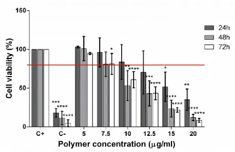
11. Cell viability assay for cytotoxicity evaluation
The cytotoxicity of the polymer and polyplexes was evaluated up to 72 hours, with controls as follows: cells in standard cell culture conditions and cells treated with a latex solution, as positive (blank) and negative controls, respectively. Cell viability was determined using the MTT (3-[4,5-dimethylthiazol-2-yl]2,5-diphenyltetrazolium bromide, Sigma-Aldrich) assay as described by [83].
For these experiments, HEK293 and D407 cells were seeded at a density of 2x10⁴cells/well (48-well plate). After 24h, cells were incubated with increasing concentrations of the free polymer (5, 7.5, 10, 12.5, 15 and 20 μg/mL). For the new polymer, D407 cells were incubated in a larger range of polymer concentrations (1, 3, 5, 7.5, 10, 12.5, 15, 20, 50, 100 μg/mL). To prepare each of the polymer solutions, 1 mg/ml polymer stock solution was diluted in cell culture medium and added to each well.
For the polyplexes formed with either the 354kDa PDMAEMA and the new PDMAEMA, we have tested three concentrations for each polymer:DNA ratio. In the cytotoxicity evaluation we have chosen the final concentration of 0,1 µg, 0,2 µg and 0,5 µg of DNA per 2x10⁴cells to reflect future transfection conditions. The polyplexes were diluted in cell culture medium and added to each well. At each time point (24, 48 and 72 hours), 25 μL MTT solution (5mg/mL in PBS) were added to each well and incubated for 4 hours. The solution was removed and replaced with 250μL of isopropanol-HCl (0.04N) to dissolve the blue formazan crystals produced and left to incubate further for 1 hour at 37ºC, 5% CO2. Absorbance at 570nm and 630nm were measured for each well using an
infiniteM200 (TECAN) microplate reader. The relative cell viability (%) was calculated by [abs]sample/[abs]controlx100 [84], with cells from the positive control
38
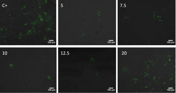
12. Transfection efficiency assay
For the determination of the gene transfer efficiency of 354kDa PDMAEMA, cells were plated at 1x10⁵ cells/well in clear 6-well tissue culture plates. FuGENE HD® (Promega, USA) was used as the transfection control and according to the manufacturer instructions. PDMAEMA-DNA polyplexes were added to the cells at a ratio of 1µg of pDNA per 1x10⁵ cells and further incubated for 72 h. Cells cultured in standard culture conditions were used as a non transfection control.
Transfection efficiency was evaluated qualitatively by fluorescence microscopy and quantitatively by flow cytometry. Imaging was performed in an inverted fluorescence microscope (LEICA DMIL-LEICA DC500). Green fluorescent protein (GFP) positive cells were scored by flow cytometry (FACSCalibur, BD Biosciences, USA) using the FL-1H green channel. A total of 1x10⁴ events were counted for each sample and the percentage of positive events corresponds to the gated events minus the negative control, for transfection (non-transfected cells).
For the determination of the gene transfer efficiency of the newly synthetized PDMAEMA, cells were plated at 1,5x10⁵ cells/well in clear 6-well tissue culture plates. Cells were plated in higher number than before to ensure that we had enough cells to acquire a bigger number of events in the flow cytometry. FuGENE HD® (Promega, USA) was used as the transfection control and according to the manufacturer instructions. PDMAEMA: DNA polyplexes were added to the cells at a ratio of 1µg of pDNA per 1x10⁵ cells in two conditions: directly added (non-diluted) and diluted in 100 μL of medium without FBS. After 2 min complete medium was added and further incubated for 72 h. Cells cultured in standard culture conditions were used as a non transfection control. Transfection efficiency was evaluated qualitatively by fluorescence microscopy and quantitatively by flow cytometry. Imaging was performed in an inverted fluorescence microscope (Axiovert 40 CFL, Zeiss). Green fluorescent protein (GFP) positive cells were scored by flow cytometry (FACScan, BD Biosciences, USA) using the FL-1H green channel. A total of 1x10⁵ events were counted for
39 each sample and the percentage of positive events corresponds to the gated events minus the negative control.
13. Animals
C57BL/6 mice (3 months old) were used as experimental animals, and housed in controlled temperature and 12 hour light/dark cycle with food and water ad
libitum. All experimental procedures were carried out according to the
Portuguese and European Union regulations (FELASA) for the use of animals and the Association for Research in Vision and Ophthalmology (ARVO) for the use of animals in ophthalmic and vision research. All procedures were performed under anesthesia with 2,2,2 tribromoethanol (Sigma-Aldrich) administered by intraperitoneal injection (250 mg/Kg dose). Animals were humanely sacrificed by anesthesia and death was confirmed by cervical dislocation.
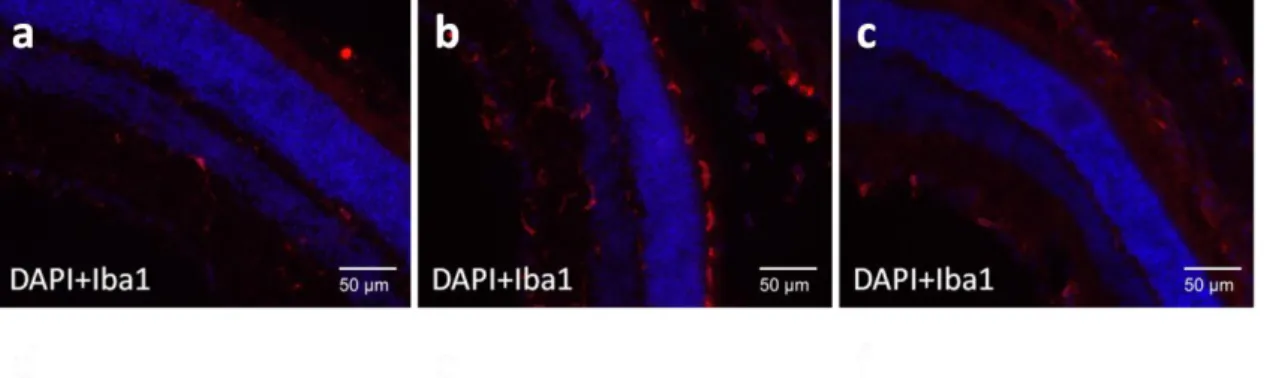
14. Activation of microglia after intravitreous injection in
C57/BL6 mice
In order to evaluate the compability of the PDMAEMA:DNA polyplexes in vivo, 3 month old C57BL/6 mice were injected with polyplexes with a N/P ratio of 12.5 by intravitreous injection in the right eye, under a stereomicroscope (Nikon Stereoscopic Microscope). The left eye was use as control. A injection procedure without administration of any vehicle was also performed (sham injection). The mice were sacrificed 14 days post-injection, the eyes enucleated and fixed in PFA 4%( Sigma-Aldrich), immersed in sucrose 30% (Sigma-Aldrich), and after 24h were included in Optimal Cutting Temperature (OCT) compound (VWR). To eliminate the autofluorescence of the retina, sections were first washed with PBS 0.1% TritonX for 5 minutes, incubated in 0.25%
40 KMnO4 in PBS for 15 minutes, rinsed with PBS twice, and then incubated in
0.1% oxalic acid in PBS for 20 minutes at room temperature. Treated sections were washed in PBS 0.1% TritonX three times. An immunofluorescence assay was performed, using an antibody against Iba-1, a marker of microglia activation. The primary antibody (Iba-1, Wako Pure Chemical Industries) was incubated overnight at 4ºC at a dilution of 1/1000. A secondary antibody, Alexa Fluor 594 (1/2000, Invitrogen), was used for detection of the signal. The secondary antibody was incubated for 1h at room temperature, and the samples were visualized in an Axio Observer Z2 Fluorescence microscope (Zeiss).
15. Statistical analysis
Statistical analysis was performed between the control and the tested conditions by a one-way ANOVA followed by a post hoc Dunnett’s Multiple Comparison Test for the cell viability and transfection assays, between the tested conditions an unpaired t-test was performed. For the polyplexes, analyses between different ratios were determined with one-way ANOVA followed by a post hoc Benferroni’s Multiple Comparison Test.
41
Results and Discussion
This section is divided in two parts: in part one, we have characterized a previously synthesized PDMAEMA with 354kDa and in part two, we describe the synthesis and characterization of a smaller Mw PDMAEMA (that was found
to have 103.3kDa).
Part one:
1. Characterization of 354kDa PDMAEMA-DNA polyplexes
1.1 PDMAEMA-DNA polyplexes have characteristics compatible
with gene therapy
Characterization of the polyplexes included the determination of size, polydispersity and zeta potential, as shown in table I.
Ideally, polyplexes should present a size below 500 nm, an optimum polydispersity of 0.1, and a positive surface charge, which constitute favorable requisites for cellular uptake.
Table I: Characterization of polyplexes by size, PdI and zeta potencial
Formulation Size (nm) Polydispersity Index (PdI) Surface Charge (+mV) PDMAEMA 5 129.96±4.28 0.310±0.062 42.32±6.25 PDMAEMA 7.5 123.0±15.36 0.301±0.090 37.26±9.06 PDMAEMA 10 141.58±28.2 0.298±0.055 44.82±7.17 PDMAEMA 12.5 164.4±49.83 0.290±0.048 25.74±5.79 PDMAEMA 20 266.52±32.64 0.383±0.071 24.06±4.83
42 *There are only significant size differences between the ratio 20/1 and the other ratios
*The ratios of 5/1, 7.5/1 and 10/1 are significantly different from the ratios 12.5/1 and 20/1 in terms of zeta potential
*There is no significant difference in the polydispersity index
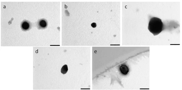
Figure 9- A analysis by transmission electron microscopy of PDMAEMA:DNA
polyplexes at different ratios: 5(a), 7.5(b), 10(c), 12.5(d) and 20(e). Scale bar represents 200nm.
In table I it is possible to observe that the size of PDMAEMA/pDNA polyplexes increases with the increase in N/P ratio from 5 to 20, but all ratios have diameters smaller than 300 nm. This trend correlates well with the morphological analysis performed by transmission electron microscopy (TEM) (figure 9). The polydispersity index, which evaluates the homogeneity, is below 0.4, indicating a homogeneous population of particles, which is desirable for consistency of results regarding cellular penetration. As expected, the polyplexes have positive charge since there is an excess of polymer to DNA.
However, whilst there is a clear trend of increase in size with increasing N/P ratio, zeta potential values present quite a random variation. In the literature, two opposite trends in size with N/P ratio, depending on Mw, were observed for
43 our polymer was observed, with a random but not very significant variation in polyplex diameter for small N/P ratios, and a significant increase when going from N/P = 16 to N/P = 20 [73]. PDMAEMA with 43 kDa referred in that study presented a completely different behavior from one of comparable Mw (39 kDa)
described in a different study [85]. The former showed a size reduction with the increase of the N/P ratio, and the latter a random variation in size and a decrease in zeta potential.
It seems, therefore, that size and surface charge are very sensitive to polymer molecular weight and, probably, to polyplex preparation conditions, rendering it difficult to establish a correlation between those parameters and the N/P ratio. In fact, there are indications that the polymer molecular weight may affect transfection more than the size of polyplexes [73].
1.2 PDMAEMA efficiently complexes pDNA
The electrophoretic mobility shift assay is based on the behavior of pDNA in an agarose gel, which migrates from the negative to the positive end. Upon complexation with a cationic polymer, the DNA is retained in the well, unable to migrate through the gel. As observed in figure 10, for all N/P ratios the PDMAEMA polyplexes were able to complex and retain the DNA, and this is evidenced by the absence of DNA bands in the gel lanes, with all DNA retained in the wells. As expected, free DNA (lane 2) migrates towards the positive pole and PDMAEMA polymer (lane 3) shows no signal in the gel.
44
Figure 10- Electrophoretic mobility shift assay for polyplexes of different N/P ratios
analyzed in a 1% agarose gel, with pDNA visualized by GreenSafe Direct Load: Lane 1: Marker 1kb; Lane 2: Free pDNA; Lane 3: Free PDMAEMA; Lane 4: NPs 5/1; Lane 5: NPs 7,5/1; Lane 6: NPs 10/1; Lane 7: NPs 12,5/1; Lane 8: NPs 20/1
1.3
PDMAEMA
polyplexes
protect
DNA
from
DNAse
degradation
In addition to the capacity of DNA complexation by PDMAEMA, it is also important to assess if the polyplexes can protect DNA from degradation, both in the extracellular and intracellular millieu. The presence of nucleases is an important barrier that the polyplexes must overcome. In order to evaluate protection of DNA from degrading enzymes, PDMAEMA polyplexes were incubated with DNase for 5 min and as shown in figure 11, able to protect DNA from degradation, as evidenced by the DNA retained in the wells when compared with free DNA subjected to the action of DNAse (lane 3), which has been totally degraded, as expected
45
Figure 11- The PDMAEMA polyplexes can effectively protect the pDNA against the
DNAse I as analyzed in a 1% agarose gel, with pDNA visualized by GreenSafe Direct Load: Lane 1: Marker 1kb; Lane 2: Free pDNA; Lane 3: Free pDNA with DNAse I; Lane 4: NPs 5/1; Lane 5: NPs 5/1 with DNAse I; Lane 6: NPs 7,5/1; Lane 7: NPs 7,5/1 with DNAse I; Lane 8: NPs 10/1; Lane 9: NPs 10/1 with DNAse I; Lane 10: NPs 12,5/1; Lane 11: NPs 12,5/1 with DNAse I; Lane 12: NPs 20/1; Lane 13: NPs 20/1 with DNAse I.
46
1.4 Stability assay of polyplexes over time
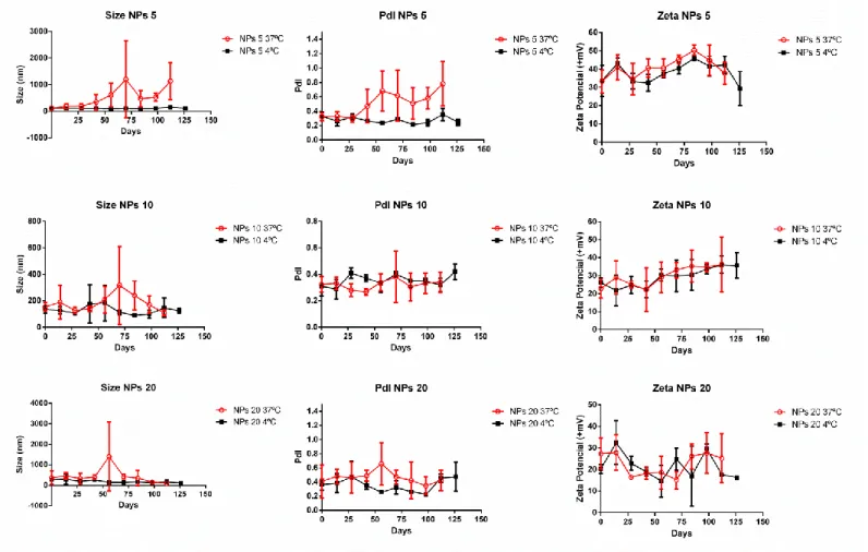
In order to assess the stability in storage (4ºC) and physiological conditions (37ºC), the polyplexes (N/P 5/10/20) were measured in terms of size, PdI and zeta potencial every 14 days (2 weeks) (figure 12). For the NPs 5 ratio in terms of size, at 37ºC the size appears to be pretty stable until around 25 days, after that the size starts to increase. At 4ºC, the polyplexes were very stable until 125 days after preparation. The polyplexes are stable until 32 days at 37ºC, from this point the PdI increased. At 4ºC the polyplexes were stable up to 125 days. The zeta potential in both conditions remains stable.
Figure 12- Measurements of size, PdI and zeta potencial of the stability assay of three
polyplexes ratios (5/10/20). The polyplexes were aged in two conditions: 37ºC (red open circles) and 4ºC (black closed squares).
47 The increased size and PdI can be explained by the higher temperature, which increases the Brownian motion leading to the increased collision of the polyplexes, causing aggregation. This behavior was also observed with PDMAEMA polyplexes aged in 10% sucrose/aqueous solutions by Cherng et al. After 6 months of aging, the size of these polyplexes increased at 40ºC in comparison with lower temperatures of 4ºC and 20ºC [86]. This was also observed for other polymer, chitosan, that at a higher temperature (45ºC), its polyplexes also increased their size [87].
The ratios 10 and 20 were shown to maintain their size stable up to 125 days in both temperature conditions. In terms of PdI and zeta potential no major changes were observed great alterations. This could indicate that higher ratios of polymer/pDNA are more stable, which follows a logical trend that higher amounts of polymer to DNA can increase the complexation of DNA and thus promote stronger interaction and as consequence, resistance to temperature induced changes.
2. Evaluation of the cytotoxicity of the PDMAEMA polymer and
PDMAEMA-DNA polyplexes
A critical feature in all materials to be used for biomedical applications, including gene therapy, is the biocompatibility of the material. The latter is first evaluated
in vitro by testing the cytotoxicity on a cell culture setup. We have evaluated the
cytotoxic profile of both the polymer and polymer-DNA complexes using two cell lines: a commonly used cell line for transfection studies - the HEK293 cells - and a retinal pigment epithelium cell line, D407, which is used as an in vitro model for evaluating potential gene therapy vectors for the retina.
48 We firstly evaluated the effect on cellular viability of the PDMAEMA-DNA polyplexes, as shown in figures 13 and 14. The results are presented in terms of mass of DNA, and for all the conditions tested (different polymer:DNA ratios and different quantities of polyplexes, there was no deleterious effect for cell viability both for HEK293 and D407 cells. Therefore, the polyplexes, even at concentrations more than two times higher than those to be used for transfection, are not cytotoxic for either cell line.
´
Figure 13- HEK293 cell viability upon challenge with PDMAEMA:DNA polyplexes at
different concentrations, in function of mass of DNA, up to 72h. Lowest acceptable threshold of cell viability indicated by red bar (80% cell viability). Statistical significance was determined with one-way ANOVA followed by a post hoc Dunnett’s Multiple Comparison Test and the statistical diference indicated by the star (*) symbol (* p<0.05, **p<0.01, *** p<0.001 and ****p<0.0001).
![Figure 3 - Schematic of cationic lipids (grey)/DNA (blue) complexes, adapted from [35]](https://thumb-eu.123doks.com/thumbv2/123dok_br/18921193.937471/22.892.347.560.241.469/figure-schematic-cationic-lipids-grey-dna-complexes-adapted.webp)
![Figure 7- Schematic representation of PDMAEMA monomer [70].](https://thumb-eu.123doks.com/thumbv2/123dok_br/18921193.937471/27.892.225.424.802.1035/figure-schematic-representation-pdmaema-monomer.webp)
![Figure 8- Proposed RAFT polymerization Mechanism as proposed by [79]. Briefly, initiation and propagation (a); RAFT pre-equilibrium (b); re-initiation (c); main RAFT equilibrium (d) and termination (e)](https://thumb-eu.123doks.com/thumbv2/123dok_br/18921193.937471/29.892.103.791.357.786/polymerization-mechanism-initiation-propagation-equilibrium-initiation-equilibrium-termination.webp)