Interstitial and Langerhans’ dendritic
cells in chronic periodontitis and
gingivitis
Abstract: The aim of the present study was to compare quantitatively the distribution of dendritic cell subpopulations in chronic periodontitis and gingivitis. Fourteen biopsies from patients with chronic periodontitis and ifteen from patients with gingivitis were studied. An immunoperoxidase technique was used to quantify the number of Langerhans’ cells (CD1a) and interstitial dendritic cells (factor XIIIa) in the oral and sulcular and junctional/pocket epithelia and in the lamina propria. A greater number of factor XIIIa+ dendritic cells in the lamina propria and CD1a+ den-dritic cells in the oral epithelium were observed in gingivitis compared to the periodontitis group (p = 0.05). In the sulcular and junctional/pocket epithelia and in the lamina propria, the number of CD1a+ dendritic cells was similar in the gingivitis and periodontitis groups. In conclusion, the number of Langerhans’ cells in the oral epithelium and interstitial den-dritic cells in the lamina propria is increased in gingivitis compared to periodontitis, which may contribute to the different pattern of host re-sponse in these diseases.
Descriptors: Periodontitis; Gingivitis; Dendritic cells; CD1a; Factor XIIIa.
Patricia Ramos Cury(a) Cristiane Furuse(b)
Ana Elisa Amaro Rodrigues(c) José Alexandre Barbuto(d) Vera Cavalcanti de Araújo(b) Ney Soares de Araújo(e)
(a)PhD, Professor, Department of
Microbiology; (b)PhDs, Professors,
Department of Oral Pathology (c)Master’s
Degree Student – São Leopoldo Mandic Dental Reseach Center, Campinas, SP, Brazil.
(d)PhD, Professor, Department of Immunology,
Institute of Biomedical Sciences, University of São Paulo, São Paulo, SP, Brazil.
(e)PhD, Professor and Chairman, Department
of Oral Pathology, School of Dentistry, University of São Paulo, São Paulo, SP, Brazil.
Corresponding author:
Patricia Ramos Cury
Laboratório de Cultura de Células, Centro de Pesquisa Odontológica São Leopoldo Mandic
Rua José Rocha Junqueira, 13, Ponte Preta Campinas - SP - Brazil
CEP: 13045-610
E-mail: [email protected]
Introduction
Gingivitis and periodontitis are characterized by distinct host responses. Although a great deal of information is available, the mechanism which regulates this difference is still poorly understood. Variation in host response against periodontopatho-gens is associated with genetics and environmental factors;1 however, whether and how gingivitis will
progress to periodontitis still has to be elucidated. It has been suggested that the progression is associ-ated with more abundant Th2 cells than Th1 cells in chronic periodontitis,2 or with an increase in the
number of B cells and plasma cells in periodontitis and predominance of T cells in gingivitis.3
Dendritic cells (DCs) represent a large family of antigen-presenting cells that circulate through the bloodstream and are scattered in nearly all tissues of the body. DCs serve as a connection between an innate immune system and an adaptive immune response, being the most potent antigen-present-ing cells. They capture microbes and their antigens while in the immature state, and stimulate a T-cell response to these antigens in their mature state. DCs are the only antigen-presenting cells capable of prim-ing naive helper/cytotoxic T cells to undergo clonal expansion, thus initiating an adaptive immune re-sponse.4 In their immature state (CD1a positive
Langerhans’ cells), DCs are eficient antigen capture cells and, as they mature, they undergo phenotypic changes that facilitate migration toward lymphoid organs and enable their ability to prime naive T cells in the context of MHC-II molecules.4 Besides
stim-ulating T cell responses, a different subset of DCs (factor XIIIa positive interstitial DCs) also directly stimulate naive B cells to differentiate into plasma cells in vitro.5
The number of Langerhans’ cells in gingival tis-sues is a topic of much speculation. Studies have reported increased numbers in periodontitis,6
de-creased numbers in gingivitis and periodontitis compared to healthy periodontal tissues,7 and no
quantitative change in periodontitis compared to gin-givitis and healthy periodontal tissues.8 On the other
hand, little is known about the distribution of factor XIIIa+ DCs and their possible role in periodontitis and gingivitis. Jotwani, Cutler9 (2003), evaluating 7
biopsies of chronic periodontitis and 5 biopsies of healthy gingival tissue, demonstrated that the num-ber of factor XIIIa+ DCs is increased in periodonti-tis compared to healthy gingival periodonti-tissue.
The present study aimed to compare quantita-tively the distribution of Langerhans’ cells (CD1a+) and interstitial DCs (factor XIIIa+) in chronic peri-odontitis and gingivitis.
Material and Methods
The study protocol was approved by the Ethics Committee, School of Dentistry, University of São Paulo, Brazil.
Biopsies were retrieved from the iles of the De-partment of Oral Pathology, São Leopoldo Man-dic Dental Research Center, and from those of the School of Dentistry, University of São Paulo. Fourteen biopsies from cases diagnosed as chronic periodontitis (mean age = 34.5 ± 9.2 years; 10 fe-male and 4 fe-male) and ifteen from cases diagnosed as gingivitis (mean age = 32.9 ± 8.9 years; 9 female and 6 male), involving systemically healthy patients, were analyzed. The diagnoses were obtained based on clinical and histological examinations. For his-tological diagnosis, the sections were stained with hematoxylin and eosin.
Serial sections of 3 µm in thickness were ob-tained from formalin-ixed parafin-embedded tis-sue. Dewaxed sections were incubated with primary antibody. Clone, pre-treatment for antigen retrieval, dilutions, and incubation time are given in Table 1. Immunohistochemistry was performed on the sections
Table 1 - Antibodies, clones, target retrieval, dilutions, incubation times, and cell specificity of the primary antisera.
Antibody Clone Antigen retrieval Dilution Incubation time Cell specificity CD1a 010* Citrate buffer 0.1 M, 95°C, 30 minutes 1:100 40 minutes Langerhans’ cells Factor XIIIa Ab-1** No treatment 1:700 40 minutes Interstitial dendritic cells
using the biotin-streptavidin-peroxidase method, fol-lowed by developing in a diaminobenzidine chromo-gen solution. Finally, the sections were counterstained with Mayer’s hematoxylin. All incubations were per-formed in a moist chamber at room temperature.
Omission of the primary antibody constituted the negative control. For the CD1a antibody, ton-sil tissue was used as a positive control; for factor XIIIa, dermatoibroma was used.
The specimens were photographed with a micro-scope (Axioskop 2 Plus, Zeiss, Gottingen, Germany) at 400 X magniication interfaced to a computer; the areas were delimited with mouse and measured with a speciic software (Microsoft AxioVision 4.2, Carl Zeiss Vision GmbH, Gottingen, Germany).
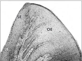
Cell counts were performed in duplicate through-out the entire sections by two masked examiners. The number of immunolabeled cells per area unit (cell number/mm²) was calculated for each primary antibody. Counts of CD1a and factor XIIIa positive cells were restricted to immunolabeled cells exhibit-ing a well-deined cell nucleus and body with at least two well-visualized dendrites. Three regions were evaluated: oral epithelium, sulcular and junctional/ pocket epithelia, and lamina propria (Figure 1).
The mean value of the counts of the two examin-ers was used for statistical analysis. Differences in the number of immunolabeled cells between the gin-givitis and periodontitis groups were analysed using
the Mann-Whitney U test. Statistical signiicance was deined as p ≤ 0.05. Calculations were per-formed using a statistical software package (SPSS Inc., Chicago, IL, USA).
Results
Table 2 presents the mean number ± standard deviation of CD1a+ and factor XIIIa+ cells/mm² in the oral epithelium, junctional/sulcular epithelia, and lamina propria in the gingivitis and periodon-titis groups.
In the oral epithelium, the number of CD1a+ den-dritic cells/mm² was signiicantly greater (p = 0.05) in gingivitis than in periodontitis (137.69 ± 90.36 and 74.09 ± 74.75 respectively) (Figures 2A and 2B). In the sulcular and junctional/pocket epithelia and lamina propria, the counts of CD1a+ dendritic cells were similar in both groups.
A greater number of factor XIIIa+ dendritic cells in the lamina propria was observed in gingi-vitis compared to the periodontits group (p = 0.05) (209.49 ± 101.26 and 104.27 ± 95.52, respectively) (Figures 2C and 2D). No factor XIIIa+ dendritic cell was observed in the epithelia.
Discussion
The present study reveals signiicant differences in the number of CD1a+ cells/mm² in the oral epi-thelium and of factor XIIIa+ cells/mm² in the lami-na propria between periodontitis and gingivitis, be-ing the numbers increased in gbe-ingivitis.
The lower number of DCs in periodontitis com-pared to gingivitis may decrease the defense response
SE
OE
JE
LP
Table 2 - Mean number ± Standard deviation of CD1a+ and factor XIIIa+ cells/mm² in the oral epithelium (OE), junctional/sulcular epithelia (JE), and lamina propria (LP) in the gingivitis and periodontitis groups.
Antibody Counting Zone
Group Gingivitis
(N = 15)
Periodontitis (N = 14)
p value
CD1a
OE 137.7 ± 90.4 74.1 ± 74.8 0.05 JE 174.3 ± 129.6 116.8 ± 137.9 0.11 LP 259.9 ± 109.8 278.6 ± 180.9 0.86 Factor XIIIa LP 209.5 ± 101.3 104.3 ± 95.5 0.05
to periodontal bacteria, thus explaining the different patterns of disease. The differences in the number of DCs might be associated with several factors: 1 - DC differentiation may be inhibited by certain peri-odontal bacteria. DC differentiation and maturation is inhibited by many infectious agents. It has been demonstrated that human monocytes cultured with granulocyte-macrophage colony-stimulating factor (GM-CSF) and interleukin-4 (IL-4) after phagocy-tosis of Candida albicans yeast do not differentiate into DCs; they retain CD14 expression, do not ac-quire CD1a expression, and are unable to express the maturation markers CD83 and CCR7. On the other hand, monocytes that had phagocytosed mycelial forms differentiate into mature CD83+ and CCR7+ cells.10 Similarly, M. tuberculosis down-regulates
CD1a expression on DCs,11 and plasmodium
infec-tion is also characterized by inhibiinfec-tion of DC func-tion in vivo and in vitro.11 2 - DCs have captured the
antigens and matured. Porphyromonas gingivalis li-popolysaccharide triggers maturation of DCs,12 thus
immature DCs (CD1a+) in periodontitis may have captured the antigens (Porphyromonas gingivalis) and maturated to present the antigens,6 and then a
lower number of CD1a+ cells was observed in peri-odontitis. 3 - Patients with periodontitis may present a lower differentiation of DCs compared to patients with gingivitis, and this may increase the risk of pro-gression of gingivitis to periodontitis. Further stud-ies are required to evaluate these hypotheses.
The Langerhans’ cell network seems to change markedly with aging13 and smoking habits.14 In this
study the groups were age-matched. However, the information concerning the smoking habits was not Figure 2 - CD1a+ dendritic cells in the oral epithelium. A - chronic periodontitis (Immunohistochemistry, 100 X); B - gingivitis (Immunohistochemistry, 200 X). In the oral epithelium, the number of CD1a+ dendritic cells/mm² was significantly greater in gin-givitis than in periodontitis. Factor XIIIa+ dendritic cells in the lamina propria. C - chronic periodontitis (Immunohistochemistry, 400 X); D - gingivitis (immunohistochemistry, 400 X). A greater number of factor XIIIa+ dendritic cells in the lamina propria was observed in gingivitis compared to the periodontitis group.
A B
available and further studies are required to assess the role of smoking.
It has been previously reported that although DC heterogeneity has been observed in humans, their lineage origins, maturation stages and functional differences have not been clearly established. Inter-stitial DCs, also known as dermal DCs, and Lang-erhans’ cells are originated from a myeloid lineage.15
They are “immature” cells, i.e., are unable to stimu-late T cells. Although these DCs lack the requisite accessory signals for T-cell activation, such as CD40, CD54 and CD86, they are extremely well equipped to capture antigens. After antigen capture, full mat-uration and mobilization of DCs are induced. In the epithelium, immature DCs known as Langerhans’ cells are labeled for CD1a+. Langerhans’ cells take up particles and microbes by phagocytosis,16 mature
and move into lymphoid tissues17 or lamina propria6
in search of T cells for antigen presentation.
In the present study, the number of CD1a+ DCs/ mm² in the oral epithelium was signiicantly higher in gingivitis compared to periodontitis. On the other hand, in the sulcular and junctional/pocket epithelia and lamina propria, the counts of CD1a+ DCs were similar in periodontitis and gingivitis. These results might be associated with a limited T cell count in periodontitis as compared to gingivitis.3 However,
contrasting results have been shown in the litera-ture regarding DC counts in periodontal diseases. Increased numbers in periodontitis,6 decreased
numbers in gingivitis and periodontitis compared to healthy periodontal tissues,7 and no quantitative
change in periodontitis compared to gingivitis and healthy periodontal tissues8 have been reported. The
number of DCs changes markedly with aging and smoking habits, which may explain such divergent results.13,14
Cells that express CD34 contain progenitors for two discrete DCs populations: the epidermal Lang-erhans’ cells and dermal or interstitial type of DCs.5
Factor XIIIa+ cells represent interstitial DCs4 which
have major effects on B-cell growth and immuno-globulin secretion.4 Naïve B cells respond uniquely
to the interstitial, non-Langerhans’ cell type of DCs, and by secretion of soluble factors, the interstitial type of DCsstimulate the prodution of antibodies directly. One important functional difference be-tween CD1a+ and factor XIIIa+ cells is that only interstitial-type DCs directly stimulate naïve B cells to make antibodies.5,18 In the present study, the
number of factor XIIIa+ cells was greater in gin-givitis than in periodontitis, indicating that factor XIIIa-positive cells may also play an important role in the periodontal disease pathogenesis. Jotwani, Cutler9 (2003) have shown that the number of factor
XIIIa+ DCs is increased in periodontitis compared to healthy gingival tissue.
Conclusion
In conclusion, the number of Langerhans’ cells in the oral epithelium and interstitial DCs in the lam-ina propria is increased in gingivitis compared to periodontitis, which may contribute to the different pattern of host response in these diseases. However, the immune process is complex, and further studies controlling smoking habits and other host variables are required to reach a better understanding of the role of DCs in periodontal disease.
References
1. Macêdo TCN, Costa MCN, Gomes-Filho IS, Vianna MIP, Santos CT. Factors related to periodontal disease in a rural population. Braz Oral Res. 2006;20(3):257-62.
2. Kinane DF, Lappin DF. Immune processes in periodontal disease: a review. Ann Periodontol. 2002;7(1):62-71. 3. Seymour GJ, Greenspan JS. The phenotypic characterization
of lymphocyte subpopulations in established human periodon-tal disease. J Periodonperiodon-tal Res. 1979;14(1):39-46.
4. Banchereau J, Steinman RM. Dendritic cells and the control of immunity. Nature. 1998;392(6673):245-52.
5. Caux C, Massacrier C, Vanbervliet B, Dubois B, Durand I, Cella M et al. CD34+ hematopoietic progenitors from human cord blood differentiate along two independent dendritic cell pathways in response to granulocyte-macrophage colony-stimulating factor plus tumor necrosis factor alpha: II. Func-tional analysis. Blood. 1997;90(4):1458-70.
7. Seguier S, Godeau G, Leborgne M, Pivert G, Brousse N. Quantitative morphological analysis of Langerhans cells in healthy and diseased human gingiva. Arch Oral Biol. 2000;45(12):1073-81.
8. Gemmell E, Carter CL, Hart DN, Drysdale KE, Seymour GJ. Antigen-presenting cells in human periodontal disease tissues. Oral Microbiol Immunol. 2002;17(6):388-93.
9. Jotwani R, Cutler CW. Multiple dendritic cell (DC) subpopu-lations in human gingiva and association of mature DCs with CD4+ T-cells in situ. J Dent Res. 2003;82(9):736-41. 10. Torosantucci A, Romagnoli G, Chiani P, Stringaro A, Crateri
P, Mariotti S et al. Candida albicans yeast and germ tube forms interfere differently with human monocyte differen-tiation into dendritic cells: a novel dimorphism-dependent mechanism to escape the host’s immune response. Infect Im-mun. 2004;72(2):833-43.
11. Cutler CW, Jotwani R, Pulendran B. Dendritic cells: immune saviors or Achilles’ heel? Infect Immun. 2001;69(8):4703-8. 12. Kanaya S, Nemoto E, Ogawa T, Shimauchi H.
Porphyromo-nas gingivalis lipopolysaccharides induce maturation of den-dritic cells with CD14+CD16+ phenotype. Eur J Immunol. 2004;34(5):1451-60.
13. Zavala WD, Cavicchia JC. Deterioration of the Langerhans cell network of the human gingival epithelium with aging. Arch Oral Biol. 2006;51(12):1150-5.
14. Robbins CS, Dawe DE, Goncharova SI, Pouladi MA, Drannik AG, Swirski FK et al. Cigarette smoke decreases pulmonary dendritic cells and impacts antiviral immune responsiveness. Am J Respir Cell Mol Biol. 2004;30(2):202-11.
15. Banchereau J, Briere F, Caux C, Davoust J, Lebecque S, Liu YJ et al. Immunobiology of dendritic cells. Annu Rev Immunol. 2000;18:767-811.
16. Svensson M, Stockinger B, Wick MJ. Bone marrow-derived dendritic cells can process bacteria for MHC-I and MHC-II presentation to T cells. J Immunol. 1997;158(9):4229-36. 17. Adema GJ, Hartgers F, Verstraten R, de Vries E,
Mar-land G, Menon S et al. A dendritic-cell-derived C-C che-mokine that preferentially attracts naive T cells. Nature. 1997;387(6634):713-7.