Two new species of Odontostilbe historically hidden under
O. microcephala (Characiformes: Cheirodontinae)
Junior Chuctaya
1,2, Cristina M. Bührnheim
3,4and Luiz R. Malabarba
1Specimens historically identified as Odontostilbemicrocephala from the upper rio Paraná and Andean piedmont tributaries of the río Paraguay are reviewed and split in three species. We found that the distribution of O.microcephala is restricted to the Andean slope of the río Paraguay basin. The species is distinguished from congeners with subterminal mouth by the elongate body, usually 10-12 gill rakers on upper branch and smaller horizontal orbital diameter (24.6-32.8 % HL, mean 28.7%). Specimens from upper rio Paraná constitute two new species, diagnosed from other Cheirodontinae by the presence of mesopterygoid teeth, grouped on median portion and forming a continuous row. The new species are distinguished from each other by having premaxillary teeth with five cusps vs. nine cusps and by the number of lamellae in left and right sides of central median raphe of olfactory rosette with 20-21 vs. 11-12.
Keywords: Biodiversity, Characidae, La Plata River basin, Mesopterygoid teeth, Taxonomy.
Espécimes historicamente identificados com Odontostilbe microcephala do rio Paraná e tributários do río Paraguay, foram revisados e separados em três espécies. A distribuição de O. microcephala é restrita ao sopé andino da bacia do río Paraguay. A espécie é distinta das congêneres com boca subterminal pela forma alongada, geralmente 10-12 rastros branquiais no ramo superior e menor diâmetro horizontal da órbita (24,6-32,8 % CC, média 28,7%). Espécimes do alto rio Paraná constituem duas espécies novas diagnosticadas de outros Cheirodontinae pela presença de dentes no mesopterigoide, agrupados em sua porção média e formando uma fileira continua. As novas espécies distinguem-se por ter dentes premaxilares com cinco cúspides vs. nove cúspides e pelo número de lamelas nos lados esquerdo e direito da rafe central da roseta olfativa com 20-21 vs. 11-12.
Palavras-chave: Bacia do rio da Prata, Biodiversidade, Characidae, Dentes do Mesopterigoide, Taxonomia.
1Programa de Pós-Graduação em Biologia Animal, Departamento de Zoologia, Universidade Federal do Rio Grande do Sul, Av. Bento
Gonçalves 9500, 91501-970 Porto Alegre, RS, Brazil. (JC) junior.chuctaya@gmail.com, https://orcid.org/0000-0002-8876-4675 (corresponding author), (LRM) malabarb@ufrgs.br
2Laboratorio de Ictiología, Museo de Historía Natural, Universidad Nacional Mayor de San Marcos, Av. Arenales 1256, Lima 14, Peru. 3Universidade do Estado do Amazonas, ENS, Av. Djalma Batista, Chapada 2470, 69050-010 Manaus, AM, Brazil. cmbuhrn@yahoo.com.br 4Instituto de Ciências Biológicas, Departamento de Biologia, Coleção Zoólogica Prof. Paulo Bührnheim, Universidade Federal do
Amazonas, Minicampus (Setor Sul). Av. Rodrigo Octávio Jordão Ramos, 6200, Coroado I, 69080-900, Manaus, AM, Brazil. Introduction
Species of Cheirodontinae are found in most river drainages of Central and South America, occurring from Costa Rica to central Chile and Argentina, in both Atlantic and Pacific drainages of the Andes (Malabarba, 2003). The subfamily includes 18 genera and 66 valid species, of which almost half (8) of the genera and almost one third (19) of the species were described in the last twenty years (Malabarba, 1998, 2003; Malabarba, Bertaco, 1999; Malabarba, Weitzman, 1999, 2000; Weitzman, Malabarba, 1999; Malabarba etal., 2004; Bührnheim, Malabarba, 2006, 2007; Bührnheim et al., 2008; Malabarba, Jerep, 2012, 2014; Jerep, Malabarba, 2014; Zarske, 2012; Vari et al., 2016; Jerep etal., 2016).
Odontostilbemicrocephala was described by Eigenmann in Eigenmann, Ogle (1907) based on two specimens from río Pilcomayo, Bolivia, a tributary of the río Paraguay. Later, the species was redescribed by Eigenmann (1915) in his monograph on Cheirodontinae, with the examination of additional material from the rio Tietê, expanding the distribution of the species to the upper rio Paraná. After Eigenmann, several studies referred O. microcephala from both río Paraguay and/or upper rio Paraná drainages (e.g., Uj, 1987; Miquelarena etal., 2008; Oyakawa, Menezes, 2011).
The examination of the specimens from Eigenmann (1907, 1915) along with additional new material proved the specimens from upper rio Paraná to belong to two new species, restricting the occurrence of Odontostilbe microcephala to tributaries of río Paraguay. Herein we provide a redescription of Odontostilbe microcephala and the description of those two new species of Odontostilbe following the definition of Malabarba (1998).
Material and Methods
Counts and measurements follow Fink, Weitzman (1974). Measurements corresponding to parts of the head were taken in stereomicroscope. Measurements from lateral side of body were taken from projections on the main axis of the body, with digital caliper and expressed to the nearest 0.1 mm. Head length is the distance between the tip of the snout and the posterior end of subopercle, which is slightly posterior to the margin of the opercle (Bührnheim, Malabarba, 2006). Measurements are presented in tables as percent of the standard length (SL), except for subunits of the head, which are presented as percent of the head length (HL).
Precaudal, caudal, and total vertebral counts include the four vertebrae of the Weberian apparatus, and the terminal “half centrum” as a single element (Malabarba, Weitzman 1999). The gill raker at the junction of ceratobranchial and epibranchial is counted jointly with the gill rakers on the lower branch as in Bührnheim, Malabarba (2006). The last two anal-fin rays are counted as a single element for being supported by the same pterygiophore. For the description of secondary sexual characters (presence of bony-hooks in fins), the fin is divided into three regions, proximal (from the base of the ray to its first bifurcation), median (between the first and second bifurcation of ray) and distal (from second bifurcation to the distal tip of the ray).
Specimens were cleared and stained (c&s) according to Taylor, Van Dyke (1985) and used for counting vertebrae, teeth, gill rakers, denticulation in gill rakers, supraneurals and proximal pterygiophores. Radiographs of type specimens of Odontostilbemicrocephala were made by The California Academy of Sciences-Ichthyology Section, and were used for counting vertebrae and fin rays. Scanning Electron Microscope (SEM) images were obtained from dentition and denticulation of gill rakers. Pictures of supraneurals were taken from cleared and stained specimens with a camera attached to a stereomicroscope. Nomenclature of bones
follows Weitzman (1962).
The following institutions provided material for the study: ANSP, The Academy of Natural Sciences, Philadelphia, USA; CAS, California Academy of Sciences, San Francisco, USA; FMNH, Field Museum of Natural History, Chicago, USA; MHNG, Muséum d’histoire naturelle, Genève, Switzerland; INPA, Instituto Nacional de Pesquisas da Amazônia, Manaus, Brazil; INHS, Illinois Natural History Survey, Illinois, USA; MCP, Museu de Ciências e Tecnologia da PUCRS, Porto Alegre, Brazil; MZUSP, Museu de Zoologia da Universidade de São Paulo, São Paulo, Brazil; USNM, National Museum of Natural History, Washington D.C., USA. UMSS, Universidad Mayor de San Simon, Cochabamba, Bolivia; UFRGS, Universidade Federal do Rio Grande do Sul, Departamento de Zoologia, Porto Alegre, Brazil.
Statistical analyses. The standard length was not used as a variable and the remaining morphometric variables were standardized as the ratio against the standard length and coded as follows: M1 (snout to anal-fin origin), M2 (snout to dorsal-fin origin), M3 (snout to pelvic-dorsal-fin origin), M4 (snout to pectoral-fin origin), M5 (dorsal-fin origin to caudal-fin origin), M6 (orbit to dorsal-fin origin), M7 (anal-fin base length), M8 (length of caudal peduncle), M9 (depth of caudal peduncle), M10 (body depth at dorsal fin), M11 (dorsal-fin length), M12 (pelvic-fin length), M13 (pectoral-fin length), M14 (head length), M15 (snout length), M16 (upper jaw length), M17 (horizontal orbit diameter) and M18 (interorbital width).
polar coordinates and bivariate randomization test show plots obtained with VARSEDIG and the variables that had better discrimination between two compared species were selected. A density plot was depicted for the quantitative variable with lower overlap between both groups and, thus, the highest discrimination capacity. For two compared species, the scatterplot of polar coordinates using variables selected with highest discrimination capacity was performed. Two figures were obtained with the results of a bivariate randomization test, one with the value of the individual of species 1 (red point), with the higher probability (p-value) of belonging to species 2 and the second with value of the individual of species 2 (red point) with the highest p-value of belonging to species 1, respectively. If when comparing group 1 with group 2 and group 2 with 1, the p-value is close to, or lower than, 0.05 for the X or Y polar coordinates, it is concluded that the selected variables are significantly contributing to discriminate between both species.
Quantitative variables were represented by graphical a boxplot. A version of this type of graphic is to add an element, called “notch” representing approximate confidence intervals 95% for median: two medians are different (the groups are different) if intervals or notches corresponding boxes do not overlap.
All statistical analyses were performed using R (R Development Core Team, 2016), with the exception of Principal Components Analysis (PCA) which were calculated in Past 3.x version 2016 (Hammer etal., 2001). The boxplot graph was performed using “sm” package (Bowman, Azzalini, 2013). VARSEDIG was implemented with the aid of the following R packages: adehabitatHR, adehabitatHS, adehabitatLT and adehabitatMA packages (Calenge, 2006); ade4 package (Chessel etal., 2004; Dray et al., 2007; Dray et al., 2015); sp package (Pebesma, Bivand, 2005; Bivand etal., 2013); deldir package (Turner, 2016); CircStats package (Lund, Agostinelli, 2012); MASS package (Venables, Ripley, 2002); boot package (Davison, Hinkley, 1997; Canty, Ripley, 2016); kulife package (Ekstrom et al., 2013) and car package (Fox, Weisberg, 2011). Geographic distribution map was performed in the Quantum GIS version 2.8 software (Quantum GIS Development Team, 2016).
Results
Odontostilbeweitzmani, new species
urn:lsid:zoobank.org:act:B3D5A4C7-DF4C-482A-83B9-0147BE3C60FE
Figs. 1-5
Odontostilbe microcephala non Eigenmann, 1907. -Eigenmann, 1915 [in part]: 94-95 [three smaller specimens from CM 6854a-c (FMNH 131317, 35.9-38.8 mm SL) and ten spe6854a-cimens from CM 6855 (CAS 60508, 3, 25.0-39.4 mm SL) and FMNH 57872
(7, 21.4-23.9 mm SL)] from rio Tietê at Salto Avanhandava above the falls, São Paulo, upper rio Paraná basin]. -Uj, 1987: 132 (key), 138 (description), 154 (osteological description of
skull, dorsal and anal fins, generic position), 160 (distribution).
Odontostilbe sp. - Santos et al., 2017: e20160196, 4, fig. 3(t)
[checklist].
Holotype. MZUSP 121648, 42.5 mm SL, Itirapina, São Paulo State. Lapa stream near the mouth and along the seawall of rock and the bridge on the road, 22°22′ 35.5″ S 47°46′55.6″W, 31 Jan 2002, E. N. Fragoso.
Paratypes. All from Brazil, upper rio Paraná basin. São Paulo State: CAS 60508, 3, 25.0-39.4 mm SL, rio Tietê at Salto Avanhandava below the falls, São Paulo, 21°10′3.5″ S 50°7′3.22″W, 14 Sep 1908, J. D. Haseman. FMNH 131317, 3, 35.9-38.8 mm SL, rio Tietê at Salto Avanhandava below the falls 21°10′03″S 50°7′03″W, 15 Sep 1908, J. D. Haseman. FMNH 57872, 7, 21.4-23.9 mm SL, rio Tietê at Salto Avanhandava below the falls, São Paulo, 21°10′03″S 50°7′03″W, 15 Sep 1908, J. D. Haseman. MCP 32277, 2, 37.9-42.3 mm SL, rio Piracicaba, Piracicaba, tributary of rio Tietê, 22°43′19″S 47°39′21″W, 29 Jan 2001, C. Lucena, J. Silva, E. Pereira & A. Cardoso. MCP 12108, 2, 42.3-43.8 mm SL, Ilha Solteira, rio Paraná, W, 19 Oct 1969, Expedition Department of Zoology USP leg. MZUSP 42657, 186, 33.3-43.4 mm SL, rio Mogi Guaçu, Emas, Pirassununga, 21°55′00″S 47°23′00″W, EEBP-Emas. MZUSP 16851, 13, 36.8-41.8 mm SL, rio Mogi Guaçu, Emas, Pirassununga, 21°55′00″S 47°23′00″W, 22 Oct 1963, H. A. Britski. MZUSP 87947, 10, 42.5-50.5 mm SL, Itirapina, stream of Lapa near the mouth and along the seawall of rock and the bridge on the road, 22°15′00″S 47°51′48″ W, 31 Jan 2002, E. N. Fragoso. Mato Grosso do Sul State: MCP 12105, 2, 34.2-37.2 mm SL, rio Paraná, in front of Jupiá, Três Lagoas, 11-13 Oct 1969, Expedition Department of Zoology USP leg. MZUSP 4011, 2199, 25.3-40.0 mm SL, rio Paraná, in front of Jupiá, Três Lagoas,21°07′10″S 51°45′00″W, 4 Dec 1960, P. E. Vanzolini & S. Saiar. Goiás State: MCP 27814, 9, 39.5-50.8 mm SL, rio São Marcos, road Catalão/ Davinópolis, Catalão, 18°06′15″S 47°41′35″W, 23 Jan 2001, C. Lucena, J. Silva, E. Pereira & A. Cardoso. MCP 26001, 1, 40.3 mm SL, rio Corumbá, downstream UHE Corumbá, rio Paranaíba, Caldas Novas, 17°29′00″S 48°23′00″W, 15 Oct 1996, Nupelia. MCP 20337, 50, 36.8-54.2, rio Corumbá, affluent of rio Paranaíba. region of the municipalities of Caldas Novas, Corumbaína, Pires do Rio and Ipameri, 18°00′00″S 49°00′00″W, 07 Sep 1996, Nupelia.
Diagnosis. Odontostilbeweitzmani differs from all species of the genus, except from Odontostilbe avanhandava, by the presence of mesopterygoid teeth, grouped on median portion of the bone, forming a continuous row (vs. absent). Odontostilbeweitzmani is distinguished from O. avanhandava by premaxillary teeth with 5(13) or 7(3) cusps [usually 5], with central cusp wider and larger than lateral cusps (vs. premaxillary teeth bearing 7-11 [usually 9] cusps, all approximately with the same size), and by the presence of 20-21 lamellae in left and right sides of central median raphe of olfactory rosette (vs. 11-12). Additionally O. weitzmani is distinguished from O. dierythrura, O. euspilurus, and O. microcephala by the terminal mouth (vs. subterminal mouth); from O. pulchra and O. ecuadorensis
by the presence of hooks on first to seventh, [mostly fifth
to sixth] branched anal-fin rays of males (vs. hooks on first to 22nd branched anal-fin rays of males in O. pulchra, first
to sixteenth in O. ecuadorensis); from O. splendida by the origin of adipose fin positioned at vertical through last anal-fin ray insertion (vs. origin of adipose fin, at vertical
through last 2 or 3 anal-fin rays insertion); from O. pao and O. parecis by the presence of 5-7 [mostly 5] cusps in premaxillary teeth (vs. 8-10 cusps in O. pao and O. parecis); from O. pequira by anterior dentary teeth with 5 cusps, being central cusp larger and longer than lateral cusps (vs. dentary with 4 anterior teeth with 3 large and equally longer compressed cusps and 2-3 lateral small cusps); and from O. paraguayensis by lacking fusion of supraneurals (vs. fusion of supraneurals, wich are projected dorsal to musculature and skin of dorsum).
Description. Morphometric data on Tab. 1. Body elongate and slightly compressed (Fig. 1). Greatest body depth at vertical through dorsal-fin origin. Snout slightly elongated and round. Dorsal profile of head convex from snout to vertical through anterior bony orbital margin, slightly concave from that point to distal tip of supraoccipital bone. Predorsal profile slightly convex from posterior end of supraoccipital to dorsal-fin origin. Dorsal-fin origin located anteriorly at midlength of SL. Body profile in base of dorsal fin straight to slightly convex and slightly convex from there to origin of adipose fin. Body profile between adipose-fin base and first rays of caudal fin slightly concave. Ventral profile of head slightly convex from snout to origin of pelvic fins and slightly convex from there to origin of anal fin. Body profile along base of anal fin straight or slightly convex. Ventral profile of caudal peduncle straight to slightly concave. Caudal peduncle slightly longer than deep.
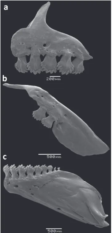
Head small relative to body length, 22.8-27.7% SL. Mouth terminal, and opening below horizontal line through middle of eye pupil. Upper and lower jaws with approximately same length. Maxilla short, posteroventrally angled, posterior tip surpassing vertical through anterior border of eye and surpassing horizontal through ventral border of eye. Teeth pedunculated distally expanded, all similar in shape. Premaxilla with 5 teeth aligned in a single row, with 5(13) or 7(3) cusps [mostly 5]. Maxilla with 2 teeth bearing 5(7) cusps. Dentary teeth with 9 gradually decreasing in size posteriorly; anterior 6, larger, bearing 5-7 cusps; followed posteriorly by smaller teeth with 1-3 cusps (Fig. 2). All jaw teeth with central cup larger and wider than lateral cusps. Ventral surface of mesopterygoid with 18-19 teeth with 1-3 cusps grouped on median portion forming a continuous row (Fig. 3).
Dorsal-fin rays ii, 9(60). First unbranched dorsal-fin ray about half-length of second unbranched dorsal-fin ray; branched rays gradually decreasing in size posteriorly. Dorsal fin with 10(5) pterygiophores. Presence of small ossification associated with first proximal dorsal-fin radial (character 124, described by Zanata, Vari, 2005). Proximal radial of first pterygiophore of dorsal fin posterior to neural spine of ninth precaudal vertebra. Medial radial fused with distal radial from first to fourth pterygiophores; as separate bony elements from fifth to tenth pterygiophores. Proximal radial of first to ninth pterygiophore with lateral projections, absent in tenth dorsal pterygiophore.
Adipose-fin origin posterior to vertical through base of last anal-fin ray. Unbranched anal-fin rays v(3); branched anal-fin rays 17(3), 18(20), 19(30), or 20(6) with 1 row of scales on base of anterior anal-fin rays. Profile of distal margin of anal fin distinctively concave forming an angle in tenth branched ray. Anal fin with 18(1), 20(3), or 21(1) pterygiophores. Medial radial fused with distal radial in first to sixth pterygiophore and no fused on remaining pterygiophores. Proximal radial of first pterygiophore in contact with hemal spine of first caudal vertebra.
Fig. 2. Dentition of Odontostilbeweitzmani, paratype MCP 20337 male: (a) left side premaxilla, (b) maxilla, and (c) dentary, lateral view. Scanning electron micrographs (SEM).
Pectoral fin i(59), 11(22), 12(30), or 13(7). Tip of extended pectoral fin not reaching pelvic-fin origin. Pelvic-fin origin anterior to vertical through dorsal-Pelvic-fin origin. Pelvic-fin rays i, 7(60). Principal caudal-fin rays 19(58). Dorsal procurrent caudal-fin rays 8(7), 9(2), 11(8), or 12(1) and ventral procurrent caudal-fin rays 7(2), 8(5), 9(15), or 10(4).
Fig. 3. Mesopterygoid of Odontostilbe weitzmani paratype, male (MCP 20337) ventral view: (a) mesopterygoid with teeth forming a row, (b) detailed image of the teeth of mesopterygoid with 1, 2 and 3 cusps. Scanning electron micrographs (SEM).
Supraneurals 4(3) without anterior and posterior projections, 5(3) upper gill rakers and 10(3) lower gill rakers (2 on hypobranchial). Upper gill rakers with 2-5 denticles on anterolateral border, and 0-2 on posterolateral border, mainly on basal portion of upper gill rakers. Lower gill rakers with 3 or 6 denticles on anterolateral border, and none on posterolateral border. Gill raker at junction of ceratobranchial and epibranchial with 3-4 denticles on dorsolateral and 3 on ventrolateral borders (Fig. 4). Olfactory rosette slightly oval with 20(2) or 21(1) lamellae in left and right sides of central median raphe. Precaudal vertebrae 16(3); caudal vertebrae 18(3); total vertebrae 34(3).
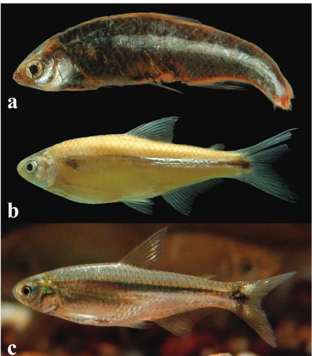
Color in alcohol. Overall body coloration yellowish. Dorsal surface of head from snout to posterior limit of frontal darker with scattered light grey chromatophores. Darker in region of parietal and supraoccipital. Opercular apparatus and infraorbital region of beige silver color extending to cleithrum. Ventral region between pectoral and pelvic fins with a lighter yellow area. Humeral region with a darkened triangular area due to muscular hiatus of pseudotympanum. Scales of dorsal portion of body from supraoccipital to caudal peduncle with small dots dim-gray concentrated in posterior margin.Posterior region of pseudotympanum to caudal peduncle with a silvery lateral thin band above lateral line, top edge of chromatophores dim-gray forming a dark band reaching caudal spot. A conspicuous horizontally oval black caudal spot continued on base of middle caudal-fin rays (Fig. 1).
Tab. 1. Morphometric data for Odontostilbeweitzmani. Number of individuals (N), mean, minimum (min), maximum (max) and standard deviation (SD) include values of the holotype (male).
Holotype Females Males Unsexed
N Min Max Mean SD N Min Max Mean SD N Min Max Mean SD
Standard length (mm) 42.5 37 29.9 50.8 39.3 - 17 29.8 43.9 38.0 - 9 21.4 25.2 23.5
-Percents of Standard Length
Snout to anal-fin origin 62.3 36 61.6 67.3 63.5 1.2 15 60.6 63.9 62.4 0.9 7 63.3 66.6 64.4 1.2
Snout to dorsal-fin origin 46.1 36 45.3 51.7 47.9 1.1 15 45.4 49.9 47.2 1.5 7 47.9 52.1 49.9 1.5
Snout to pelvic-fin origin 43.9 36 43.3 49.3 45.4 1.2 15 43.3 45.5 44.6 0.6 7 45.5 49.5 47.3 1.4
Snout to pectoral-fin origin 21.9 36 20.7 25.8 23.1 1.1 15 21.9 26.6 23.1 1.2 7 24.1 26.4 25.5 0.8
Dorsal to caudal-fin origin 51.9 36 47.7 54.2 50.9 1.7 15 47.3 53.8 51.6 1.8 7 48.1 51.8 49.9 1.1
Orbit to dorsal-fin origin 32.1 36 31.3 36.1 33.0 1.2 15 30.0 34.7 32.2 1.4 7 29.6 34.9 32.9 1.8
Anal fin base length 23.2 36 20.9 27.9 23.8 1.2 15 21.9 27.4 23.9 1.3 7 22.4 26.0 23.8 1.2 Length of caudal peduncle 13.9 36 10.8 14.5 12.5 1.0 15 11.3 14.1 12.9 0.8 7 12.5 14.6 13.6 0.9 Depth of caudal peduncle 10.5 36 9.3 11.1 10.3 0.4 15 9.5 11.0 10.2 0.5 7 9.6 11.7 10.4 0.7
Body depth at dorsal fin 28.8 36 23.1 32.9 27.9 2.0 15 23.6 30.9 26.3 1.9 7 27.3 31.0 28.2 1.3
Dorsal fin length 27.2 36 23.5 27.2 25.4 1.1 15 23.5 29.9 26.6 2.2 7 23.5 25.7 25.0 0.9
Pelvic fin length 17.1 36 14.0 18.5 16.1 1.0 15 15.0 21.9 17.5 2.0 7 13.7 17.2 15.3 1.3
Pectoral fin length 22.3 36 18.6 21.8 20.1 0.7 15 19.6 22.6 21.0 0.9 7 16.4 20.8 18.0 2.1
Head length 23.2 36 23.1 25.3 24.0 0.6 15 23.2 26.4 24.6 0.8 7 25.8 27.4 26.7 0.6
Percents of Head Length
Snout length 23.8 36 21.1 29.2 24.2 1.7 15 20.9 27.5 24.7 1.7 7 22.8 28.2 25.0 2.2
Upper Jaw length 35.3 36 30.7 37.9 34.0 1.5 15 31.0 36.9 34.0 1.7 7 31.5 35.0 33.3 1.4
Dorsal fin with scattered black chromatophores slightly darker and more numerous on mid-distal portion of dorsal fin, extending from second unbranched to eighth branched dorsal-fin rays. Pectoral and pelvic fins with dark grey chromatophores scattered mainly in unbranched rays. Anal fin with dark grey dots slightly scattered in all rays, concentrated in basal region. Adipose fin unpigmented.
Color in life. Caudal spot black, not reaching upper and lower margin of caudal peduncle and extending to base of central rays of caudal-fin, followed by silvery pigments in each lobe. Basal part of anal fin gold silvery. Body silvery, with scattered dark chromatophores on scales and fins (Fig. 5).
Sexual dimorphism. Mature males have second unbranched ray of dorsal fin and unbranched ray of pelvic fin slightly longer than remaining rays, differing from immature and females. Mature males with small hooks on pelvic and anal–fins rays. Pelvic–fin with small hooks in all branched rays, with 1 retrorse bony hook per segment on medioventral border of both lepidotrichia in median and distal length of pelvic-fin rays. Tip of bony hooks not reaching proximal border of segment of lepidotrichia where inserted. Anal-fin rays with one pair of small retrorse bony hooks per segment symmetrically arranged (exceptionally two pairs of hooks), present from last unbranched ray to fourth or seventhbranched rays, decreasing in number posteriorly. Hooks mostly distributed along middle third of anal-fin ray length in posterolateral border. Distal tip of bony hooks not reaching proximal border of segment of lepidotrichia where inserted.
Males bearing gill gland (Fig. 4) similar in size and number of modified filaments (8) to those observed in other externally fertilized species of Cheirodontinae (Oliveira etal., 2012).
Fig. 5. First gill arch of Odontostilbeweitzmani: (a) left side, lateral view, showing gill gland (arrow) on anteriormost portion of lower branch of MCP 20337 male. In detail (b) gill rakers near the junction of ceratobranchial and epibranchial, and (c) gill rakers on lower branch of gill arch. Scanning electron micrographs (SEM).
Geographic distribution: Odontostilbeweitzmani is known to inhabit tributaries of the upper rio Paraná, in the states of Goiás, Mato Grosso do Sul, Minas Gerais, and São Paulo, in Brazil (Fig. 6).
Fig. 6. Southern South America showing the distribution of Odontostilbe avanhandava (green diamonds), O. microcephala (yellow squares) and O. weitzmani (red circle). Type localities represented by star of respective colors.
Etymology. The specific epithet is named after Stanley H. Weitzman, in recognition for his work on the systematics of Neotropical characiforms, particularly of the characid subfamily Cheirodontinae.
Conservation status. Odontostilbe weitzmani is abundant where it occurs. This species may be categorized as Least Concern (LC), according to the IUCN criteria for evaluation on threatening status, version 13 (IUCN, 2017).
Remarks. Besides the type specimens from the upper rio Paraná basin, on which we based the description, we analyzed additional lots of Odontostilbeweitzmani from two localities along the rio Corumbá, Goiás (MCP 25999 and MCP 26000- female, 55.1-56.8 mm SL). These specimens have the same number of cusps on premaxillary teeth, number of branched anal-fin rays and other morphometric characters as the type specimens, but can be differentiated in the length of caudal peduncle (9.9-11.8% SL, mean 10.6% SL vs. 10.8-14.5% SL, mean 12.5% SL in O. weitzmani type specimens) and body depth at dorsal-fin (35.0-37.1% SL, mean 36.0, vs. 23.1-32.9% SL, mean 27.9% SL in O. weitzmani type specimens). We suggest that this difference is related to the larger size of the individuals from rio Corumbá. Stretches of the rivers where these specimens were collected have been changed from lotic to lentic environments by the construction of hydroelectric power dams, and this may possibly influence the body size, body depth and caudal peduncle length of studied populations.
Several specimens of Odontostilbe weitzmani collected in the upper rio Paraná basin are found among the material that Eigenmann (1915) used to redescribe Odontostilbe microcephala (Fig. 1c; CM 6854a-c, now FMNH 131317; CM 6855a-p, now CAS 60508 and FMNH 57872), and since then, specimens of O. weitzmani from the upper rio Paraná have been erroneously identified as O. microcephala.
Odontostilbeavanhandava, new species
u r n : l s i d : z o o b a n k . o rg : a c t : 1 C 9 6 3 F 2 2 2 5 B 9 4 5 4 3 B 0 C 0 -EFA18386A8B2
Figs. 7-9
Odontostilbemicrocephalanon Eigenmann, 1907. -Eigenmann, 1915 [in part]: 94-95 (the largest specimen from CM 6854a-c (FMNH 57871) from rio Tietê at Salto Avanhandava above the falls, São Paulo, Brazil, upper rio Paraná basin).
Holotype. LIRP 3239, 48.7 mm SL, female, ribeirão da Batalha, farm Batalha (Pedro Quaresma), rio Paranaíba, Paracatu, Minas Gerais State, Brazil, 17°25′25″S 47°27′11″W, 27 Apr 2002, C. A. A. Figueiredo & E. S. S. Rego.
Paratypes. All from Brazil, upper rio Paraná basin. São Paulo State: FMNH 57871, 1, 62.1 mm SL, rio Tietê at Salto Avanhandava below the falls, 14 Sep 1908, J. Haseman [material reviewed by Eigenmann (1915)]. LIRP 00314, 2, 47.4-51.1 mm SL, rio Pardo, Ribeirão Preto, 15 Oct 1991, W. E. Alvear. MCP 12103, 1, 56.1 mm SL, Ilha Solteira, rio Paraná, Set 1965. MCP 26004, 2, 39.3-57.9 mm SL, rio Paranapanema, at Jurumirim dam reservoir, Itatinga, 23°06′00″S 48°36′00″W, UNESP de Botucatu. Goiás State: MCP 26003, 1, 43.8 mm SL, rio do Peixe (tributary of rio Corumbá), rio Paranaíba, Caldas Novas, 17°36′00″S 48°27′00″W, 19 Oct 1996, Nupelia. MCP 26002, 1, 57.7 mm SL, rio do Peixe, nex to mouth (tributary of rio Corumbá), rio Paranaíba, Caldas Novas, 17°36′00″S 48°27′00″W, 21 Sep 1996, Nupelia. MNRJ 19718, 1, 54.2 mm SL, rio São Bento, a tributary of the left margin of the rio São Marcos, downstream of the future AHE Serra dam, Paranaíba basin, Divinópolis, 18°10′10″S 47°38′07″W, 27 Sep 1999, C. A. Figueiredo, F. A. Bockmann & A. P. R. Pires. MZUSP 80112, 2, 66.4-77.7 mm SL, Ensecadeira da Usina Hidroeletrica de Corumbá IV, rio Corumbá, Luziânia, H. L. R. Silva. Mato Grosso do Sul State: MCP 12101, 1, 30.2 mm SL, lakes along of the margin of rio Paraná, in front of Jupiá, 11-23 Sep 1964.
Diagnosis. Odontostilbe avanhandava differs from its congeners, except of O. weitzmani, by the presence of mesopterygoid teeth, grouped on median portion forming a continuous row, covering half of mesopterygoid bone (vs. absent). Odontostilbeavanhandava is distinguished from O. weitzmani by the presence de 11-12 lamellae in left and right sides of central median raphe of olfactory rosette (vs. 20-21) and from all species of the genus, except O. fugitiva, O. splendida, O. parecis, and O. pao by the presence of teeth with 9 or 11 cusps in the premaxilla, mostly 9, occasionally 7 cusps (vs. 3-7 cusps; teeth with 9 or 11 cusps always absent). Odontostilbeavanhandava is distinguished from O. fugitiva, O. splendida, and O. pao by the subterminal mouth (vs. terminal mouth); additionally from O. parecis and O. fugitiva by the presence of 16-18 branched anal-fin rays, mostly 17 (vs. 20-21 in O. parecis and 19-24, mostly 21-22 in O. fugitiva); from O. splendida by the anterior position of the adipose fin, positioned at vertical through last anal-fin ray insertion (vs. at vertical through last 2nd or 3rd
anal-fin ray insertions); from O. pao by the presence of 5 scale rows between lateral line and dorsal-fin origin (vs. 6-7, mostly 7).
Description. Morphometric data on Tab. 2. Body elongate and slightly compressed (Fig. 7). Greatest body depth at vertical through dorsal-fin origin. Snout elongated and round. Dorsal profile of head convex from snout to posterior margin of frontal bone, slightly concave from there to distal tip of supraoccipital bone. Predorsal profile slightly convex from posterior end of supraoccipital to dorsal-fin origin. Dorsal-fin origin located at midlength of SL. Body profile in base of dorsal fin straight and slightly convex from that point to origin of adipose fin. Body profile between adipose-fin base and dorsal procurrent caudal-fin rays slightly concave. Ventral profile of head slightly convex from snout to origin of pelvic fins and convex from there to origin of anal fin. Body profile along base of anal fin straight or slightly convex. Ventral profile of caudal peduncle slightly concave. Caudal peduncle slightly longer than deep.
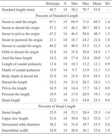
Tab. 2. Morphometric data for Odontostilbeavanhandava, number of individuals (N), mean, minimum (min), maximum (max) and standard deviation (SD) include values of holotype (female).
Holotype N Min Max Mean SD
Standard length (mm) 48.7 14 30.2 70.7 53.0
Percents of Standard Length
Snout to anal-fin origin 67.1 14 66.9 71.6 68.3 1.4
Snout to dorsal-fin origin 47.2 14 46.2 50.7 48.3 1.4
Snout to pelvic-fin origin 47.2 14 46.5 50.8 48.3 1.3
Snout to pectoral-fin origin 21.1 14 18.7 24.2 21.6 1.8
Dorsal to caudal-fin origin 49.2 14 48.9 53.5 51.3 1.6
Orbit to dorsal-fin origin 32.4 14 31.8 38.0 34.9 1.7
Anal-fin base length 18.2 14 17.4 22.4 20.0 1.5 Length of caudal peduncle 12.4 14 10.3 13.2 12.1 0.9
Depth of caudal peduncle 15.9 14 9.7 15.9 11.1 1.5
Body depth at dorsal-fin 25.4 14 25.4 32.9 29.2 2.3
Dorsal-fin length 24.2 14 21.6 26.5 24.2 1.3
Pelvic-fin length 16.5 14 14.4 17.7 16.1 0.9
Pectoral-fin length 19.5 14 17.9 20.9 19.1 1.0
Head length 22.2 14 21.3 24.0 22.5 0.8
Percents of Head Length
Snout length 25.5 14 23.3 28.4 25.5 1.4
Upper Jaw length 31.0 14 30.8 36.3 32.8 1.7
Horizontal orbit diameter 38.3 14 31.0 39.7 35.5 2.4
Interorbital width 34.9 14 30.9 36.1 33.6 1.9
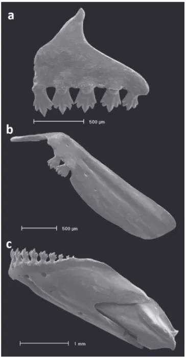
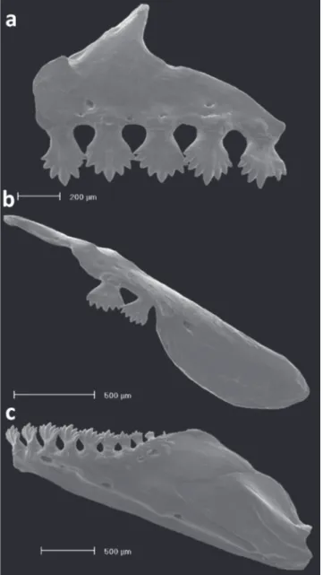
surpassing horizontal through ventral border of eye. Two or 3 maxillary teeth, with 7-9 [mostly 9] cusps nearly of equal size. Premaxilla with a single row of 5-6 teeth slightly inclined to inside mouth (Fig. 8), bearing 7-11 [mostly 9] cusps nearly of equal size. Dentary with 7 or 8 teeth, anterior 6 teeth larger, bearing 7 cusps nearly equal in size, followed posteriorly by 1 or 2 smaller conical teeth, rarely tricuspidate. Dentary teeth inclined anteriorly. Presence of mesopterygoid teeth, grouped on median portion of bone forming a continuous row, covering half-length of ventral surface of mesopterygoid.
Fig. 8. Dentition of Odontostilbeavanhandava, MCP 26004, paratype: (a) premaxilla, (b) maxilla, and (c) dentary, left side, lateral view. Scanning electron micrographs (SEM).
Dorsal-fin rays ii,9(14) in all examined specimens. Dorsal-fin origin at vertical of pelvic-fin insertion. Profile of distal margin of dorsal fin slightly concave. First unbranched dorsal-fin ray less that half-length of second, following branched rays gradually decreasing in size posteriorly. Presence of small ossification associated with first proximal dorsal-fin radial (character 124, described by Zanata, Vari, 2005). First unbranched ray of dorsal-fin inserted in first pterygiophore and last two branched rays inserted in tenth pterygiophore. Proximal radial of first pterygiophore in contact with neural spine of eleventh precaudal vertebra. Dorsal fin with medial radial fused with distal radial from first to fourth pterygiophore and individualized from fifth to tenth pterygiophore. Proximal radial of first to ninth pterygiophores with lateral projections.
Adipose-fin origin posterior to vertical through base of last anal-fin ray. Unbranched anal-fin rays iv(1); branched anal-fin rays 16(4), 17(8), 18(2), with two rows of scales covering base of anterior 6 branched rays. Anal-fin origin posterior to vertical through base of last ray of dorsal fin. Profile of distal margin of anal fin concave. Anal fin with all unbranched rays associated with first pterygiophore, except last unbranched ray associated with second pterygiophore. Medial radial fused with distal radial from first to fifth pterygiophores, visible from that point to last pterygiophore. Proximal radial of first pterygiophore in contact with hemal spine of first caudal vertebra.
Pectoral fin rays i(14), 11(2), 12(1), 13(5), 14(4), or 15(2). First pectoral-fin ray reaching anterior tip of pelvic bone. Pelvic fin i,7(14) rays; insertion located approximately at vertical through origin of dorsal fin. In mature males, first unbranched ray prolonged in filament, usually surpassing origin of anal fin. Principal caudal fin rays 19(14). Procurrent caudal-fin rays: dorsal 7(3), 8(9), 9(1), or 11(1) rays and ventral with 6(2), 7(4), 8(3), 9(4), or 10(1) rays.
Cycloid scales; pored scales on lateral line 36(1), 37(4), 38 (4), or 39(2). Predorsal scales arranged in a regular series with 11(2) or 12(6) scales; scale rows between lateral line and dorsal line origin 5(14); scales rows between lateral line and pelvic-fin origin 4(14); scale rows around caudal peduncle 14(11).
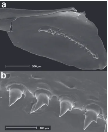
Fig. 9. (a) First gill arch of Odontostilbe avanhandava, MCP 26004, paratype, left side, lateral view. In detail (b) gill rakers on upper branchial branch, and (c) gill rakers on lower branchial branch. Scanning electron micrographs (SEM).
Color in alcohol. Overall body coloration yellowish. Dorsal surface of head from snout to posterior limit of frontal with chromatophores scattered giving a dark yellow coloration. Dark red chromatophores noticeably more concentrated in region of parietal and supraoccipital. Opercular apparatus and infraorbital region silver beige. Branchiostegal rays silver beige. Ventral region between pectoral and pelvic fins with a lighter yellow area. Humeral region with a darkened triangular area due to muscular hiatus of pseudotympanum.
Scales of dorsal portion of body from supraoccipital to caudal peduncle with small dark dots concentrated on posterior margin. Region posterior of pseudotympanum to caudal peduncle with thin silvery band, dorsal and ventral edges of gray color, chromatophores forming a black band reaching caudal spot. A conspicuous oval horizontally black caudal spot overlapping base of middle caudal fin rays.
Dorsal fin with scattered black chromatophores slightly darker and more numerous on mid-distal portion of dorsal fin, extending from first unbranched dorsal-fin ray to ninth branched fin ray. Pectoral and pelvic fins hyaline. Anal fin with sparse dark dots scattered over all rays, more concentrated in the basal region. Adipose fin unpigmented.
Sexual dimorphism. Mature males with small hooks on pelvic and anal fins. Pelvic–fin with small hooks in all branched rays, with one retrorse bony hook per segment, on mesoventral border of both lepidotrichia in median and distal portions of pelvic fin. Tip of bony hooks not reaching proximal border of segment of lepidotrichia where inserted. Anal–fin rays with one pair of small retrorse bony hooks per segment symmetrically placed, on last unbranched ray and first to fourth branched rays, decreasing in number posteriorly. Bony hooks on median portion of posterior branch (never on anterior branch). Tip of bony hooks not reaching proximal border of segment of lepidotrichia where inserted.
Geographic distribution. Odontostilbe avanhandava is known to inhabit tributaries of the upper rio Paraná basin, in the states of Mato Grosso do Sul, Goiás, Minas Gerais, and São Paulo, Brazil (Fig. 6).
Etymology. The epithet avanhandava, refers to the “Salto do Avanhandava” falls of the rio Tietê, the locality of collection of the oldest known specimen, collected by John D. Haseman in 1908. The toponym Ava - Nhandava means: “Man who speaks the Nhandeva dialect”. The “Salto do Avanhandava” does not exist anymore and was flooded by the Nova Avanhandava Hydroelectric Power Dam. A name in apposition.
Conservation status. Odontostilbe avanhandava presents the same distribution of Odontostilbe weitzmani. This species is not abundant in scientific collections, but represents a wide distribution in upper rio Paraná basin. Odontostilbe avanhandava may be categorized as Least Concern (LC), according to the IUCN criteria for evaluation on threatening status, version 13 (IUCN, 2017).
Odontostilbemicrocephala Eigenmann, 1907
Figs. 10-12
Odontostilbe microcephalus Eigenmann, in Eigenmann, Ogle, 1907:10 (original description; type locality: rio Pilcomayo, Bolivia). -Fowler, 1940:61-62 (color description in alcohol; collected río Pilcomayo, tributary of the río Paraguay, Villa Montes, Department of Tarija).
Odontostilbemicrocephala. -Eigenmann, 1910:429 (cited, habitat: Pilcomayo).-Eigenmann, 1915 (in part): 94-95 (type and
paratype from río Pilcomayo, Bolivia); 94 (figure maxilla,
premaxilla and mandible). -Ringuelet et al., 1967: 94 (key diagnosed), 95-96 (brief description).-Géry, 1972:70-71 (comparative material). -Géry, 1977:558 (key).-Casciotta
et al., 1992 (comparative material). -Malabarba, 2003:218 (distribution: rio Pilcomayo basin, Bolivia). -Bührnheim, Malabarba, 2006:172 (comparative material). -Bührnheim, Malabarba, 2007:5 (comparative material). -Miquelarena etal., 2008: 57-58 (Key), 82-83 (description, distribution).-Mirande, Aguilera, 2009:181 (description, distribution) -Mirande, 2010:557 (comparative material).
Diagnosis. Odontostilbe microcephala is distinguished from all species of the genus by the subterminal mouth (vs. terminal mouth, except in Odierythrura, O. euspilurus, and O. avanhandava); usually 10 gill rakers on upper branch and 14-15 on lower branch (vs. 11-12 on lower branch in O. dierythrura, 5-6 on upper branch and 9-10 on lower branch in O. euspilurus). Odontostilbe microcephala differs from O. avanhandava in the horizontal orbit diameter (24.6-32.8 % HL, mean 28.7% vs. 31.0-39.7% HL, mean 35.5 %), number of scales rows between lateral line and dorsal-fin origin (6 vs. 5 in O. avanhandava). Odontostilbe microcephala further differs from O. weitzmani by the presence of 12-13 lamellae in left and right sides of central median raphe of olfactory rosette with (vs. 20-21) and by the horizontal orbit diameter (24.6-32.8 % HL, mean 28.7% vs. 34.0-40.9% HL, mean 37.0 %). Additionally O. microcephala is distinguished from O. avanhandava and O. weitzmani by the absence of mesopterygoid teeth (vs. presence).
Description. Morphometric data in Tab. 3. Body relatively elongate and compressed (Fig. 10). Greatest body depth at or immediately anterior to dorsal-fin origin. Dorsal profile of head relatively convex from snout to vertical through posterior border of nares, straight or slightly
convex from that point to rear of supraoccipital spine. Predorsal profile of body slightly convex from posterior end of supraoccipital to dorsal-fin origin located at vertical of midlength of standard length, dorsal-fin base straight to slightly convex. Dorsal profile of body between last dorsal-fin ray and adipose-dorsal-fin slightly convex, slightly concave from that point to caudal-fin origin. Caudal peduncle slightly longer than deep. Ventral profile of head slightly convex from mouth to pectoral-fin origin; convex from that point to ventral-fin origin, with an obtuse angle in the prepelvic region. Ventral profile straight from pelvic-fin origin to anal-fin origin. Anal-fin base straight to slightly concave. Caudal-peduncle ventral profile straight.
Head small. Mouth subterminal, upper jaw and snout extending anteriorly beyond lower jaw tip. Maxilla short, positioned obliquely at angle inferior of 45 degrees relative to body axis. Premaxillary dentition in single row with 5 teeth, slightly inclined to inside mouth, bearing 7 cusps, exceptionally 9; central cusp slightly longer and wider than lateral cusps. Maxilla with two teeth bearing 7 cusps, similar in shape to premaxillary teeth. Dentary with 8 or 9 teeth, anterior 7 teeth larger, bearing 7 cusps, with central cusp slightly longer and wider than lateral cusps, followed posteriorly by 1 or 2 smaller and conical teeth. Dentary teeth inclined anteriorly (Fig. 11).
Tab. 3. Morphometric data for Odontostilbemicrocephala. Males range, females range, unsexed range, number of individuals (N), mean, minimum (min), maximum (max) and standard deviation (SD) include values of the paratype CAS 59791 and Standard length of holotype CAS 59790 (male).
Holotype CAS59790
Paratype CAS59791
Females Males
N Min Max Mean SD N Min Max Mean SD
Standard length (mm) 32.7 33.7 39 29.7 52.0 38.8 - 22 30.4 43.6 36.4
-Percents of Standard Length
Snout to anal-fin origin 67.1 36 61.6 69.1 64.2 1.4 18 61.7 64.7 63.4 1.0
Snout to dorsal-fin origin 48.1 36 45.5 50.8 48.3 1.1 18 45.7 50.1 48.0 1.0
Snout to pelvic-fin origin 446.0 36 42.7 48.8 45.7 1.2 18 44.0 49.2 45.9 1.4
Snout to pectoral-fin origin 21.1 36 20.1 23.0 21.7 0.7 18 20.7 23.8 22.3 0.9
Dorsal to caudal-fin origin 50.5 36 48.0 56.8 52.1 1.9 18 48.7 55.0 52.2 1.7
Orbit to dorsal-fin origin 39.5 36 33.7 39.6 37.1 1.4 18 32.8 38.6 36.2 1.4
Anal fin base length 23.2 31 20.1 24.4 21.9 1.0 18 20.2 25.2 22.6 1.2
Peduncle length 14.9 32 11.5 16.9 14.4 1.2 18 11.2 16.2 14.0 1.4
Peduncle depth 10.4 32 9.8 12.5 10.8 0.7 18 8.6 11.7 10.5 0.9
Body depth at dorsal fin 34 24.3 34.0 28.2 2.4 18 24.7 30.7 27.5 2.0
Dorsal fin length 27 22.8 28.1 24.6 1.2 17 23.8 32.9 27.2 3.1
Pelvic fin length 29 13.9 17.4 16.0 0.9 18 16.3 21.4 18.3 1.7
Pectoral fin length 29 17.3 25.9 20.2 1.6 18 17.8 23.2 20.9 1.4
Head length 23.4 36 20.5 23.9 22.4 0.8 18 21.2 23.6 22.7 0.6
Percents of Head Length
Snout length 22.8 36 18.8 24.8 22.6 1.4 18 20.1 25.1 23.2 1.4
Upper Jaw length 30.4 33 29.0 35.7 31.5 1.9 18 29.6 35.8 32.1 2.0
Horizontal orbit diameter 25.3 36 25.3 32.8 28.6 1.9 18 24.6 31.8 28.8 2.0
Dorsal-fin rays ii,9(23). Dorsal-fin origin posterior to vertical through pelvic-fin insertion. Profile of distal margin of dorsal fin slightly concave. First unbranched dorsal-fin ray about half-length of second ray, following branched rays gradually decreasing in size posteriorly. In mature males, second unbranched ray is elongated in filament; its length three times longer than first unbranched ray, reaching origin of adipose fin in some specimens (Fig. 12b). First unbranched ray of dorsal fin inserted in first pterygiophore and last 2 branched rays inserted in tenth pterygiophore. Proximal radial of first pterygiophore in contact with neural spine of 11th or 12th precaudal vertebrae. Dorsal fin with medial radial fused with distal radial from first to fifth pterygiophore and not fused from sixth to ninth pterygiophore. All proximal radials with lateral projections. Origin of adipose fin anterior to vertical through base of last anal-fin ray.
Fig. 11. Dentition of Odontostilbe microcephala, paratype USNM 32473, left side, lateral view: (a) premaxilla, (b) maxilla, and (c) dentary. Scanning electron micrographs (SEM).
Unbranched fin rays iv(1), or v(3); branched anal-fin rays 16(1), 17(6), 18(8), 19(7), with two rows of scales covering base of anterior 5 branched rays. Anal-fin origin posterior to vertical through base of last dorsal-fin ray. Profile of distal margin of anal fin concave. Anal fin with 20 pterygiophores, anterior 3 or 4 unbranched rays associated with first pterygiophore. First 4 to 7 pterygiophores with medial radial fused with distal, not fused from that point to last pterygiophore. Proximal radial of first pterygiophore in contact with hemal spine of first caudal vertebra.
Pectoral-fin rays i(23), 10(1), 11(11), 12(9), or 13(2). Longest pectoral-fin ray reaching anterior edge of pelvic bone, reaching origin of pelvic fin in mature males. Pelvic fin i,7(23) rays; fin insertion slightly anterior to vertical through origin of dorsal fin. In mature males, first unbranched ray prolonged in filament, usually surpassing origin of anal fin. Elongated thin hooks present on all branched pelvic-fin rays in mature males. Principal caudal-fin rays 19(23). Procurrent caudal-caudal-fin rays: dorsal 9(5), 10(9), 11(5), or 12(3) and ventral 6(1), 7(1), 8(7), 9(10), or 10(3).
Cycloid scales; lateral line with 35(3), 36(4), or 37(8) pored scales. Predorsal scales arranged in regular series with 11(4), 12(5), or 13(1) scales; scale rows between lateral line and dorsal-fin origin 6(15); scale rows between lateral line and pelvic-fin origin 4(14), exceptionally 3(1); scale rows around caudal peduncle 14(23).
Supraneurals 5(11) or 6(3). Precaudal vertebrae 17(4) or 18(5); caudal vertebrae 17(4), 18(4), or 19(1); total vertebrae 34(1), 35(6), 36(1), or 37(1). Upper gill rakers 10, lower 14-15 (2 on hypobranchial). Upper gill rakers with 3 or 4 denticles on anterolateral border, and 2 or 3 denticles on posterolateral border. Lower gill rakers with 4 denticles on anterolateral border, and 1 or 2 denticles on posterolateral border. Denticulation mainly on basal portion of gill rakers. Olfactory rosette circular in shape and with 12 (2) or 13 (1) lamellae in left and right sides of central median raphe.
Color in alcohol. Overall body color pale yellow to light brown. Dorsal surface of head from snout to anterior limit of frontal pale yellow, black chromatophores noticeably more concentrated in region of parietal and supraoccipital bones. Region of third to fourth infraorbitals and opercular apparatus silver. Dorsum from posterior limit of supraoccipital to caudal peduncle with dark gray chromatophores more concentrated on scales border. Ventral region between pectoral and pelvic fins light yellow. Humeral region with slightly darkened triangular area due to muscular hiatus of pseudotympanum. Body with silver lateral band between pseudotympanum and caudal peduncle. Caudal peduncle with brown rhomboid spot, not reaching dorsal and ventral borders of caudal peduncle, covering basal portion of central rays of caudal fin.
first unbranched pectoral-fin ray. Pelvic fin hyaline. Anal fin hyaline with light gray chromatophores in basal portion of anterior rays. Adipose fin hyaline with light gray chromatophores basally. Caudal-fin ray hyaline with basal portion covered by caudal peduncle spot.
Coloration of paratype CAS 59791 badly preserved, characterized by long silver lateral band extending from region immediately posterior to pseudotympanum to near caudal-fin base.
Color in life. Caudal spot black, not reaching upper and lower margin of caudal peduncle and extending to base of caudal fin, followed by silvery pigments in each lobe. Medial part of dorsal and anal fins gold silvery. Lateral band dark green with golden line on upper margin, extending from pseudotympanum to caudal spot. Body silvery, with scattered dark chromatophores on scales and fins.
Sexual dimorphism. Sexually mature males with small hooks on anal and pelvic fins. Anal-fin rays (Fig. 12c, d) with one pair of retrorse bony hooks per ray segment symmetrically distributed, with robust base and curved or straight tips, present on last unbranched ray and anterior 6 to 8 branched rays, decreasing in number posteriorly. Bony hooks present on median portion of posterior branch, absent on anterior branch. Tip of bony hooks not reaching proximal border of segment of lepidotrichia where inserted. Pelvic-fin rays (Fig. 12e,f) with small hooks on all branched rays; hooks retrorse with robust base and curved tips inserted on median and distal partions of fin rays, some very small hooks on distal border of proximal portion. One bony hook, per segment on posterolateral border of anterior and posterior lepidotrichia in median portion and on posterolateral border of posterior lepidotrichia in distal portion. Tip of bony hooks not reaching proximal border of segment of lepidotrichia where inserted.
Geographic distribution. Odontostilbe microcephala is known from río Pilcomayo, río Paraguay basin, in Bolivia and Argentina and río Masicuri, headwaters of rio Mamoré, Amazon basin, in Bolivia (Fig. 6). Carvalho, Albert, (2011) discuss the distribution of Odontostilbe microcephala between the Paraguay and Mamoré basins that is located mainly in the Chaco Plain and traversed by several major rivers (i.e., Parapetí, Grande, and Pilcomayo rivers), where typical taxa from the La Plata and upper Madeira basins occurs, due possibly to an exchange of water during the rainy season. O. microcephala is also distributed in the río Bermejo, río Juramento and río Sali-Dulce basins, in Northwestern Argentina (Mirande, Aguilera, 2009) and arheic basin of río Horcones a tributary of the río Salado. The first two basins are parallel to the Pilcomayo and drains into the Paraguay and Paraná rivers. The Sali-Dulce basin is endorheic.
Ecological notes. Examination of gut contents of two specimens of Odontostilbemicrocephala (USNM 305484) revealed the presence of insect larvae, composed of a high number of nymphs of Ephemeroptera and pupae of Diptera, followed by larvae of Trichoptera and allochthonous adults of Hymenoptera (Vespidae).
Rondineli et al. (2011) described the species as insectivorous based on specimens collected from the rio Corumbataí (São Paulo, Brazil). The material examined by Rondineli and co-authors is possibly Odontostilbe avanhandava or O. weitzmani since O. microcephala does not occur in the upper rio Paraná. These authors, however, do not provide any descriptive information or photo of the fish he examined, not allowing confirmation of the identifications.
Conservation status. Odontostilbe microcephala is categorized as Least Concern (LC), according to the IUCN criteria for evaluation on threatening status, version 3.1 (Reis, Lima, 2009), due to its wide distribution and the lack of any known major threats across its range. For this categorization, Reis, Lima (2009) not considered the individuals of the upper part of the Paraná basin (currently O. weitzmani and O. avanhandava), due to this their current categorization of threat does not vary.
Remarks. Odontostilbemicrocephala was briefly described by Eigenmann (in Eigenmann, Ogle, 1907) based on two specimens from the río Pilcomayo in Bolivia. Eigenmann (1915) redescribed the species with more detail based on the type specimens plus some specimens collected in the rio Tietê, São Paulo, Brazil (CAS 60508, FMNH 57871, FMNH 57872, FMNH 131317), and extended its distribution to the upper rio Paraná basin. Specimens examined by Eigenmann (1915), however, were analyzed herein and belong to the two new above described species, and no specimens of O. microcephala were found among the material available from the upper rio Paraná basin. Due to the redescription of Eigenmann (1915), the name of O. microcephala has been
cited for the upper rio Paraná basin in different studies (e.g. Oyakawa, Menezes, 2011). This species is now restricted to the Andean slope of the río Paraguay and edorheic río Salí basin (Argentina and Bolivia).
Material examined. Holotype CAS 59790, 1, 46.0 mm SL,
río Pilcomayo, Bolivia, tributary of río Paraguay, 21°11′32″S 63°47′32″W, 1900-1901. Paratype CAS 59791, 1, 45.0 mm SL,
río Pilcomayo, Bolivia, tributary of río Paraguay, 20°43′30″S 64°12′55″W, 1900-1901. Non-type specimens: MCP 38311, 4, 38.9-43.6 mm SL, endorheic of río Uruena, drainage of Bajo Paraná,
Rosario de la Frontera, Salta, Argentina, 25°00′00″S 64°30′00″W, 2
Mar 2001, G. Monasterio de Gonzo & M. Mosqueira. MCP 38310, 4, 39.8-55.3 mm SL, arheic basin of río Horcones, Salta, Rosario
de la Frontera, Argentina, 25°00´00″S, 64°30´00″W, 03 Mar
2001, G. Monasterio de Gonzo & M. Mosqueira. USNM 176033,
1, 48.9 mm SL, río Dulce, endorheic, afluent Mar Chiquita Lake Argentina, 29°38′47″S 62°52′19″W, 1 Aug 1933, T. Marini. UMSS
473, 6, 37.4-43.0 mm SL, río Masicurí, farm Piraymiri, río Grande,
río Mamoré, río Madera, río Amazonas, Bolivia, 18°50′36″S 63°45′32″W, 22 nov 2005, L. Cordova, M. Maldonado, M. Arraya,
USNM 306349, 2, 41.7-47.4 mm SL, rio Bermejo, 4-5 km S, Pueblo Salado, 30 air KM NW Bermejo, border of Department of Tarija,
río Paraguay, Argentina-Bolivia, 22°27′00″S 64°32′00″W, 5 Oct
1988, W. Starnes, L. Starnes, J. Sarmiento & R. Vasquez. USNM 305484, 109, 33.54-51.97 mm SL, río Pilcomayo, Department of
Tarija, at Villamontes rr Bridge, río Paraguay, Bolivia, 21°16′48″S 63°28′12″W, 1 Oct 1988, W. Starnes, L. Starnes, J. Sarmiento &
R. Vasquez. USNM 321173, 49, 29.7-44.0 mm SL (2 c&s, 30.6-44.0 mm SL), río Camatindi, 8 Km N Border Department of Tarija, 40 Km N Villamontes, Department Chuquisaca, riío Paraguay,
Bolivia, 20°59′34″S 63°23′51″W, 2 Oct 1988 Cols W. Starnes, L.
Starnes, J. Sarmiento & R. Vasquez. USNM 319279, 200, 21.96-38.04 mm SL, rio Parapeti, at Rr bridge at San Antonio, 40 Air km E Camiri, Department Santa Cruz, Amazon and Paraguay, Bolivia,
20°01′12″S 63°12′00″W, 30 Sep 1988, W. Starnes, L. Starnes, J.
Sarmiento & R. Vasquez. MHNG 2653.042, 2, 38.8-40.9 mm SL, Pazo Hondo, río Pilcomayo, Paraguay, 12 Aug 1994, C.Dlouhy.
Comparative material examined. Odontostilbefugitiva: Perú:
ANSP 178908, 2 c&s, 29.5-32.5 mm SL. Brazil: INPA 18506, 3 c&s, 24.5-32.4 mm SL. INPA 18512, 1 c&s, 34.9 mm SL.
Ecuador: MZUSP 77844, 2 c&s, 36.9-40.0 mm SL. Odontostilbe
pulchra:Trinidad and Tobago: INHS 40101, 3 c&s, 32.5-34.4 mm SL. INHS 40081, 1 c&s, 30.5 mm SL. Odontostilbenareuda:
Brazil: MZUSP 87759, 1 c&s, 27.5 mm SL. Bolivia: FMNH
106433, 1 c&s, 31.6 mm SL. Odontostilbedierythrura: Bolivia:
MCP 38624, 7 (2 c&s, 33.5-38.4 mm SL), 32.7-34.5 mm SL.
Odontostilbeeuspilurus: Perú: ANSP 143702, 2 c&s, 29.5-33.1 mm SL. Ecuador: MCP 38420, 13, 35.2-41.2 mm SL. Odontostilbe
paraguayensis: Brazil: MCP 35618, 54, 28.5-33.9 mm SL.
Paraguay: MCP 12031, 3, 30.92-33.90 mm SL. Odontostilbe
Morphometric comparisons. The results of the PCA of morphometric data (Fig. 13) are congruent with the morphological analysis in delimitating Odontostilbe avanhandava, O. microcephala, and O. weitzmani. Although overlapping, the boxplot graphic of lateral line scales count shows differences between O. microcephala vs. O. weitzmani and O. avanhandava vs. O. weitzmani, and that O. avanhandava and O. microcephala present a smaller overlap. Furthermore, although overlapping branched anal-fin rays count are also different between O. avanhandavavs. O. weitzmani and slight different between O. avanhandava and O. microcephala (Fig. 14).
Fig. 13. Principal Components Analysis (PCA) between the species Odontostilbe avanhandava (red) O. microcephala (light blue) and O. weitzmani (black).
Fig. 14. Box-plot graphics of Odontostilbeavanhandava, O. microcephala and O.weitzmani: lateral line series of scales (top); and branched rays of the anal fin (bottom).
The variable that best discriminate O.microcephala and O. weitzmani (Fig. 15), is M17 (horizontal orbit diameter), not overlaping between both species and presenting a high discrimination capacity (Fig. 15a). Other variable with high discrimination capacity is M6 (orbit-dorsal fin origin). The scatterplot of polar coordinates obtained for both species using M17 and M6 (Fig. 15b) shows the differences between O. microcephala and O. weitzmani. The bivariate randomization test also separates individuals of both species. The individual of O. microcephala (Fig. 15c, red point) with the higher probability of belonging to O. weitzmani, with p= 0.019 for X-axis, is not included among the specimens belonging to O. weitzmani, rejecting the null hypothesis. Consequently, the X polar coordinates of all individuals of O. microcephala are significantly different from those of O. weitzmani and, therefore, none of the individuals identified as O. microcephala could be designated as belonging to O. weitzmani. The same is found for the individual of O. weitzmani (Fig. 15d, red point) with higher probability of belonging to O. microcephala (p = 0.025 for X-axis), rejecting the null hypothesis, consequently the X polar coordinates of all individuals of O. weitzmani are significantly different from those of O. microcephala. Repeating this same analysis, variables that best discriminate O. avanhandava and O. weitzmani are M1 (snout to anal-fin origin) and M7 (anal-fin base length), from O. avanhandava and O. microcephala are M1 and M17 (horizontal orbit diameter).
Discussion
Over the course of the taxonomic history, some species of Odontostilbe have been wrongly identified, mainly due to problems in the original descriptions made at the beginning of the XX Century. It is the case of Odontostilbe microcephala described originally from Bolivia (Eigenmann in Eigenmann, Ogle, 1907), and redescribed by Eigenmann (1915) with inclusion of material from the upper rio Paraná basin. The most part of this material was herein reviewed, concluding that specimens used in the redescription belong to Odontostilbeavanhandava and O. weitzmani, endemic to the upper rio Paraná basin.
Based on our analysis, Odontostilbe avanhandava, O. microcephala, and O. weitzmani can be clearly diagnosed based in the mouth position, tooth cusp number, number of lamellae in the olfactory rosette, number of gill rakers in the upper and lower branch, horizontal orbit diameter, head size, snout to anal-fin origin distance, number of branched rays of the anal-fin, perforated scales of lateral line and form of hooks in the pelvic fin.
Jerep, 2014). The presence and morphology of these bony hooks represent an important data source for comparative phylogenetic, reproductive and behavioral studies (Bertaco, Malabarba, 2005; Vieira etal., 2016). Even though they are usually less developed and less informative in the species of Odontostilbe, their number and size were useful to diagnose O. avanhandava, O. microcephala, and O. weitzmani.
Differences in the mouth position were found among species of Odontostilbe previously described by Bührnheim, Malabarba (2006, 2007), with terminal mouth observed
in O. ecuadorensis, O. fugitiva, O. nareuda, O. pao, O. paraguayensis, O. parecis, O. pequira, O. pulchra, O. splendida, and O. weitzmani, and subterminal mouth in O. avanhandava, O. dierythrura, O. euspilurus and O. microcephala. This variation in mouth position may reflect differences in feeding habits, such as mode of foraging, orientation, or diet composition. Species that feed in midwater often have terminal mouth, while benthic feeders exhibit subterminal mouth (Keast, Webb, 1966; Langerhans etal., 2003).
Fig. 15. Morphometric comparison of Odontostilbe microcephala and O. weitzmani. (a) Density plot with the overlap of the variable M17 (horizontal orbit diameter), that best discriminates the two species. (b) Scatterplot of the polar coordinates obtained for both species using variables M17 and M6 (orbit to dorsal-fin origin); the arrows show the vector of variables. (c) Bivariate randomization test, showing the individual (red point) with a higher probability of belonging to O. weitzmani
The shape and number of gill rakers in the lower and upper branch of the first branchial arch have been described recently for some species of Odontostilbe, being a character that varies slightly between species (Bührnheim, Malabarba, 2006, 2007). Odontostilbe microcephala, however, shows the highest number of gill rakers (upper 10, lower 14-15), comparatively to other species : O. avanhandava (upper 6, lower 12), O. ecuadorensis (upper 5-6, lower 10-11), O. fugitiva (upper 6-8, lower 11-14), O. nareuda (upper 5-6, lower 10-12), O. pao (upper 6, lower 11), O. weitzmani (upper 5, lower 10) , O. parecis (upper 5-6, lower 9-10), O. pulchra (upper 6-7, lower 11-12), and O. splendida (upper 5-6, lower10-12). The number of gill rakers, may play an important role in food-particle retention, particularly with respect to zooplankton feeding (MacNeil, Brandt, 1990; Amundsen etal., 2004), and may reflect differences in the feeding habits of O. microcephala.
Description of denticles on the gill rakers was provided for the first time for species of the Cheirodontinae by Bührnheim, Malabarba (2006); this character was considered in the phylogeny of Characidae by Mirande (2010: character 201). Denticles on gill rakers of the first branchial arch are found among all groups of Characiformes, especially in Characidae, being its absence observed in some Characidae as for example the Gymnocharacinae, Axelrodia lindeae Géry 1973, and Pseudochalceus kyburzi Schultz 1966 (Mirande, 2010). When present, the denticles may be restricted to one margin or be distributed along of the entire surface of gill rakers as in Astyanaxpuka Mirande etal. (2007). The number of denticles on anterolateral border of the gill rakers presents large intraspecific variation: O. ecuadorensis (upper 1-6, lower 1-6), Odontostilbefugitiva (upper 1-5, lower 1-4), O. nareuda (upper 1-3, lower 0-3), O. pao (upper 1-2, lower 0-2), O. paraguayensis (upper 0-2, lower 4-6), O. parecis (upper 1-3, lower 0-5), O. pulchra (upper 1-4, lower 1-6), and O. splendida (upper 0-3, lower 1-4). Odontostilbemicrocephala (upper 3-4, lower 4) presents the denticulation mainly on basal portion of gill rakers, differing from O. weitzmani (upper 4-5, lower 3-6) that presents the denticles also on the lateral surfaces of gill rakers.
Mesopterygoid teeth were reported, among the Characiformes, in Hoplerythrinus unitaeniatus (Spix, Agassiz) (Weitzman, 1964), Boulengerella Eigenmann (Vari, 1995), Crenuchus Günther and Ammocryptocharax vintonae (Eigenmann) (Buckup, 1993), Acestrorhynchus Eigenmann, Kennedy and Cynodontinae (Lucena, Menezes, 1998; Toledo-Piza, 2000), and Roeboides dispar (Lucena, 2001). Mesopterygoid teeth can present interspecific and/or ontogenetic variation, being present in some individuals and absent in others within the same species (Toledo-Piza, 2000). In Odontostilbe, this character is known only in O. avanhandava and O. weitzmani.
Generic assignment of the new species. Cope (1970) diagnosed Odontostilbe based on three characters that are currently shared with other species of Cheirodontinae: the teeth spatulate and crenate in a single series on the premaxilla and dentary, the anal fin elongated and the lateral line continued to the caudal fin. Malabarba (1998) proposed two synapomorphies to diagnose Odontostilbe related to the length of the second branched ray of the dorsal fin and length of the first unbranched ray of the pelvic fin in males. More recently, Bührnheim, Malabarba (2006, 2007) used this criterion to describe five new species to this genus (O. ecuadorensis, O. nareuda, O. pao, O. parecis, and O. splendida), along with the redescription of O. pulchra and its type species Odontostilbefugitiva. Odontostilbeweitzmani and O. microcephala present the two synapomorphies proposed by Malabarba (1998) for the genus.
The elongation of second branched ray of the dorsal fin and the unbranched ray of the pelvic fin in males is not an exclusive character of Odontostilbe. It has been recorded in Cheirodontops geayi (Bührnheim, Malabarba, 2006), but that species differs from Odontostilbe by the premaxillary teeth with only 3 cusps. It is also found in Serrapinnustocantinensis Malabarba, Jerep (2014), but that species shares with other Serrapinnus the caudal peduncle conspicuously arched ventrally in mature males and several other synapomorphies of the Cheirodontini not shared with the species of Odontostilbe (Malabarba, Jerep, 2014).