Subclinical Anthracycline-Induced Cardiotoxicity in the Long- Term
Follow-Up of Lymphoma Survivors: A Multi-Layer Speckle
Tracking Analysis
Yu Kang,
1*Fei Xiao,
2*Haiyan Chen,
3*Wei Wang,
1Lijing Shen,
2Hang Zhao,
1Xuedong Shen,
1Fangyuan Chen,
2Ben He
1Department of Cardiology - Renji Hospital - School of Medicine - Shanghai Jiaotong University, Shanghai - China1
Department of Hemotology - Renji Hospital - School of Medicine - Shanghai Jiaotong University, Shanghai - China2
Department of Echocardiography - Zhongshan Hospital of Fudan University, Shanghai - China3 * Yu Kang, Fei Xiao and Haiyan Chen contribute equally to the work.
Mailing Address: Fangyuan Chen and Ben He •
Department of Hemotology - Renji Hospital - School of Medicine - Shanghai Jiaotong University. No. 1630 Dongfang Rd, 200126, Shanghai - China E-mail: chenfangyuan1027@163.com; heben1027@163.com
Manuscript received July 11, 2017, revised manuscript September 05, 2017, accepted October 06, 2017
DOI: 10.5935/abc.20180042
Abstract
Background: Anthracycline generates progressive left ventricular dysfunction associated with a poor prognosis.
Objectives: The purpose of this study was to evaluate whether layer-specific strain analysis could assess the subclinical left ventricular dysfunction after exposure to anthracycline.
Methods: Forty-two anthracycline-treated survivors of large B-cell non-Hodgkin lymphoma, aged 55.83 ± 17.92 years (chemotherapy group) and 27 healthy volunteers, aged 51.39 ± 13.40 years (control group) were enrolled. The cumulative
dose of epirubicin in chemotherapy group was 319.67 ± 71.71mg/m2. The time from last dose of epirubicin to the
echocardiographic examination was 52.92 ± 22.32 months. Global longitudinal (GLS), circumferential (GCS) and radial strain (GRS), subendocardial, mid and subepicardial layer of longitudinal (LS-ENDO, LS-MID, LS-EPI) and circumferential strain (CS-ENDO, CS-MID, CS-EPI) values were analyzed. Transmural strain gradient was calculated as differences in peak systolic strain between the subendocardial and subepicardial layers. A value of p < 0.05 was considered significant.
Results:Conventional parameters of systolic and diastolic function showed no significant difference between two groups. Compared with controls, patients had significantly lower GCS and GLS. Multi-layer speckle tracking analysis showed significant reduction of circumferential strain of subendocardial layer, transmural CS gradient and longitudinal strain of all three layers. In contrast, the two groups did not differ in transmural longitudinal strain gradient and radial strains.
Conclusions: It proved the preferential impairment of subendocardial deformation in long-term survivors after exposure to anthracycline. Multi-layer speckle tracking echocardiography might facilitate the longitudinal follow-up of this at-risk patient cohort. (Arq Bras Cardiol. 2018; 110(3):219-228)
Keywords: Cardiotoxicity; Anthracyclines; Lymphoma, Non-Hodgkin; Hematologic Neoplasias/drug therapy; Echocardiography.
Introduction
Anthracycline, a commonly used chemotherapeutic agent in the treatment of a wide spectrum of hematologic malignancies and solid tumors, is undermined by potential life-threatening cardiotoxicity.1,2 Anthracycline-induced left ventricular
dysfunction is believed to be refractory to conventional pharmacological therapy and to be associated with a poor prognosis. Therefore, detection of subclinical myocardial dysfunction is of vital importance to balance between the cardiac risk and the potential cancer treatment.
Two-dimensional speckle tracking echocardiography, based on tracking local image details from frame to frame throughout the cardiac cycle,3-6 has allowed more accurate measurements
of regional myocardial systolic performance.
It has been proved that patients treated with epirubicin-based chemotherapy experienced significant decrease in strain values while LVEF remained stable and within normal limits.7,8 Based on the 2D speckle tracking
technology, a novel offline tool is recently introduced which has a potential of measuring strains in subendocardial and subepicardial layers comparatively.
Therefore, the objectives of this study were to evaluate whether layer-specified systolic strain analysis could differentiate the subclinical left ventricular function changes in patients after exposure to anthracycline-based chemotherapy.
Methods
Study population
ECG, arrhythmia, a previous history of heart failure and/or coronary artery disease. The following data were collected including date of completion of chemotherapy, duration of follow-up, cumulative doses of anthracyclines, symptoms and signs of heart failure, and cardiac medications. Twenty-eigh age-matched and gender-matched referred to our hospital for non-specific chest pain or palpitation, but with no history of cardiovascular disease patients and with completely normal electrocardiograms, echocardiograms, treadmill stress exercises and 24-hour, continuous ambulatory electrocardiograms were selected as controls (control group).
Echocardiographic imaging
Images were obtained in the left lateral decubitus position with Vivid E9 (GE Healthcare, Horton, Norway) ultrasound systems. Standard two-dimensional images were acquired according to recommendations of the American Society of Echocardiography.9 Depth was minimized to optimize
the frame rate. At least three beats were digitally stored for offline analysis. Left ventricular ejection fraction was calculated using the modified Simpson's biplane method. Left ventricular mass index, relative wall thickness, transmitral peak early (E) and peak late (A) diastolic filling velocities were also measured. Tissue Doppler echocardiography was performed with the sample volume positioned at the basal LV free wall and septum at the mitral annular junction to obtain lateral and septal mitral annular systolic (S’) and early diastolic myocardial tissue velocities (E’).
Multi-layer speckle tracking echocardiography
Gray scale images for offline speckle tracking analysis were acquired at frame rate of 53 to 84 MHz. Echopac version 11.1 (GE Healthcare, Horton, Norway) was used for multi-layer strain analysis. The automatic tracking analysis was performed in the apical 4-chamber, 2-chamber, long-axis apical view for longitudinal strain and in the parasternal short-axis view at basal, mid-papillary and apical level for circumferential and radial strain according to the vendor's instructions. The endocardial border was manually traced at end-diastole. The ROI (region of interest) for strain analysis was adjusted manually. The locations of the tracking points were adjusted when necessary so that the region of interest extended from endocardial to epicardial borders to approximate the myocardium, which was divided into subendocardial, middle and subepicardial myocardium layers of equal thickness.
Peak circumferential (CS) and radial strain (RS) measurements were obtained from the basal, mid-segments of the septal, lateral, inferior, anterior, anteroseptal, posterior walls, apical segments of anterior, inferior, septal, lateral walls, totally 18 segments. Peak longitudinal strain (LS) measurements were obtained from the basal, mid- and apical segments of the anterior, inferior, anteroseptal, anterolateral, inferoseptal, inferolateral walls, totally 16 segments. In each segment, the subendocardial, middle and subepicardial LS and CS were calculated automatically. Regional strain values were averaged to determine global longitudinal/circumferential/radial strain (GLS, GCS, GRS), global subendocardial, middle and subepicardial
LS (LS-ENDO, LS-MID, LS-EPI) and CS (CS-ENDO, CS-MID, CS-EPI). Transmural strain gradient was calculated as differences of peak systolic strain between the subendocardial and subepicardial layers. Strain values of each level were calculated.
Reproducibility
Intra- and inter-observer reproducibility was assessed by calculating the difference between the values of 10 randomly selected patients measured by one observer twice and by a second observer.
Statistical analysis
Continuous variables with normal distribution were expressed as the mean ± standard deviation. Continuous variables with non-normal distribution were expressed as median and interquartile range. Differences between two groups were determined using independent samples t test for continuous variables with normal distribution and Kruskal Wallis test for with non-normal distribution. One sample K-S test was used in determining the normality of data. One way ANOVA test was used to compare the differences between strain values of different layers and different levels within each group. Relations between strain values and cumulative anthracycline dose were determined by Pearson correlation analysis. Interobserver and intraobserver reproducibility of strain values were assessed using intraclass correlation coefficients (ICCs) and Bland-Altman analysis. Data were analyzed by SPSS version 16.0 (SPSS, Inc, Chicago, IL, USA).
A value of p < 0.05 was considered significant.
Results
Three patients and one healthy volunteer were excluded from the analysis because of poor image quality (defined as > 2 non-visualized segments). Forty-two patients, 18 males, ranging in age from 22 to 77 years (mean age 55.83 ± 17.92 years), and 27 healthy volunteers, 14 males, ranging in age from 32 to 77 years (mean age 51.39 ± 13.40 years), were finally included in the statistical analysis. Table 1 shows the two groups clinical characteristics. In all patients, the cumulative dose of epirubicin was 319.67 ± 71.71 mg/m2 (ranging from 150.94 mg/m2 to
440.00 mg/m2). Patients have not received radiotherapy or other
cardiotoxic agents. No patient complained of cardiovascular related symptoms. EKG remained normal in all patients. The time from last dose of epirubicin to the echocardiographic examination was 52.92 ± 22.32 months (ranging from 24 months to 104 months).
Conventional echocardiographic parameters
Table 1 – Clinical characteristics of two groups
Normal Chemotherapy p value
Number 27 42
Male (n/%) 12 (44.44) 18 (42.86) 0.84
Age (y) 50.39 ± 13.40 55.83 ± 17.92 0.16
Hypertension (n/%) 0(0) 4(9.52)
ACEI (n/%) 0(0) 1(2.38)
ARB (n/%) 0 (0) 1(2.38)
CCB (n/%) 0(0) 0(0)
β-blocker (n/%) 0(0) 1(2.38)
Smoker (n/%) 5(17.24) 9(21.42) 0.470
DM (n/%) 0(0) 1(2.38)
SBP (mmHg) 124.8 ± 12.6 121.6 ± 12.5 0.627
DBP (mmHg) 70.7 ± 9.3 69.5 ± 7.9 0.233
HR (bpm) 78.0 ± 11.3 81.0 ± 14.5 0.099
ACEI: angiotensin-converting enzyme inhibitors; ARB: angiotensin receptor blockers; CCB: calcium-channel blocker; DBP: diastolic blood pressure; DM: diabetes mellitus; HR: heart rate; SBP: systolic blood pressure; p values were assessed by independent samples t test
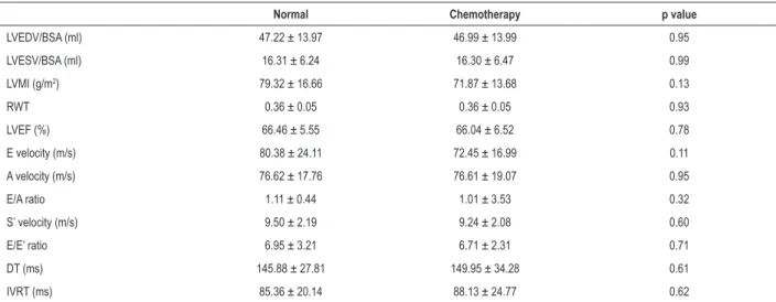
Table 2 – Conventional echocardiographic parameters between two groups.
Normal Chemotherapy p value
LVEDV/BSA (ml) 47.22 ± 13.97 46.99 ± 13.99 0.95
LVESV/BSA (ml) 16.31 ± 6.24 16.30 ± 6.47 0.99
LVMI (g/m2) 79.32 ± 16.66 71.87 ± 13.68 0.13
RWT 0.36 ± 0.05 0.36 ± 0.05 0.93
LVEF (%) 66.46 ± 5.55 66.04 ± 6.52 0.78
E velocity (m/s) 80.38 ± 24.11 72.45 ± 16.99 0.11
A velocity (m/s) 76.62 ± 17.76 76.61 ± 19.07 0.95
E/A ratio 1.11 ± 0.44 1.01 ± 3.53 0.32
S’ velocity (m/s) 9.50 ± 2.19 9.24 ± 2.08 0.60
E/E’ ratio 6.95 ± 3.21 6.71 ± 2.31 0.71
DT (ms) 145.88 ± 27.81 149.95 ± 34.28 0.61
IVRT (ms) 85.36 ± 20.14 88.13 ± 24.77 0.62
BSA: body surface area; DT: deceleration time; FS: fraction shortening; LVEDV: left ventricular end-diastolic volume; LVEF: left ventricular ejection fraction; LVESV: left ventricular end-systolic volume; LVMI: left ventricular mass index; IVRT: isovolumic relaxation time; RWT: relative wall thickness; p values were assessed by independent samples t test
Multi-layer speckle tracking echocardiography
In both groups, longitudinal and circumferential strains were highest in the apical region and decreased significantly from apical to basal level (Table 3,4). The left ventricular longitudinal and circumferential strains of different myocardial layers in patients and controls are shown in Table 5, Figure 1. Transmural strain gradients in LS and CS were demonstrated in both patients and controls, with strain values decreasing from the subendocardial to subepicardial layers. GCS was significantly decreased in chemotherapy group respect to control group
(-27.73% ± 3.37% vs -24.94% ± 4.14%, p = 0.004). The reduction of GCS was attributable to significantly reduced CS-ENDO but preserved CS-EPI strain in patients compared with controls. The longitudinal strain values of global left ventricle and all three layers were significantly decreased in chemotherapy group. However, the two groups did not differ in transmural longitudinal strain gradient. In contrast, there was no statistic difference in radial strains between the two groups.
Table 3 – Three-layer circumferential strain values between two groups stratified by levels
CS-Endo(%) CS-Mid(%) CS-Epi(%)
Control Chemotherapy P value Control Chemotherapy P value Control Chemotherapy p value
Basal level -33.48 ± 5.10 -26.26 ± 4.34 0.000 -23.95 ± 4.26 -22.37 ± 4.28 0.149 -17.58 ± 4.03 -16.85 ± 3.93 0.453
Mid level -34.29 ± 4.21 -31.25 ± 5.39 0.014 -24.45 ± 3.46 -22.57 ± 3.67 0.053 -17.54 ± 3.21 -16.85 ± 3.94 0.063 Apical level -44.31 ± 6.14 -41.13 ± 9.47 0.038 -30.32 ± 4.46 -28.91 ± 6.34 0.316 -21.77 ± 3.95 -19.81 ± 5.39 0.105
P value 0.000 0.000 0.000 0.000 0.000 0.000
CS-ENDO: subendocardial circumferential strain; CS-EPI: subepicardial circumferential strain; CS-MID: middle circumferential strain. P values were analyzed by one way ANOVA test.
Table 4 – Three-layer longitudinal strain values between two groups stratified by levels
LS-Endo(%) LS-Mid(%) LS-Epi(%)
Control Chemotherapy P value Control Chemotherapy P value Control Chemotherapy p value
Basal level -18.51 ± 2.55 -16.82 ± 2.36 0.006 -17.74 ± 2.50 -16.49 ± 2.11 0.027 -17.07 ± 2.50 -15.63 ± 2.00 0.009
Mid level -22.76 ± 2.72 -21.04 ± 2.87 0.014 -20.82 ± 2.39 -19.45 ± 2.49 0.028 -19.23 ± 2.56 -16.87 ± 2.26 0.000 Apical level -34.36 ± 3.23 -32.29 ± 5.69 0.101 -26.20 ± 3.06 -25.08 ± 4.23 0.623 -20.8 ± 2.55 -19.53 ± 5.12 0.538
P value 0.000 0.000 0.000 0.000 0.000 0.000
LS-ENDO: subendocardial longitudinal strain; LS-EPI: subepicardial longitudinal strain; LS-MID: middle longitudinal strain. P values were analyzed by one way ANOVA test.
Inter and intra-observer variation
Inter-observer measurement showed ICC = 0.91 for CS-ENDO, 0.83 for CS-MID, 0.91 for CS-EPI, 0.95 for GCS, 0.61 for RS, 0.87 for LS-ENDO, 0.85 for LS-MID, 0.90 for LS-EPI, 0.91 for GLS. Similarly, intra-observer measurement showed ICC = 0.96 for CS-ENDO, 0.89 for CS-MID, 0.97 for CS-EPI, 0.97 for GCS, 0.73 for RS, 0.86 for LS-ENDO, 0.85 for LS-MID, 0.82 for LS-EPI, 0.94 for GLS, indicating satisfactory
reproducibility by speckle-tracking-derived multilayer analysis of circumferential and longitudinal strain values. Bland-Altman curves of strain values were shown on Figure 2.
Discussion
Globally, cancer is diagnosed in 12.7 million people annually, with its incidence projected to increase by 40% in high-income countries from 2008 to 2030.10
Table 5 – Strain values between two groups.
Control Chemotherapy p value
GLS (%) -21.86 ± 2.38 -20.36 ± 2.58 0.016*
GCS (%) -27.73 ± 3.37 -24.94 ± 4.14 0.004*
GRS (%) 31.44 ± 12.98 26.89 ± 9.75 0.118
LS-ENDO(%) -25.21 ± 2.72 -23.38 ± 3.11 0.014*
LS-MID(%) -21.53 ± 2.36 -20.35 ± 2.58 0.029*
LS-EPI(%) -18.83 ± 2.19 -17.35 ± 2.48 0.013*
LS gradient (%) -6.38 ± 1.28 -6.03 ± 2.07 0.439
CS-ENDO(%) -37.37 ± 3.79 -32.88 ± 5.23 0.000*
CS-MID(%) -26.24 ± 2.98 -24.62 ± 4.13 0.073
CS-EPI(%) -19.56 ± 4.45 -17.32 ± 4.13 0.066
CS gradient(%) -17.80 ± 3.69 -15.55 ± 4.59 0.0*
Endo MId Epi Trans Global
Endo MId Epi Trans Global
Control Chemotherapy
Control Chemotherapy
Control Chemotherapy 0
–10
–20
–30
0
–10
–20
–30
–40
–30
Longitudinal strain (%)
Circumference strain (%)
Radial strain (%)
50
40
30
20
10
0
*
* *
* *
*
Figure 1 – Strain values between two groups. *:p < 0.05.
Anthracyclines are powerful cytotoxic agents, available to treat a wide spectrum of hematologic malignancies and solid tumors. However, life altering cardiac sequelae from anthracyclines remain a problem, with a range of 5-23% of patients developing late-onset heart failure secondary to anthracycline induced-cardiotoxicity.11
Reliable, sensitive and non-invasive methods in detecting cardiac function are of vital importance in these patients. The present study demonstrated that subclinical cardiotoxicity existed in long-survivors after receiving anthracycline therapy albeit normal conventional echocardiographic findings, implicating the more sensitive nature of these parameters in monitoring anthracycline cardiotoxicity.
Recently tagged magnetic resonance imaging provided a detailed quantitative analysis of left ventricular transmural differences in myocardial deformation.12
Echocardiographic speckle-tracking strain analysis, which is angle-independent, provides a noninvasive method to assess left ventricular mechanics, thus translating clinically relevant aspects of cardiac performance from “bench to bedside”. Furthermore, the echocardiographic speckle tracking derived transmural gradients has recently been validated against sonomicrometry crystal in a sheep model.13
As proved by previous studies,14-16 our observations showed
great interobserver and intraobserver agreement, suggesting reasonable reproducibility of the speckle tracking-derived multi-layer strain parameters.
The present study confirmed the presence of transmural and translevel gradient in myocardial circumferential and longitudinal strains, with higher values in the subendocardial myocardial layer and in the apical level in both normal subjects and patients exposed to anthracycline, as is improved by Shi et al.16 The difference in amplitude of myocardial
contraction between the subendocardial and subepicardial regions was related to the orientation pattern of myocardial fiber in the heart. It has been described that in normal heart, contraction is greater in the subendocardial myocardium layer than in the subepicardial myocardium layer.17 However, with
greater contraction and higher energy requirements, subendocardial layer was more susceptible to injury, which can be detected by multi-layer speckle tracking strain analysis. Beck et al.18 has demonstrated that a multi-layer
analysis of myocardial deformation is highly accurate in the differentiation between different degrees of scar transmurality as assessed by MRI. In particular, multi-layer strain analysis provided higher accuracy to discriminate nontransmural versus noninfarction or trasmural versus nontransmural infarction compared with global strain. Altiok et al.19 has also
2.0 1.5 1.0 0.5 0.0 –0.5 –1.0 –1.5 –2.0
Difference of GLS (%)
Intraobserver
Average of GLS (%)
+1.96 SD 1.70 –1.96 SD –1.32 Mean 0.19 –24
–25 –23 –22 –21 –20 –19 –18
2.0 1.5 1.0 0.5 0.0 –0.5 –1.0 –1.5 –2.0 –3.0 –2.5
Difference of GLS (%)
Interobserver
Average of GLS (%)
+1.96 SD 1.59 –1.96 SD –2.12 Mean –0.26
–26 –24 –22 –20 –18 –16
Difference of LS-ENDO (%)
Intraobserver
Average of LS-ENDO (%) 3 2 1 0 –1 –2 –3 +1.96 SD 2.6 –1.96 SD –2.5 Mean 0.0
–26 –24 –22 –20
–28 –30
Difference of LS-ENDO (%)
Interobserver
Average of LS-ENDO (%) 3 2 1 0 –1 –2 –3 –4 +1.96 SD 2.6 –1.96 SD –2.5 Mean 0.1
–26 –24 –22 –20 –28
–30 –18
Difference of LS-MID (%)
Intraobserver
Average of LS-MID (%) 3 4 2 1 0 –1 –2 –3 +1.96 SD 3.1 –1.96 SD –2.4 Mean 0.4 –24
–25 –23 –22 –21 –20 –19 –18
Difference of LS-MID (%)
Interobserver
Average of LS-MID (%) 3 2 1 0 –1 –2 –3 +1.96 SD 2.3 –1.96 SD –2.3 Mean 0.0
–23 –22 –21 –20
–24
–25 –19 –18 –17
Difference of LS-EPI (%)
Intraobserver
Average of LS-EPI (%) 3 4 2 1 0 –1 –2 –3 +1.96 SD 3.0 –1.96 SD –2.6 Mean 0.2
–20 –18 –16
–22 –14
Difference of LS-EPI (%)
Interobserver
Average of LS-EPI (%) 2 1 0 –1 –2 –3 –4 +1.96 SD 1.2 –1.96 SD –3.0 Mean –0.9
–22 –21 –20
–23 –19 –18 –17 –16 –15
Difference of GCS (%)
Intraobserver
Average of GCS (%) 2 1 0 –1 –2 –3 +1.96 SD 1.4 –1.96 SD –2.7 Mean –0.7
–34 –32 –30 –28 –26 –24
–36
–38 –22
Difference of GCS (%)
Interobserver
Average of GCS (%) 3 2 1 0 –1 –2 –3 –4 +1.96 SD 3.4 –1.96 SD –3.0 Mean 0.2 –30 –25 –35 –40 –20
Difference of CS-ENDO (%)
Interobserver
Average of CS-ENDO (%) 1.5 1.0 0.5 0.0 –1.5 –1.0 –0.5 –2.0 –2.5 –3.0 +1.96 SD 1.20 –1.96 SD –2.45 Mean –0.63
–40 –35 –30 –25 –45
–50
Difference of CS-ENDO (%)
Intraobserver
Average of CS-ENDO (%) 5 4 2 3 1 0 –1 –2 –3 –4 +1.96 SD 4.0 –1.96 SD –3.1 Mean 0.4
–40 –35 –30
–45 –50
Difference of CS-MID (%)
Intraobserver
Average of CS-MID (%) 3 4 2 1 0 –1 –2 –3 –4 –5 –6 +1.96 SD 2.6 –1.96 SD –4.7 Mean –1.0
–30 –25 –20
–35 –40
Difference of CS-MID (%)
Interobserver
Average of CS-MID (%) 4 6 2 0 –2 –4 –6 +1.96 SD 5.3 –1.96 SD –5.0 Mean 0.2
–26 –24 –22 –20
–28 –30 –32 –34
Difference of CS-EPI (%)
Interobserver
Average of CS-EPI (%) 8 6 4 2 0 –2 –4 –6 +1.96 SD 5.9 –1.96 SD –5.0 Mean 0.4
–30 –25 –20 –15
–35
–40 –10
Difference of CS-EPI (%)
Intraobserver
Average of CS-EPI (%) 3 2 1 0 –1 –2 –3 –4 +1.96 SD 1.9 –1.96 SD –3.6 Mean –0.9
–30 –25 –20 –15
–35
–40 –10
accurate distinction between segments with non-transmural infarction vs those with no infarction and between segments with transmural vs non-transmural infarction as defined by late gadolinium enhancement cardiovascular magnetic resonance. In the present study, we adopted a multi-layer strain approach in analyzing layer-specific ventricular deformation and observed the decrease of subendocardial circumferential strain values and transmural circumferential gradient in long-term survivors exposed to anthracycline. It has been proved in animal models of anthracycline cardiotoxicity that severe myocytolysis mainly involved the subendocardium of the ventricle.20 Moreover, Perel et al.21 observed a regional
and diffuse pattern of subendocardial enhancement using cardiac magnetic resonance imaging in patients with anthracycline-induced cardiomyopathy. Hence, the findings in our study of reduction of subendocardial circumferential strain values and transmural circumferential gradient but preserved subepicardial circumferential strain was consistent with the same hypothesis of subendocardial injury induced by anthracycline. Moreover, it has been proved22 that in patients
with chronic ischemic cardiomyopathy, subendocardial circumferential strain was a powerful predictor of cardiac events and appeared to be a better parameter than LVEF and other strain variables analyzed by echocardiography. Therefore, we believed that further importance may need to be attached to the changes of subendocardial circumferential strains.
We observed that after anthracycline exposure, longitudinal strains of all the three layers decreased significantly. However, transmural longitudinal strain gradient did not show any difference compared to normal group. It is reported that the subendocardium is predominantly composed of longitudinal myocardial fiber. The subendocardial deformation is greatest in the longitudinal direction and verifies the endo-epicardial gradient in normal left ventricles on magnetic resonance imaging.23,24 Hence, the longitudinal left ventricular mechanics
are predominantly governed by the subendocardial region of the myocardium, which probably accounts for our findings of the reduction of all three layers of longitudinal strain values and the absence of difference in longitudinal transmural gradient.
The lack of difference in radial strain between two groups in our study was perhaps not surprising, which was concordance with some previous studies.25,26 It was recently published that
peak radial strain differed largely between different software and algorithms, and small changes in width can change large RS differences.27 In the present study, the interobserver variation
did not show satisfactory reproducibility of RS measurement. Hence, it may suggest that indices of radial deformation are not as sensitive as circumferential and longitudinal strains in detecting subclinical left ventricular dysfunction.
The absence of associations between strain parameters and cumulative anthracycline indicated lack of a safe dose that was free of cardiotoxicity. It has been proved that even children who have received a cumulative doxorubicin dose as low as 45mg/m2 have reduced left ventricular mass28 and
anthracycline damage to all cardiac structures may begin with the first anthracycline dose.29
Limitation
Several limitations to this study warranted comment. This was a cross-sectional study of a relatively small patient cohort, which did not provide information on the value of myocardial deformation parameters in prognostication. Further out-come studies with hard clinical endpoints will be required to determine the clinical significance of our findings. Secondly, although speckle tracking echocardiography allows interrogation of global strain, these parameters are not entirely load dependent and need to be interpreted with caution when an alteration of cardiac status with acute changes in load is anticipated.
Conclusion
Despite normal left ventricular ejection fraction, subtle abnormalities in myocardial systolic function were present in long-term survivors after anthracycline exposure. It provided the evidence of preferential impairment of subendocardial deformation in long-term survivors after exposure to anthracycline. Multi-layer speckle tracking echocardiography, a potential non-invasive tool for the detection of subclinical anthracycline-induced myocardial abnormalities, might facilitate the longitudinal follow-up of this at-risk patient cohort.
Author contributions
Conception and design of the research: Kang Y, Shen X; Acquisition of data: Kang Y, Xiao F Xiao F, Chen H, Wang W, Zhao H; Analysis and interpretation of the data: Kang Y, Chen H, Wang W; Statistical analysis: Kang Y, Xiao F, Chen H, Shen L, Zhao H; Obtaining financing: Chen F, He B; Writing of the manuscript: Kang Y, Chen H, Shen L; Critical revision of the manuscript for intellectual content: Chen F, He B.
Potential Conflict of Interest
No potential conflict of interest relevant to this article was reported.
Sources of Funding
This work was supported by grants from the National Nature Science Foundation of China (81401411) and Nature Science Foundation of Shanghai (14ZR1425200, 16ZR1420600).
Study Association
This study is not associated with any thesis or dissertation work.
Ethics approval and consent to participate
1. Lipshultz SE, Lipsitz SR, Sallan SE, Dalton VM, Mone SM, Gelber RD, et al. Chronic progressive cardiac dysfunction years after doxorubicin therapy for childhood acute lymphoblastic leukemia. . 2005;23(12):2629-36. doi: 10.1200/JCO.2005.12.121.
2. Van Dalen EC, van den Brug M, Caron HN, Kremer LC. Anthracycline-induced cardiotoxicity: comparison of recommendations for monitoring cardiac function during therapy in paediatric oncology trials. . 2006;42(18):3199-205. doi: 10.1016/j.ejca.2006.08.002.
3. Amundsen BH, Helle-Valle T, Edvardsen T, Torp H, Crosby J, Lyseggen E, et al. Noninvasive myocardial strain measurement by speckle tracking echocardiography: validation against sonomicrometry and tagged magnetic resonance imaging. . 2006;47(4):789-93. doi: 10.1016/j.jacc.2005.10.040. 4. Cho GY, Chan J, Leano R, Strudwick M, Marwick TH. Comparison of two-dimensional speckle and tissue velocity based strain and validation with harmonic phase magnetic resonance imaging . Am J Cardiol. 2006;97(11):1661-6. doi: 10.1016/j.amjcard.2005.12.063.
5. Helle-Valle T, Crosby J, Edvardsen T, Lyseggen E, Amundsen BH, Smith HJ, et al. New noninvasive method for assessment of left ventricular rotation: speckle tracking echocardiography. . 2005;112(20):3149-56. doi: 10.1161/ CIRCULATIONAHA. 104.531558.
6. Reisner SA, Lysyansky P, Agmon Y, Mutlak D, Lessick J, Friedman Z. Global longitudinal strain: a novel index of left ventricular systolic function. . 2004;17(6):630-3. doi: 10.1016/j.echo.2004.02.011
7. Kang Y, Xu X, Cheng L, Li L, Sun M, Chen H, et al. Two-dimensional speckle tracking echocardiography combined with high sensitive cardiac troponin T in early detection and prediction of cardiotoxicity during epirubicine-based chemotherapy. Eur J Heart Fail. 2014:16(3):300-8. doi: 10.1002/ejhf.8. 8. Kang Y, Cheng L, Li L, Chen H, Sun M, Wei Z, et al. Early detection of
anthracycline-induced cardiotoxicity using two-dimensional speckle tracking echocardiography. Cardiol J. 2013:20(6):592-9. doi: 10.5603/CJ.2013.0158. 9. Lang RM, Bierig M, Devereux RB, Flachskampf FA, Foster E, Pellikka PA, et al. Recommendations for cardiac chamber quantification by echocardiography in adults: an update from the American Society of Echocardiography and the European Association of Cardiovascular Imaging. Eur Heart J Cardiovasc Imaging. 2015;16(3):233-70. doi: 10.1093/ehjci/jev014
10. Cardinale D, Colombo A, Lamantia G, Colombo N, Civelli M, De Giacomi G, et al. Anthracycline-induced cardiomyopathy: clinical relevance and response to pharmacologic therapy. 2010;55(3):213-20. doi: 10.1016/j. jacc.2009.03.095
11. Delfino JG, Fornwalt BK, Eisner RL, Leon AR, Oshinski JN. Determination of transmural, endocardial, and epicardial radial strain and strain rate from phase contrast MR velocity data. J Magn Reson Imaging. 2008;27(3):522-8. doi: 10.1002/jmri.21211.
12. Ishizu T, Seo Y, Enomoto Y, Sugimori H, Yamamoto M, Machino T, Kawamura R, et al. Experimental validation of left ventricular transmural strain gradient with echocardiographic two-dimensional speckle tracking imaging. Eur J Echocardiogr. 2010;11(4):377-85. doi: 10.1093/ejechocard/jep221 13. Zhang Q, Fang F, Liang YJ, Xie JM, Wen YY, Yip GW, et al. A novel multi-layer
approach of measuring myocardial strain and torsion by 2D speckle tracking imaging in normal subjects and patients with heart disease. Int J Cardiol. 2011;147(1):32-7. doi: 10.1016/j.ijcard.2009.07.041
14. Ozawa K, Funabashi N, Takaoka H, Kamata T, Kanaeda A, Saito M, et al. Characteristic myocardial strain identified in hypertrophic cardiomyopathy subjects with preserved left ventricular ejection fraction using a novel multi-layer transthoracic echocardiography technique. Int J Cardiol. 2015 Apr 1;184:237-43. doi: 10.1016/j.ijcard.2015.01.070
15. Götte MJ, Germans T, Rüssel IK, Zwanenburg JJ, Marcus JT, van Rossum AC, et al. Myocardial strain and torsion quantified by cardiovascular magnetic resonance
tissue tagging: studies in normal and impaired left ventricular function. J Am Coll Cardiol. 2006;48(10):2002-11. doi: 10.1016/j.jacc.2006.07.048
16. Shi J, Pan C, Kong D, Cheng L, Shu X. Left Ventricular longitudinal and circumferential layer-specific myocardial strains and their determinants in healthy subjects. Echocardiography. 2016;33(4):510-8. doi: 10.1111/echo.13132. 17. Tsutsui H, Uematsu M, Yamagishi M, Haruta S, Shimakura T, Miyatake K.
Usefulness of the subendocardial myocardial velocity gradient in low-dose dobutamine stress echocardiography. Heart Vessels. 2000;15(1):11-7. PMID: 11001480.
18. Becker M, Ocklenburg C, Altiok E, Füting A, Balzer J, Krombach G, et al. Impact of infarct transmurality on layer specific impairment of myocardial function. Eur Heart J. 2009;30(12):1467-76. doi: 10.1093/ eurheartj/ehp112
19. Altiok E, Neizel M, Tiemann S, Krass V, Becker M, Zwicker C, et al. Layer-specific analysis of myocardial deformation for assessment of infarct transmurality: comparison of strain encoed cardiovascular magnetic resonance with 2D speckle tracking echocardiography. Eur Heart J Cardiovasc Imaging. 2013;14(6):570-8. doi: 10.1093/ehjci/jes229. 20. Milei J, Boveris A, Llesuy S, Molina HA, Storino R, Ortega D, et al.
Amelioration of adriamycin-induced cardiotoxicity in rabbits by prenylamine and vitamins A and E. Am Heart J. 1986;111(1):95-102. Doi: http://dx.doi. org/10.1016/0002-8703(86)90559-4
21. Perel RD, Slaughter RE, Strugnell WE. Subendocardial late gadolinium enhancement in two patients with anthracycline cardiotoxicity following treatment for Edwig’s sarcomea. J Cardiovasc Magn Reson. 2006;8(6):789-91. doi: 10.1080/10976640600737664.
22. Hamada S, Schroeder J, Hoffmann R, Altiok E, Keszei A, Almalla M, et al. Prediction of outcomes in patients with chronic ischemic cardiomyopathy by layer-specific strain echocardiography: a proof of concept. J Am Soc Echocardiogr. 2016;29(5):412-20. doi: 10.1016/j.echo.2016.02.001. 23. Hashimoto I, Li X, Hejmadi Bhat A, Jones M, Zetts AD, Sahn DJ. Myocardial
strain rate is a superior method for evaluation of left ventricular subendocardial function compared with tissue Doppler imaging. J Am Coll Cardiol. 2003;42(9):1574-83. Doi: https://doi.org//10.1016/j.jacc.2003.05.002 24. Moore CC, Lugo-Olivieri CH, McVeigh ER, Zerhouni EA. Three-dimensional
systolic strain patterns in the normal human left ventricle: characterization with tagging MR imaging. Radiology. 2000;214(2):453-66. doi: 10.1148/ radiology.214.2.r00fe17453
25. Cheung YF, Hong WJ, Chan GC, Wong SJ, Ha SY. Left ventricular myocardial deformation and mechanical dyssynchrony in children with normal ventricular shortening fraction after anthracycline therapy. Heart. 2010;96(14):1137-41. doi: 10.1136/hrt.2010.194118.
26. Tassan-Mangina S, Codorean D, Metivier M, Costa B, Himberlin C, Jouannaud C, et al. Tissue Doppler imaging and conventional echocardiography after anthracycline treatment in adults: early and late alterations of left ventricular function during a prospective study. Eur J Echocardiogr. 2006;7(2):141-6. doi: 10.1016/j.euje.2005.04.009. 27. Biaggi P, Carasso S, Garceau P, Greutmann M, Gruner C, Tsang W, et
al. Comparison of two different speckle tracking software systems: does the method matter? Echocardiography. 2011;28(5):539-47. doi: 10.1111/j.1540-8175.2011.01386.x.
28. Lipshultz SE, Lipsitz SR, Sallan SE, Dalton VM, Mone SM, Gelber RD, et al. Chronic progressive cardiac dysfunction years after doxorubicin therapy for childhood acute lymphoblastic leukemia. . 2005;23(12):2629-36. doi: 10.1200/JCO.2005.12.121.
29. Lipshultz SE, Rifai N, Sallan SE, Lipsitz SR, Dalton V, Sacks DB, et al. Predictive value of cardiac troponin T in pediatric patients at risk for myocardial injure. Circulation. 1997;96(8):2641-8. Doi: https://doi.org/10.1161/01.CIR.96.8.2641