Regulation of PKM2 and Nrf2-ARE
Pathway during Benzoquinone Induced
Oxidative Stress in Yolk Sac Hematopoietic
Stem Cells
Jie Zhu1,2,3., Zhuoyue Bi4., Tan Yang1,2,3, Wei Wang1,2,3, Zhen Li1,2,3, Wenting Huang1,2,3, Liping Wang5, Shaozun Zhang1,2,3, Yanfeng Zhou1,2,3, Ningna Fan1,2,3, YuE Bai1,2,3, Wentao Song6, Chunhong Wang1,2,3, Hong Wang1,2,3*, Yongyi Bi1,2,3*
1.School of Public Health, Wuhan University, Wuhan, Hubei, P.R. China,2.Hubei Key Laboratory of Allergy and Immune-related Diseases, Wuhan, Hubei, P.R. China,3.Hubei Biomass-resource Chemistry and Environmental Biotechnology Key Laboratory, Wuhan University, Wuhan, Hubei, P.R. China,4.Hubei Provincial Key Laboratory for Applied Toxicology (Hubei Provincial Academy for Preventive Medicine), Wuhan, P.R. China,5.School of Public Health, Kunming Medical University, Chenggong District, Kunming, P.R. China,6.Nanchang Center for Disease Control and Prevention, Nanchang, P.R. China
*[email protected] (HW);[email protected] (YYB)
.These authors contributed equally to this work.
Abstract
Benzene is an occupational toxicant and an environmental pollutant that is able to induce the production of reactive oxygen species (ROS), causing oxidative stress and damages of the macromolecules in target cells, such as the hematopoietic stem cells. We had previously found that embryonic yolk sac hematopoietic stem cells (YS-HSCs) are more sensitive to benzene toxicity than the adult bone marrow hematopoietic stem cells, and that nuclear factor-erythroid-2-related factor 2 (Nrf2) is the major regulator of cytoprotective responses to oxidative stress. In the present report, we investigated the effect of PKM2 and Nrf2-ARE pathway on the cellular antioxidant response to oxidative stress induced by benzene metabolite
benzoquinone (BQ) in YS-HSC isolated from embryonic yolk sac and enriched by magnetic-activated cell sorting (MACS). Treatment of the YS-HSC with various concentrations of BQ for 6 hours induces ROS generation in a dose-dependent manner. Additional tests showed that BQ is also capable of inducing expression of NADPH oxidase1 (NOX1), and several other antioxidant enzymes or drug-metabolizing enzymes, including heme oxygenase 1 (HMOX1), superoxide dismutase (SOD), catalase and NAD(P)H dehydrogenase quinone 1 (NQO1). Concomitantly, only the expression of PKM2 protein was decreased by the treatment of BQ but not the PKM2 mRNA, which suggested that BQ may induce PKM2 degradation. Pretreatment of the cells with antioxidant N-acetylcysteine
OPEN ACCESS
Citation:Zhu J, Bi Z, Yang T, Wang W, Li Z, et al. (2014) Regulation of PKM2 and Nrf2-ARE Pathway during Benzoquinone Induced Oxidative Stress in Yolk Sac Hematopoietic Stem Cells. PLoS ONE 9(12): e113733. doi:10.1371/ journal.pone.0113733
Editor:Dean G. Tang, The University of Texas M.D Anderson Cancer Center, United States of America
Received:June 20, 2014
Accepted:October 30, 2014
Published:December 1, 2014
Copyright:ß2014 Zhu et al. This is an
open-access article distributed under the terms of the
Creative Commons Attribution License, which permits unrestricted use, distribution, and repro-duction in any medium, provided the original author and source are credited.
Data Availability:The authors confirm that all data underlying the findings are fully available without restriction. All relevant data are within the paper and its Supporting Information files.
Funding:The project was supported by Chinese National Natural Science Foundation to YYB (no. 81372961) and Chinese National Natural Science Foundation to HW (grant no. 81102104,
http://www.nsfc.gov.cn/). The funders had no role in study design, data collection and analysis, decision to publish, or preparation of the manuscript.
(NAC) decreased ROS generation and prevented BQ-induced PKM2 degradation, suggesting involvement of ROS in the PKM2 protein degradation in cellular response to BQ. These findings suggest that BQ is a potent inducer of ROS generation and the subsequent antioxidant responses of the YS-HSC. The accumulated ROS may attenuate the expression of PKM2, a key regulator of the pyruvate metabolism and glycolysis.
Introduction
Benzene is a known human leukemogen. Epidemiological studies revealed that occupational or environmental exposure to benzene during pregnancy is associated with increased risk for the development of childhood malignancies, such as leukemia [1–7]. Experiments using animal models showed an increased frequency of chromatid breaks and DNA recombination exchange in fetal hematopoietic cells following benzene treatment [8,9]. Despite extensive studies in the past years, relatively little is known about the detailed carcinogenic mechanism of benzene. Accumulating evidence suggests that one of the
underlying mechanisms of benzene-induced carcinogenesity is oxidative damage caused by its metabolites.
Benzene metabolized primarily in the liver involving cytochromes P450-catalyzed oxidation of benzene to benzene oxide by addition of oxygen atom to the benzene ring. Benzene oxide then further biotransformed to phenol (PH), hydroquinone (HQ) and 1,4-benzoquinone (BQ). BQ is the most potent benzene metabolites in inducing generation of the reactive oxygen species (ROS), such as hydrogen peroxide (H2O2), superoxide anion radicals (O2.-) and hydroxyl
radicals (HO.) [10]. The increased ROS are able to cause oxidative damage of the cellular macromolecules in the target cells [11–13]. Although several biological sources have been linked to ROS generation, the most important source of ROS in cellular response to BQ is the NADPH oxidase 1 [14]. ROS are highly capable of disrupting normal hematopoiesis, which may attribute to the utero-initiated benzene toxicity [9,15,16].
in cancer cells [22]. A recent study showed that acute ROS accumulation caused inhibition of pyruvate kinase M2 (PKM2), spontaneously transfer glucose flux into the pentose phosphate pathway and thereby generate sufficient anti-oxidant responses for the detoxification of ROS [23].
We had previously established an experimental system for assessment of the hematopoietic toxicity and leukemogenicity of benzene and its metabolites during mouse embryonic development. We found that the cytotoxic and apoptotic effects of benzene metabolites were much pronounced in embryonic YS-HSCs than in adult BM-HSCs [24]. The present study was focused on the possible involvement of ROS generation and the activation of the anti-oxidant defense system in embryonic YS-HSCs treated with BQ. We provided evidence showing that BQ is highly capable of inducing ROS, along with an increased elevation of NOX1 protein, nuclear accumulation of Nrf2 and the activation of Nrf2-ARE pathway in YS-HSCs. The expression of PKM2 protein was decreased by treatment of BQ. Inhibition of PKM2 may play an important role in determining the biological effects of benzene-induced ROS in yolk sac hematopoietic cells. Pretreatment of YS-HSCs with antioxidant NAC reversed PKM2 reduction, suggesting BQ-induced ROS generation contributed to PKM2 degradation.
Materials and Methods
Ethics Statement
As described in detail previously [24], all experiments were approved by the Institutional Animal Care and Use Committee of Wuhan University (No. 10061), and the animal procedures were performed in accordance with institutional and national guidelines. All surgery was performed under anesthesia with ketamine and lidocaine, and all efforts were made to minimize suffering.
Embryonic YS-HSCs isolation and proliferation
Special pathogen free (SPF) Kunming mice were purchased from the Central Animal Facility of Wuhan University as described previously [24]. Kunming mice were originated from Swiss mice and were brought to Kunming, China, from the Indian Haffkine Institute in 1944. This mice line has been widely utilized in pharmacological, toxicological, medicinal and biological research and testing because of its’ high disease resistance, good adaptive capacity, high breeding coefficient, and good survival rate.
After separation of yolk sac cells, the cells were analyzed for surface expression of hematopoietic markers. Cells were washed and fluorescently stained with mouse CD117 antibodies conjugated to PE (#130-0991-730), CD41-FITC or CD45-FITC (eBioscience, San Diego, CA, USA). Cells were incubated for 10 minutes in the dark on ice. Finally, cells were washed and resuspended in phosphate buffered saline (PBS) containing 0.5% bovine serum albumin (BSA). Flow cytometric analysis was carried out with a flow cytometer (Epics Altra II, Beckman, USA).
As described in detail previously [24], the yolk cells was first depleted of lineage positive (Lin+) cells using magnetic columns and was then positively selected by
using anti-murine CD117 antibodies conjugated to mini-magnetic beads (Miltenyi Biotec, Inc., Auburn, CA, http://www.miltenyibiotec.com ). Up to 3.26106 of lineage2 CD117+ cells were isolated from eight yolk sac cell
suspensions. Isolated YS-HSC cells were cultured at 37
˚
C with 5% humidified CO2in the Serum-Free Expansion Medium (Stem Cell Technologies, Inc,Vancouver, BC, Canada) with the addition of cytokines SCF (100 ng/ml), IL-3 (20 ng/ml), and Flt-3 (100 ng/ml) (Peprotech USA). Cell were grown in
polystyrene 6-well plates in a volume of 2 ml per well up to 0.66106cells/ml and
kept in fresh culture medium for 12 h before treatments.
Analysis of intracellular ROS
To assess the generation of ROS in YS-HSCs cells, freshly isolated YS-HSCs cells (56105/ml) were incubated for 6 hours with 10mM and 20 mM concentrations of
BQ with or without 2 hours pretreatment of N-acetylcysteine (Sigma). Samples of the cultures were loaded with 20 mM DCF-DA (Sigma) and incubated at 37
˚
C for 45 min. The peak excitation wavelength for oxidized DCF was 488 nm. We used a flow cytometer (Epics Altra II, Beckman , USA) to measure ROS generation by the fluorescence intensity. Quantitative assay of ROS generation was performed by normalization to the control group. For fluorescence microscopic analysis, the cells were washed with PBS, mounted, and observed under an inverted phase-contrast and fluorescence microscope. (OLYMPUS IX71, Japan).Nuclear and cytoplasmic extraction
Cells were seeded in 6-well plates at density of 36106/well and treated with 0, 5,
10, 20, 40 mM BQ for 6 hours. After treatment, nuclear and cytoplasmic proteins were extracted using a Nuclear and Cytoplasmic Protein Extraction Kit (Beyotime Biotechnology) according to the manufacturer’s recommendations. Cells were washed twice with ice-cold PBS. Cell suspension was made by addiing 200 ml ice-cold Cytoplasmic Extraction Reagent I (CER I) to the cell pellets, vortexing and incubating the tube on ice for 10 minutes. The Cytoplasmic Extraction Reagent II (CER II, 10 ml) was then added to the tube, vortexed for 5 seconds on the highest setting, incubated on ice for 1 minute and centrifuged (14,0006g) the tube for
Reagent and incubated on ice for a total of 40 minutes, vortexing for 15 seconds every 10 minutes and centrifuged the tube for 10 minutes at 4
˚
C. The supernatant (nuclear extract) was collected. The extracts were stored at 280˚
C until use.Whole-cell protein extraction
Cells (3.023.56106/ml) were washed twice with ice-cold PBS and then lysed in a
lysis buffer (2 mM sodium orthovanadate, 50 mM NaF, 20 mM HEPES, pH 7.5, 150 mM NaCl, 1.5 mM MgCl2, 5 mM sodium pyrophosphate, 10% glycerol,
0.2%Triton X-100, 5 mM EDTA, 1 mM PMSF, 10 mg/ml leupeptin, and 10 mg/ ml aprotinin) on ice for 30 min. Insoluble materials were pelleted at 14,0006g for
20 min at 4
˚
C. The supernatant (whole-cell extract) was collected and stored at280
˚
C until use.Immunoblotting
Cell lysates was mixed with the SDS sample buffer, and was boiled for 5 min. Equal amounts of protein (30 mg) were electrophoresed on 10% SDS/PAGE and were electroblotted onto PVDF membranes and identified with rabbit polyclonal antibody to NOX1, SOD2 (Abcam), SOD1, Catalase, PKM2 (Cell Signaling Technology), HMOX1, NQO1, GAPDH (Bioworld Technology). Secondary goat anti-rabbit IgG–HRP (Bioworld Technology) was used according to the species of primary antibody. Chemiluminescence was performed using SuperSignal West Dura (Pierce) and the FluorChem 8900 visualization system (Alpha Innotech). The relative density of detected signals was analyzed by using the image analysis software (Image 1.56, NIH).
Statistical analysis
SPSS 17.0 was used for data analysis. Data were expressed as means¡ SDs. The statistical analysis was determined by one-way analysis of variance (ANOVA) followed by Least Significant Difference test (LSD) and Dunnett’s T3.Pvalues less than 0.05 were considered significant.
Results and Discussion
Characterization of hematopoietic surface marker expression in
E8-10 (embryonic day 8–10) murine yolk sacs
CD117 [24]. Whereas the percentage of CD41- and CD45-positive cells is relatively lower, only about 2.98¡1.23% and 2.7¡0.44%, respectively.
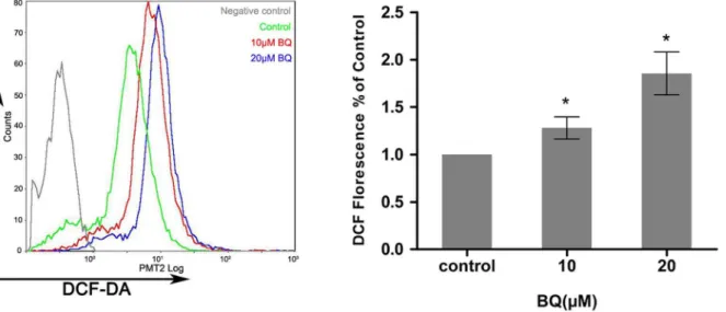
Exposure of YS-HSCs to BQ increased ROS generation
To examine the capability of BQ in the induction of ROS generation in embryonic primitive hematopoietic cells, YS-HSCs were treated with different concentrations (10 mM, 20mM) of BQ for 6 hours. ROS generation was examined by flow cytometry and fluorescence microscopy detecting the DCF-DA signals. Levels of ROS were significantly elevated by BQ treatment, as evidenced by right shifted fluorescence intensity (Figure 2). Since intracellular ROS generation is chiefly regulated by NADPH oxidase, the activation status of NADPH oxidase was determined following the treatment of the YS-HSC with BQ. An increased protein level of NOX1 was observed in the cells treated with BQ, suggesting that BQ is a potential activator of NOX1 that is the key subunit of NADPH oxidase linked to ROS generation (Figure 3).
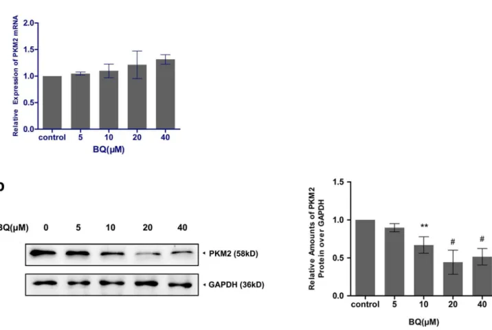
Degradation of PKM2 by BQ exposure
To examine the effect of BQ on PKM2 expression in YS-HSC, the mRNA levels and protein levels of PKM2 were determined by quantitative real–time PCR and Figure 1. Expression of hematopoietic surface markers in E8-10 (embryonic day 8–10) murine yolk sacs.Representative flow cytometric data from more than three independent analyses were shown.
doi:10.1371/journal.pone.0113733.g001
Table 1.Expression of hematopoietic surface makers in E8-10d yolk sac cells.
Maker Positive cells, % CD41 2.98¡1.23%(n511) CD45 2.7¡0.44%(n53)
Western blotting, respectively. PKM2 gene expression levels exhibited no statistically significant differences among control and all BQ treated groups in YS-HSC (Figure 4a). On the other hand, exposure to various concentrations of Figure 2. Exposure of YS-HSCs to BQ increased ROS generation.YS-HSCs cells were isolated and treated with 10mM BQ, 20mM BQ, or without BQ. The intracellular ROS concentrations were measured by DCF-DA staining. Illustrated overlays on the left panel reflected the ROS generation.
Quantifications of ROS generation are displayed on the right pannel. Data are expressed as the mean¡standard deviation (n.3), *p,0.01, as compared to the control group.
doi:10.1371/journal.pone.0113733.g002
Figure 3. The ROS-generating NADPH oxidase NOX1 was induced by BQ.Immunoblotting data were shown in the upper panel. GAPDH was measured as loading control. Densitometry of immunoblotting results was shown in the lower panel. Values are presented as means¡SDs (n5325). *,P,0.05; **,P,0.01;#, P,0.001 compared with controls.
BQ for 6 h resulted in decreased expression levels of PKM2 proteins. As shown in
Figure 4b, BQ reduced the expression of PKM2 protein expression in a concentration-dependent manner in YS-HSC. A notable inhibition PKM2 was observed in the cells treated with 10 to 40 mM BQ (,0.5-fold). BQ only reduced PKM2 protein but not the PKM2 mRNA, suggesting that BQ may induce PKM2 degradation.
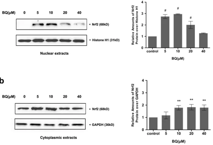
Exposure to BQ upregulated Nrf2 protein expression and caused
Nrf2 nuclear accumulation
As the fact that intracellular ROS metabolism is predominantly regulated by Nrf2, changes in Nrf2 protein expression in response to BQ were examined in YS-HSCs. Because Nrf2 nuclear accumulation is a marker of Nrf2 activation, nuclear and cytoplasmic extracts were prepared to detect Nrf2 protein expression. As shown in
Figure 5, a significant elevation of Nrf2 protein expression were observed in both Figure 4. Degradation of PKM2 by BQ exposure.(a)Effect of BQ on PKM2 mRNA expression. Cells were treated with BQ at increasing concentrations and expression of PKM2 mRNA was measured by quantitative real–time PCR. The relative expression of target genes was calculated using the 2-DDCt method. Data represent means¡SDs of at least three individual experiments. (b)The expression of PKM2 protein was decreased by treatment of BQ. Immunoblotting of PKM2 was shown in the left panel. GAPDH was measured as loading control. Densitometry of immunoblot results was shown in the right panel. Values are presented as means¡SDs (n5325). *,P,0.05; **P,0.01;#,P,0.001 compared with controls.
nuclear and cytoplasmic extracts collected from the cells treated with various concentrations of BQ for 6 hours. These results suggest that exposure to BQ promoted Nrf2 nuclear accumulation and activation in YS-HSCs.
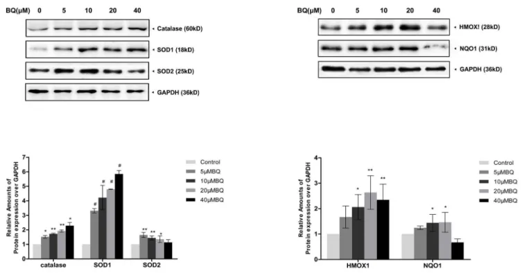
Activation of Nrf2-ARE pathway in the BQ-treated YS-HSCs
To further investigate the effect of BQ on Nrf2 antioxidant program, the
expression of various target proteins involved in the Nrf2-ARE pathway, including detoxification enzymes (NQO1) and antioxidant enzymes (catalase, SOD, and HO-1), were examined by Western Blotting. Increased expression of Nrf2 target proteins were observed following 6 hours BQ treatment (Figure 6). These data demonstrated BQ treatment activates the Nrf2-ARE pathway in the YS-HSCs. Figure 5. Exposure to BQ upregulated Nrf2 protein expression and caused Nrf2 nuclear accumulation.Western Blot analysis of nuclear and cytoplasmic extracts from YS-HSCs treated with increasing concentrations of BQ for 6 hours. Histone H1 and GAPDH antibodies were used as loading controls for nuclear and cytoplasmic extracts, respectively. Left panels: representative blotting results. Densitometry of the immunoblotting results was shown in the right panels. Values are presented as means¡SDs (n5325). *P,0.05, **P,0.01,#P,0.001.
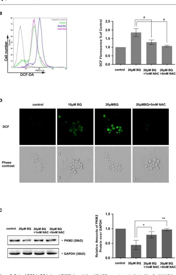
ROS contribute to the BQ-induced PKM2 degradation
To test whether ROS generation is relevant for PKM2 degradation induced by BQ, we measured the effects of ROS inhibition on PKM2 protein expression levels. YS-HSC cells were treated with 20 mM BQ for 6 hours with or without pretreatment of the cells with N-acetylcysteine (NAC), a free radical scavenger, for 2 hours. As expected, 20 mM BQ treatment enhanced DCF staining substantially.
Pretreatment of YS-HSC cells with the antioxidant NAC almost completely suppressed DCF staining (Figure 7a & 7b). Thus, pretreatment of NAC inhibited BQ-induced ROS generation. Notably, the degradation of PKM2 protein was almost completely inhibited by 5 mM NAC pretreatment (Figure 7c). These results confirmed that generation of ROS stimulated by BQ is required for BQ-induced PKM2 degradation.
ROS are important molecules in normal cell signaling. However, excessive generation of ROS can affect the intracellular redox states, leading to mutagenic changes and thereby promote oncogenesis. Cellular ROS are generated from various biological reactions. In the present study, the expression of NOX1 protein was induced by BQ exposure, accompanied by an increased ROS generation. These data indicated that NADPH oxidase is the major source of ROS in the cellular response to BQ.
Figure 6. Increased expression of Nrf2-ARE pathway proteins were observed in the BQ-treated YS-HSCs.Induction of catalase, SOD1, SOD2, HMOX1 and NQO1 protein levels by BQ. Immunoblotting data were shown in the upper panel. GAPDH was measured as loading control. Densitometry of immunoblot results was shown in the lower panel. Values are presented as means¡SDs (n5325). *,P,0.05; **,P,0.01;#,P,0.001 compared with controls.
We had characterized the primary regulatory molecules and cell signaling pathway of antioxidant system. BQ exposure induced Nrf2 nuclear accumulated and activated the downstream proteins of Nrf2-ARE signaling pathways.
Activation of Nrf2-ARE signaling pathways may play an important role in regulating the cellular redox state (Figure 8).
Cancer cells take up more glucose and produce increasable lactate regardless of the concentration of oxygen, this phenomenon is called Warburg effect [25]. PKM2 is a key enzyme in the glycolytic pathway and is found in all proliferative tissue cells, including tissue stem cells, in particular hematopoietic stem cells, and glycolytic metabolism also present in these cells [26]. Inactivation of PKM2 promote glucose metabolism and generate NADPH, to maintain glutathione in the reduced state, which plays a critical role in detoxification of ROS and oxidative stress damage repair process [27,28] (Figure 8). . We observed the PKM2 protein expression was inhibited in response to BQ in YS-HSCs. The effect of BQ in reducing PKM2 expression may be a new mechanism that mediates the toxicity or carcinogenicity of the ROS in YS-HSCs. Pretreatment of antioxidant reduced PKM2 degradation, suggesting that the production of ROS by BQ
Figure 8. BQ induced ROS and two possible pathways involved in the ROS detoxification.Exposure to BQ causes NOX1 up-regulation, resulting in ROS generation in YS-HSCs. Intracellular ROS elevation promotes two possible pathways: (a) Nrf2 nuclear accumulation and activation of Nrf2-ARE pathways, including detoxification enzymes and antioxidant enzymes; (b) PKM2 degradation and promoting glucose flux into the pentose phosphate pathway (PPP), leading to generate NADPH responsible for the maintain of reducing glutathione, which plays a critical role in detoxification of ROS.
doi:10.1371/journal.pone.0113733.g008
western blotting. Immunoblotting of PKM2 was shown in the left panel. GAPDH was measured as loading control. Densitometry of immunoblot results was shown in the right panel. Values are presented as means¡SDs (n5325). *,P,0.05; **P,0.01;#,P,0.001 represent significant differences between the experiments.
contributes to BQ-induced PKM2 degradation. However, the mechanism by which ROS induced PKM2 degradation is still unknown. In human lung cancer cells, high levels of ROS cause inactivation of PKM2 by oxidation of Cys358 [23]. Whether oxidation of PKM2 involved in the BQ-induced PKM2 degradation in YS-HSCs still need further experimental verification.
Supporting Information
Table S1. Expression of hematopoietic surface makers in E8-10d yolk sac cells. doi:10.1371/journal.pone.0113733.s001 (XLS)
Acknowledgments
We thank Dr. Weihuang Liu from the School of Basic Medical Sciences of Wuhan University for her technical assistance with the flow cytometry
Author Contributions
Conceived and designed the experiments: JZ TY CHW HW YYB. Performed the experiments: JZ WW ZL WTH SZZ YFZ NNF YEB. Analyzed the data: JZ LPW ZYB WTS. Contributed reagents/materials/analysis tools: ZYB WTS. Wrote the paper: JZ ZYB.
References
1. Chatzis C, Alexopoulos EC, Linos A(2005) Indoor and outdoor personal exposure to benzene in Athens, Greece. Sci Total Environ 349: 72–80.
2. Eden T(2010) Aetiology of childhood leukaemia. Cancer Treat Rev 36: 286–297.
3. Harrison RM, Leung PL, Somervaille L, Smith R, Gilman E(1999) Analysis of incidence of childhood cancer in the West Midlands of the United Kingdom in relation to proximity to main roads and petrol stations. Occup Environ Med 56: 774–780.
4. Smith MT(2010) Advances in understanding benzene health effects and susceptibility. Annu Rev Public Health 31: 133–148 132 p following 148.
5. Steffen C, Auclerc MF, Auvrignon A, Baruchel A, Kebaili K, et al.(2004) Acute childhood leukaemia and environmental exposure to potential sources of benzene and other hydrocarbons; a case-control study. Occup Environ Med 61: 773–778.
6. Swaen GM, Tsai SP, Burns C (2010) Meta-analysis on benzene exposure and non-Hodgkin’s lymphoma. Occup Environ Med 67: 286–287; author reply 287.
7. Zhang J, Smith KR(2003) Indoor air pollution: a global health concern. Br Med Bull 68: 209–225.
8. Lau A, Belanger CL, Winn LM(2009) In utero and acute exposure to benzene: investigation of DNA double-strand breaks and DNA recombination in mice. Mutat Res 676: 74–82.
9. Badham HJ, Winn LM(2010) In utero and in vitro effects of benzene and its metabolites on erythroid differentiation and the role of reactive oxygen species. Toxicol Appl Pharmacol 244: 273–279.
11. Abernethy DJ, Kleymenova EV, Rose J, Recio L, Faiola B (2004) Human CD34+hematopoietic progenitor cells are sensitive targets for toxicity induced by 1,4-benzoquinone. Toxicol Sci 79: 82–89.
12. Ross D(2000) The role of metabolism and specific metabolites in benzene-induced toxicity: evidence and issues. J Toxicol Environ Health A 61: 357–372.
13. Kolachana P, Subrahmanyam VV, Meyer KB, Zhang L, Smith MT(1993) Benzene and its phenolic metabolites produce oxidative DNA damage in HL60 cells in vitro and in the bone marrow in vivo. Cancer Res 53: 1023–1026.
14. Bedard K, Krause KH(2007) The NOX family of ROS-generating NADPH oxidases: physiology and pathophysiology. Physiol Rev 87: 245–313.
15. Ghaffari S(2008) Oxidative stress in the regulation of normal and neoplastic hematopoiesis. Antioxid Redox Signal 10: 1923–1940.
16. Badham HJ, Winn LM(2010) In utero exposure to benzene disrupts fetal hematopoietic progenitor cell growth via reactive oxygen species. Toxicol Sci 113: 207–215.
17. Rubio V, Zhang J, Valverde M, Rojas E, Shi ZZ(2011) Essential role of Nrf2 in protection against hydroquinone- and benzoquinone-induced cytotoxicity. Toxicol In Vitro 25: 521–529.
18. Nguyen T, Sherratt PJ, Pickett CB (2003) Regulatory mechanisms controlling gene expression mediated by the antioxidant response element. Annu Rev Pharmacol Toxicol 43: 233–260.
19. Nguyen T, Yang CS, Pickett CB(2004) The pathways and molecular mechanisms regulating Nrf2 activation in response to chemical stress. Free Radic Biol Med 37: 433–441.
20. Tong KI, Katoh Y, Kusunoki H, Itoh K, Tanaka T, et al.(2006) Keap1 recruits Neh2 through binding to ETGE and DLG motifs: characterization of the two-site molecular recognition model. Mol Cell Biol 26: 2887–2900.
21. Kwak MK, Wakabayashi N, Kensler TW(2004) Chemoprevention through the Keap1-Nrf2 signaling pathway by phase 2 enzyme inducers. Mutation Research-Fundamental and Molecular Mechanisms of Mutagenesis 555: 133–148.
22. Christofk HR, Vander Heiden MG, Harris MH, Ramanathan A, Gerszten RE, et al.(2008) The M2 splice isoform of pyruvate kinase is important for cancer metabolism and tumour growth. Nature 452: 230–233.
23. Anastasiou D, Poulogiannis G, Asara JM, Boxer MB, Jiang JK, et al.(2011) Inhibition of pyruvate kinase M2 by reactive oxygen species contributes to cellular antioxidant responses. Science 334: 1278– 1283.
24. Zhu J, Wang H, Yang S, Guo L, Li Z, et al.(2013) Comparison of toxicity of benzene metabolite hydroquinone in hematopoietic stem cells derived from murine embryonic yolk sac and adult bone marrow. PLoS One 8: e71153.
25. Warburg O(1956) On the origin of cancer cells. Science 123: 309–314.
26. Wang YH, Israelsen WJ, Lee D, Yu VWC, Jeanson NT, et al. (2013) Differential Dependence On Aerobic Glycolysis In Normal and Malignant Hematopoietic Stem and Progenitor Cells To Sustain Daughter Cell Production. Blood 122.
27. Christofk HR, Vander Heiden MG, Wu N, Asara JM, Cantley LC (2008) Pyruvate kinase M2 is a phosphotyrosine-binding protein. Nature 452: 181–U127.