Complete mitochondrial genome of the lappet moth,
Kunugia undans
(Lepidoptera: Lasiocampidae): genomic comparisons among
macroheteroceran superfamilies
Min Jee Kim, Jun Seong Jeong, Jong Seok Kim, Su Yeon Jeong and Iksoo Kim
Department of Applied Biology, College of Agriculture & Life Sciences, Chonnam National University,
Gwangju, Republic of Korea.
Abstract
The mitochondrial genome (mitogenome) characteristics of the monotypic Lasiocampoidea are largely unknown, be-cause only limited number of mitogenomes is available from this superfamily. In this study, we sequenced the com-plete mitogenome of the lappet moth,Kunugia undans (Lepidoptera: Lasiocampidae) and compared it to those of Lasiocampoidea and macroheteroceran superfamilies (59 species in six superfamilies). The 15,570-bpK. undans genome had one additionaltrnR that was located between trnA and trnN loci and this feature was unique in Macroheterocera, including Lasiocampoidea. Considering that the twotrnR copies are located in tandem with proper secondary structures and identical anticodons, a gene duplication event might be responsible for the presence of the two tRNAs. Nearly all macroheteroceran species, excluding Lasiocampoidea, have a spacer sequence (1–34 bp) at thetrnS2andND1 junction, but most lasiocampid species, including K. undans, have an overlap at the trnS2andND1 junction, which represents a different genomic feature in Lasiocampoidea. Nevertheless, a TTAGTAT motif, which is typically detected in Macroheterocera at thetrnS2andND1 junction, was also detected in all Lasiocampoidea. In summary, the general mitogenome characteristics of Lasiocampoidea did not differ greatly from the remaining macroheteroceran superfamilies, but it did exhibit some unique features.
Keywords:Kunugia undans, mitochondrial genome, Lasiocampoidea, Macroheterocera.
Received: November 17, 2016; Accepted: February 25, 2017.
Introduction
The typical metazoan mitochondrial genome (mito-genome) consists of 13 protein-coding genes (PCGs), 22 tRNAs, two rRNAs, and a major non-coding sequence re-ferred to as the A+T-rich region. The characteristic features of the mitogenome (e.g., fast evolution, low recombination rates, and multiple copies per cell) are considered benefi-cial in several biological fields (Cameron, 2014). In partic-ular, whole mitogenome sequences have been utilized for phylogenic analyses of several insect lineages (Dowtonet al., 1997; Kimet al., 2011; Luet al., 2013; Maoet al., 2014, 2015; Timmermanset al., 2014), and genomic characteris-tics have also been scrutinized to understand phylogenetic and evolutionary features of given taxonomic groups (Cameron and Whiting,2008; Wanet al., 2013; Kimet al., 2014).
Mitogenome sequences in insects have been com-piled in nearly 1,000 species that represent all insect orders and the Lepidoptera. As one of the four most species-rich
insect orders, Lepidoptera is represented by 338 mitoge-nomes in GenBank (last visited on August 14, 2016), including 37 nearly complete sequences from 23 superfa-milies. Among these, the monotypic Lasiocampoidea is represented by four species in two genera. Considering that the monotypic superfamily consists of 1,952 species with
five subfamilies (van Nieukerken et al., 2011),
mitoge-nome sequences from additional diverse taxonomic groups could be required for mitogenome-based phylogenetic stu-dies. In fact, recent large-scale mitogenome-based lepi-dopteran phylogenies only included a single genus or a single species (Timmermanset al., 2014; Ramírez-Ríoset al., 2016).
The lappet moth, Kunugia undans (Walker)
(Lepi-doptera: Lasiocampidae), is distributed in South Korea (ex-cluding the far eastern Ulleungdo Island), far eastern Russia, Japan, and Australia (Parket al., 1999; Shin, 2001). In Korea, adults are found from September to October, eggs then overwinter, and larvae hatch in the spring (Parket al.,
1999). Its host plants are Castanea crenata S. et Z.,
Quercus acutissima Carr., Quercus variabilis Bl. in
Fagaceae, and Malus pumila var. dulcissima Koidz. in
Rosaceae (Parket al., 1999). Variations in size, coloration, DOI: http://dx.doi.org/10.1590/1678-4685-GMB-2016-0298
Send correspondence to Iksoo Kim. Department of Applied Biol-ogy, College of Agriculture & Life Sciences, Chonnam National Uni-versity, Gwangju 61186, Republic of Korea. E-mail: [email protected].
and lines on the wings are present. The wingspan of the spe-cies is 56–65 mm in males and 79–92 mm in females, and forewings have a small white spot at the medial cell (Shin, 2001).
In this study, we determined the complete mitoge-nome sequence of the lappet mothK. undans, adding a new mitogenome sequence of a previously unreported genus of Lasiocampoidea. The genomic characteristics of the se-quence were compared to those of other lasiocampid spe-cies in terms of genome structure, genomic arrangement, nucleotide composition, codon usage, etc. Furthermore, to better understand the evolutionary characteristics of the
Lasiocampoidea, includingK. undans,the mitogenome
se-quences were compared to the representatives of the Macroheterocera clade, to which Lasiocampoidea belongs.
Materials and Methods
DNA extraction, PCR and sequencing
An adultK. undanswas collected from Shinan-gun in Jeollanamdo Province in Korea (34°3’60’’ N, 125°6’50’’ E) in 2009. After collection in the field, the sample was pre-pared as a dried specimen and deposited at Chonnam Na-tional University, Gwangju, Korea under the accession code KTOL-Bom-27. DNA was extracted from the hind legs using a Wizard Genomic DNA Purification Kit, in ac-cordance with the manufacturer’s instructions (Promega, Madison, WI, USA). For whole mitogenome sequencing, primers that amplify three long overlapping fragments
(LF1 fromCOIandND4, LF2 fromND5tolrRNA, and LF3
fromlrRNAtoCOI) were adapted from Kimet al.(2012). Three long fragments (LFs) were amplified using LA
TaqTM(Takara Biomedical, Tokyo, Japan) under the
fol-lowing conditions: 96 °C for 2 min; 30 cycles of 98 °C for 10 sec and 48 °C for 15 min; and a final extension step of 72 °C for 10 min. Using the LFs as templates, 26 overlap-ping short fragments (SF) were amplified using the primers
adapted from Kimet al.(2012) and AccuPower®PCR
Pre-Mix (Bioneer, Daejeon, Korea). The PCR conditions for SFs were as follows: denaturation for 5 min at 94 °C; 35 cy-cles of 1 min denaturation at 94 °C; 1 min annealing at 48–51 °C; 1 min extension at 72 °C; and a final extension of 7 min at 72 °C. Primers used to amplify and sequence the LFs and SFs are presented in Table S1. DNA sequencing
was conducted using the ABI PRISM® BigDye®
Termina-tor v3.1 Cycle Sequencing Kit and an ABI PRISMTM 3100 Genetic Analyzer (PE Applied Biosystems, Foster City, CA, USA). All products were sequenced from both direc-tions.
Gene annotation
Individual SF sequences were assembled into the
complete mitogenome using Seqman software
(DNASTAR, Madison, Wisconsin, USA). Identification, boundary delimitation, and secondary structure folding of
tRNAs were performed using tRNAscan-SE 1.21 with the search mode set as default, the Mito/Chloroplast as the searching source, the genetic code of invertebrate mito-genomes for tRNA isotype prediction, and a cove score cut-off of 1 (Lowe and Eddy, 1997). Twenty-one tRNAs were detected based on these parameters. However,trnS1,
which has a truncated DHU arm, was detected using a hand-drawn secondary structure in conjunction with an alignment of the predicted trnS1 regions of other
lasio-campid species, and the anticodon was given particular consideration (Timmermanset al., 2014; Qinet al., 2015; Kimet al., 2016). Individual PCGs were identified, and a boundary was delimited using the blastx and tblastn
pro-grams in BLAST
(http://blast.ncbi.nlm.nih.gov/BLAST.cgi). With the aid of sequences from other lasiocampid species, the start and stop codons of PCGs were confirmed using MAFFT ver. 6
(Katohet al., 2002). Two rRNAs and the A+T-rich region
were identified and delimited using the nucleotide blast al-gorithm in Blast, and it was further confirmed with the alignment of mitochondrial rRNA genes and sequences of the A+T-rich region of other lasiocampid species using MAFFT ver. 6.
Comparative analysis
For the comparative analysis of theK. undans mito-genome, available lasiocampid species and one species from each genus of the macroheteroceran superfamily were
downloaded from either GenBank or AMiGA (Feijaoet al.,
2006), resulting in 11 mitogenome sequences from four
Lasiocampidae species (includingK. undans) and 48
spe-cies from five macroheteroceran superfamilies (Bomby-coidea, Geometroidea, Noctuoidea, Drepanoidea, and Mimallonoidea). The nucleotide sequences of the PCGs were translated based on the invertebrate genetic code for mitochondrial DNA (mtDNA). Codon usage and
nucleo-tide composition were determined by MEGA 6 (Tamuraet
al., 2013), and gene overlap and intergenic-space
se-quences were hand-counted. The A/T content of each gene, whole genome, and each codon position of the PCGs were calculated with DNASTAR (Madison, USA) (Burland,
2000). The K. undans sequence data were deposited to
GenBank under accession no. KX822016.
Results and Discussion
Mitogenome organization and composition
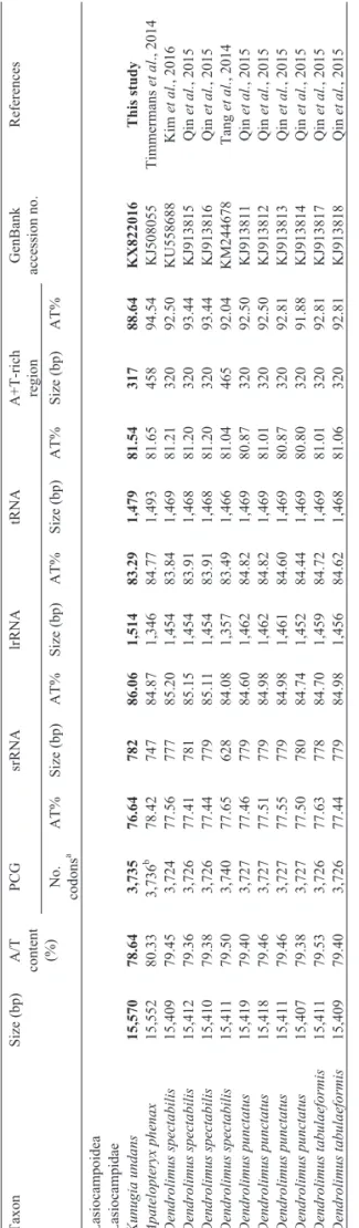
The mitogenome size ofK. undansis 15,570 bp, and
is slightly larger than that of any other lasiocampid species,
which range in size from 15,407 bp in Dendrolimus
counts of the lasiocampid species are well within the range found in macroheteroceran species, and no peculiarities as-sociated with total size and codon count were detected in Lasiocampoidea (Table 1, Table S2).
Compared to the typical sets of genes and regions found in animal mitogenomes (13 PCGs, 22 tRNAs, 2
rRNA genes, and one non-coding A+T-rich region), theK.
undansmitogenome contains one extratrnR, which is lo-cated in tandem to anothertrnR[referred to astrnR(A) for the copy located next totrnAandtrnR(B) for the copy
lo-cated next to trnN] between trnA and trnN (Figure 1).
Pairwise sequence divergence between the two tRNAs was 10.94% (7 bp). Among lasiocampid species (data not shown), pairwise sequence divergence was 3.18-7.81% and
10.94% compared totrnR(A) andtrnR(B), respectively,
indicating thattrnR(A) is more likely to be a functional copy, in that the sequence divergence range reflects the
cur-rent taxonomic hierarchy. Nevertheless, bothtrnRcopies
have an identical anticodon (TCG) that is found in all other Lasiocampoidea (Table 2, Table S3), and they exhibit the proper secondary cloverleaf structure (Figure S1). Thus, the functionality oftrnR(B) remains unknown. The tandem location of twotrnRcopies that exhibit proper secondary structures and an identical anticodon may indicate a gene duplication event rather than horizontal transfer (Higgset
al., 2003). In Lepidoptera, Coreana raphaelis
(Papilio-noidea) was the first species reported to have 23 tRNA genes instead of the usual 22 because of a tandemly
dupli-cated trnS1 between trnN and trnE (Kim et al., 2006).
Ctenoptilum vasava(Papilionoidea) was subsequently re-ported to have an extratrnS1(Kimet al., 2006; Haoet al.,
2012). However, the extratrnR found in the K. undans
mitogenome is likely unique in Macroheterocera, in that our careful reexamination of all available lasiocampid spe-cies and all Macroheterocera did not reveal extra tRNAs
(data not shown). Currently, theK. undansmitogenome is
the only availableKunugiasequence, so whether this
dupli-Table 1 -Characteristics of Lasiocampoidea mitogenomes. Taxon Size (bp) A/T content (%) PCG srRNA lrRNA tRNA A+T-rich region GenBank accession no. References No. codons a AT% Size (bp) AT% Size (bp) AT% Size (bp) AT% Size (bp) AT%
Lasiocampoidea Lasiocampidae Kunugia
undans 15,570 78.64 3,735 76.64 782 86.06 1,514 83.29 1,479 81.54 317 88.64 KX822016 This study Apatelopteryx phenax 15,552 80.33 3,736 b 78.42 747 84.87 1,346 84.77 1,493 81.65 458 94.54 KJ508055 Timmermans et al ., 2014 Dendrolimus spectabilis 15,409 79.45 3,724 77.56 777 85.20 1,454 83.84 1,469 81.21 320 92.50 KU558688 Kim et al. , 2016 Dendrolimus spectabilis 15,412 79.36 3,726 77.41 781 85.15 1,454 83.91 1,468 81.20 320 93.44 KJ913815 Qin et al. , 2015 Dendrolimus spectabilis 15,410 79.38 3,726 77.44 779 85.11 1,454 83.91 1,468 81.20 320 93.44 KJ913816 Qin et al. , 2015 Dendrolimus spectabilis 15,411 79.50 3,740 77.65 628 84.08 1,357 83.49 1,466 81.04 465 92.04 KM244678 Tang et al. , 2014 Dendrolimus punctatus 15,419 79.40 3,727 77.46 779 84.60 1,462 84.82 1,469 80.87 320 92.50 KJ913811 Qin et al. , 2015 Dendrolimus punctatus 15,418 79.46 3,727 77.51 779 84.98 1,462 84.82 1,469 81.01 320 92.50 KJ913812 Qin et al. , 2015 Dendrolimus punctatus 15,411 79.46 3,727 77.55 779 84.98 1,461 84.60 1,469 80.87 320 92.81 KJ913813 Qin et al. , 2015 Dendrolimus punctatus 15,407 79.38 3,727 77.50 780 84.74 1,452 84.44 1,469 80.80 320 91.88 KJ913814 Qin et al. , 2015 Dendrolimus tabulaeformis 15,411 79.53 3,726 77.63 778 84.70 1,459 84.72 1,469 81.01 320 92.81 KJ913817 Qin et al. , 2015 Dendrolimus tabulaeformis 15,409 79.40 3,726 77.44 779 84.98 1,456 84.62 1,468 81.06 320 92.81 KJ913818 Qin et al. , 2015 aTermination codons were excluded in the total codon count. bSequences include a few undetermined nucleotides.
Figure 1- Schematic illustration of the gene arrangement with the
cation event was species- or genus-specific is an intriguing question.
The A/T nucleotide composition of the whole
ge-nome was 78.64% inK. undans, indicating biased A/T
nu-cleotides, but it represents the lowest percentage detected in lasiocampid species (Table 1). Among macroheteroceran superfamilies, the A/T composition of the whole mito-genome in Lasiocampoidea is slightly lower than that of
any other macroheteroceran superfamily (79.47% vs
80.23-80.79%), but the difference is slight (Table S2). The
A/T content amongK. undansgenes varied between RNA
(86.06% in srRNA, 83.29% in lrRNA, and 81.54% in
tRNAs) and PCG (76.64%) genes, and the same trend was also found in other sequenced Macroheterocera, including Lasiocampoidea (Table 1, Table S2).
TheK. undansgene arrangement is identical to that of other ditrysian Lepidoptera that exhibit thetrnM-trnI-trnQ order (where the underline indicates a gene inversion) at the A+T-rich region and ND2 junction, with the exception of
the duplicatedtrnR(Table 2; Kim et al., 2011;
Timmer-manset al., 2014; Parket al., 2016; Zhaoet al., 2016). This arrangement is found in all sequenced Macroheterocera (Parket al., 2016), including Lasiocampoidea (Table 2; Ta-ble S3). However, it differs from the ancestral trnI-trnQ-trnMorder found in the majority of insects and the lepi-dopteran superfamilies Hepialoidea and Nepticuloidea, which are ancient, non-ditrysian lepidopteran groups (Cao et al., 2012; Timmermanset al., 2014). Thus, this tRNA re-arrangement has been regarded as synapomorphy for
Ditrysia. However, a new arrangement, trnI-trnM-trnQ,
was reported from a butterfly species belonging to Nym-phalidae in Papilionoidea (Xuanet al., 2016). Therefore, the latter arrangement might represent an autapomorphy, in that no other congeneric species has the arrangement (Park et al., 2016).
Genes
Twelve of the 13K. undansPCGs started with ATN,
butCOIstarted with an alternative CGA start codon, as ob-served in other moths (Figure S2). There is no typical start codon at the 5’-end oftrnYand the intergenic spacer
se-quence located betweentrnYandCOI, so CGA is the only
possible start codon forCOIinK. undans. The CGA start codon is found in all other sequenced macroheteroceran superfamilies, but some authors designate the typical ATN
codon as the start codon forCOI(Figure S2). This start
codon has been reported to be highly conserved at the start region ofCOIin other Lepidoptera, and it was confirmed in a species of Lepidoptera based on expressed sequence tag data (Margamet al., 2011; Kim et al., 2014; Parket al., 2016). Thus, the presence of a CGA start codon is now con-sidered a synapomorphic trait in Lepidoptera, although some exceptions exist. The mitochondrial PCGs available
for Lasiocampoidea, includingK. undans, ended with TAA
in the majority of PCGs, but they also infrequently ended with a single T (Table 2; Table S3). The TAG stop codon
was uniquely used inK. undansforND4andND4L, while
other lasiocampid species used a single T forND4and TAA
forND4L(Table 2; Table S3). The incomplete termination
codon is known to result in a complete TAA stop codon via posttranslational modifications that occur during the mRNA maturation process (Ojalaet al., 1981).
The biased A/T content was reflected in the form of codon usage. For instance, among the 64 available codons, the most frequently used codons [TTA (leucine), ATT
Table 2- Genomic summary ofKunugia undans.
Gene Anticodon Start codon
Stop codon
Nucleotide position (size)
trnM CAT - - 1-68 (68)
trnI GAT - - 72-135 (64)
trnQ TTG - - 136-205 (70)
ND2 ATT TAA 263-1276 (1014)
trnW TCA - - 1275-1344 (70)
trnC GCA - - 1337-1402 (66)
trnY GTA - - 1412-1479 (68)
COI CGA T-tRNA 1500-3057 (1558)
trnL2 TAA - - 3058-3125 (67) COII ATA T-tRNA 3125-3806 (682)
trnK CTT - - 3807-3877 (71)
trnD GTC - - 3879-3947 (69)
ATP8 ATC TAA 3948-4109 (162)
ATP6 ATG TAA 4103-4780 (678)
COIII ATG TAA 4787-5575 (790)
trnG TCC - - 5578-5644 (67)
ND3 ATC TAA 5645-5998 (354)
trnA TGC - - 6003-6070 (68)
trnR(A) TCG - - 6084-6147 (64)
trnR(B) TCG - - 6175-6241 (67)
trnN GTT - - 6242-6308 (67)
trnS1 GCT - - 6308-6375 (68) trnE TTC - - 6376-6440 (65)
trnF GAA - - 6471-6537 (67)
ND5 ATT T-tRNA 6538-8275 (1738)
trnH GTG - - 8276-8343 (68)
ND4 ATG TAG 8348-9682 (1335)
ND4L ATG TAG 9688-9981 (294)
trnT TGT - - 9986-10050 (65)
trnP TGG - - 10051-10115 (65)
ND6 ATA TAA 10124-10654 (531)
CytB ATG TAA 10662-11807 (1146)
trnS2 TGA - - 11809-11875 (67) ND1 ATG TAA 11869-12825 (957)
trnL1 TAG - - 12827-12892 (66)
lrRNA - - 12893-14406 (1513)
trnV TAC - - 14407-14471 (65)
srRNA - - 14472-15253 (782)
A+T–rich region
- - 15254-15570 (317)
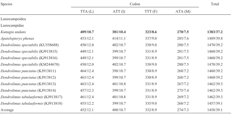
(isoleucine), TTT (phenylalanine), and ATA (methionine)] accounted for 37.2% inK. undans, and this value was the lowest frequency detected in Lasiocampoidea (Table 3). These four codons are all comprised of A or T nucleotides, thus indicating the biased usage of A/T nucleotides in
Lasiocampoidea PCGs, includingK. undans. Other
macro-heteroceran superfamilies have also shown a similar pat-tern, revealing 39.1–40.7% in Bombycoidea, 37.5–40.4% in Geometroidea, 38.0–44.6% in Noctuoidea, 40.8–40.9% in Drepanoidea, and 39.3% in Mimallonoidea (Table S4).
The nucleotide composition of the 13 concatenated
PCGs in theK. undansmitogenome was 33.5, 43.2, 11.8,
and 11.5% for adenine, thymine, cytosine, and guanine, re-spectively, indicating A/T bias (Table 4). The base
compo-sition at each codon pocompo-sition of the K. undans PCGs
indicated that the third codon position (86.5%) had a sub-stantially higher A/T content than the first (72.6%) and sec-ond (70.4%) codon positions. A similar pattern was detected in other sequenced Lasiocampoidea, with aver-ages of 77.6, 73.0, and 89.0 in the first, second, and third positions, respectively (Table 4).
Two rRNA genes inK. undans,lrRNA andsrRNA,
were of 1,514 and 782 bp, respectively, (Table 2), and the sizes of the two genes inK. undanswere larger than those of any found in other lasiocampid species, which ranged from 1,346 bp (A. phenax) to 1,452 bp (D. punctatus) in lrRNAand 747 bp (A. phenax) to 780 bp (D. punctatus) in srRNA(Table S2). tRNA sizes ranged from 64 bp (trnI) to 71 bp (trnK) in K. undans, and similar size ranges were found in other sequenced lasiocampid species (Table 2; Ta-ble S3). AllK. undanstRNAs possessed invariable lengths
of 7 bp for the aminoacyl stem, 7 bp for the anticodon loop, and 5 bp for the anticodon stem (Figure S1), and most tRNA size variation resulted from length variations in the
DHU and TC arms. For instance, trnS1has an atypical
cloverleaf secondary structure that lacked the DHU stem, but the remainingK. undanstRNAs formed the typical sec-ondary cloverleaf structure (Figure S1). The aberranttrnS1
has been reported in many metazoan species, including in-sects (Garey and Wolstenholme, 1989; Wolstenholme, 1992). The DHU stem and loop are involved in tertiary in-teractions required for the proper folding and functioning of tRNA (Rich and RajBhandary, 1976). Thus, an atypical secondary structure may hamper the functionality of tRNA, but a nuclear magnetic resonance analysis from nematodes demonstrated that the aberranttrnS1also was functionally
similar to typical tRNAs based on structural adjustments required to ensure ribosome fitting (Ohtsukiet al., 2002).
The A+T-rich region
The length of the A+T-rich region inK. undanswas
317 bp, and A/T nucleotides made up 88.64% of the se-quence (Table 2). This region contained the highest A/T
content of any region of theK. undansmitogenome (Table
1). Moreover, this region was the shortest in length, and it contained the least A/T nucleotides among lasiocampid species (Table 2, Table S3).
The insect A+T-rich region harbors signals for repli-cation and transcription initiation, so it is known to have conserved sequences in the region, which are in the form of conserved sequence blocks (Fauron and Wolstenholme, 1980; Clary and Wolstenholme, 1987; Saitoet al., 2005). In
Table 3- Frequency of the four most frequently used codons in Lasiocampoidea.
Species Codon Total
TTA (L) ATT (I) TTT (F) ATA (M)
Lasiocampoidea
Lasiocampidae
Kunugia undans 409/10.7 381/10.4 323/8.6 270/7.5 1383/37.2
Apatelopteryx phenax 453/12.1 414/11.1 337/9.0 285/7.6 1489/39.8
Dendrolimus spectabilis(KU558688) 450/12.0 402/10.7 338/9.0 280/7.5 1470/39.2
Dendrolimus spectabilis(KJ913815) 449/12.1 399/10.7 331/8.9 281/7.5 1460/39.2
Dendrolimus spectabilis(KJ913816) 449/12.1 399/10.7 331/8.9 281/7.5 1460/39.2
Dendrolimus spectabilis(KM244678) 450/12.0 402/10.7 338/9.0 280/7.5 1470/39.2
Dendrolimus punctatus(KJ913811) 464/12.4 398/10.7 330/8.9 268/7.2 1460/39.2
Dendrolimus punctatus(KJ913812) 463/12.4 399/10.7 330/8.9 268/7.2 1460/39.2
Dendrolimus punctatus(KJ913813) 463/12.4 401/10.8 331/8.9 267/7.2 1462/39.3
Dendrolimus punctatus(KJ913814) 457/12.3 399/10.7 331/8.9 275/7.4 1462/39.3
Dendrolimus tabulaeformis(KJ913817) 461/12.4 401/10.8 331/8.9 269/7.2 1462/39.3
Dendrolimus tabulaeformis(KJ913818) 455/12.2 399/10.7 335/9.0 268/7.2 1457/39.1
Average 452/12.1 400/10.7 332/8.9 274/7.3 1458/39.1
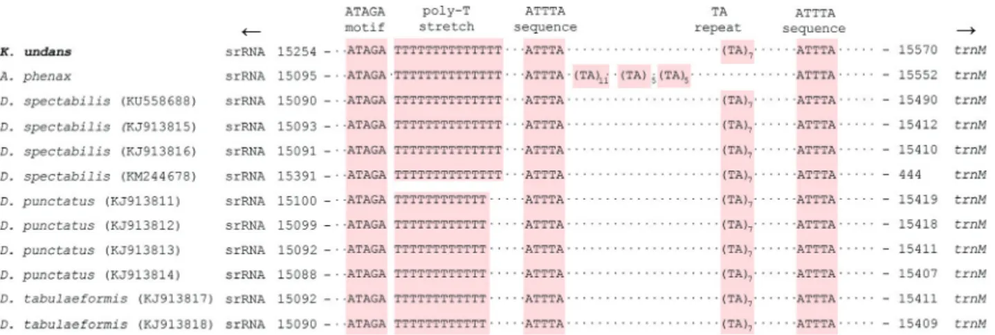
fact, previous studies revealed several conserved blocks in a substantial number of lepidopteran groups (Liaoet al., 2010; Kimet al., 2014), and a search for the A+T-rich re-gion of lasiocampid species (includingK. undans) resulted in the detection of several conserved sequences (Figure 2). The first conserved sequence, which is located close to the
5’-end of thesrRNA, is the ATAGA motif followed by a
poly-T stretch of varying length. TheK. undansA+T-rich
region contained a 14-bp T stretch that was upstream of the 5’-end of thesrRNA(Figure 2), and this poly-T stretch is well-conserved in all sequenced lasiocampid (ranging in size from 12 bp to 14 bp; Figure 2) and macroheteroceran species (Figure S3). Saitoet al.(2005) previously reported
for theBombyx morimitogenome the precise position of
the replication origin for minor-strand mtDNA, which is immediately downstream of a poly-T stretch that is located upstream of thesrRNA5’-end. Thus, this poly-T stretch is thought to function as a possible recognition site for the ini-tiation of replication of the minor mtDNA strand. Addi-tionally, another conserved motif ATAGA is located immediately downstream of the poly-T stretch, and it is very well-conserved in all sequenced lasiocampid species,
includingK. undans(Figure 2) and macroheteroceran
spe-cies (Figure S3). A previously suggested function of this motif is a regulatory role in conjunction with the poly-T stretch, but experimental data are required to support this hypothesis (Kimet al., 2009). Excluding the previously de-scribed sequences, there are only a few additional con-served sequences in the A+T-rich region of lasiocampid [e.g.,K. undans(Figure 2)] and macroheteroceran species (Figure S3), including two or more ATTTA sequences scat-tered in the A+T-rich region, a microsatellite-like TA re-peat, and a poly-T stretch. Our careful reexamination of the A+T-rich regions of macroheteroceran species resulted in the detection of repeat sequences in several species, includ-ing two of each 55-bp and 24-bp repeats inBombyx huttoni (Bombycoidea); six 26-bp and two 18-bp repeats in Phthonandria atrilineata, two 278-bp repeats inDysstroma truncata, four 24-bp repeats inOperophtera brumata (Geo-metroidea), two 16-bp repeats inAgrotis ipsilon, and two
11-bp repeats inRisoba prominens(Noctuoidea)(Yanget
al., 2009; Timmermanset al., 2014; Derkset al., 2015; Wu et al., 2015; Yanget al., 2015; Penget al., 2016). Neverthe-less, repeat sequences that were longer than 10 bp were not
detected in sequenced lasiocampid species, including K.
undans.
Non-coding sequences
Excluding the A+T-rich region, theK. undans
mito-genome has non-coding sequences that total 172 bp (with a range of 1–57 bp) and spread over 17 regions (Table 2). Comparison of available lasiocampid species indicated that intergenic spacing patterns and sizes are largely consistent in Lasiocampoidea, including those ofK. undans. In partic-ular, the 57-bp spacer found at thetrnQandND2junction
Table 4 -Codon position-based nucleotide composition of 13 concatenated Lasiocampoidea PCGs. Species Overall 1st codon position 2nd codon position 3rd codon position A T CG A T CG A T CG A T CG
Lasiocampoidea Lasiocampidae Kunugia
Figure 2- Schematic illustration of the A+T-rich region of Lasiocampoidea, includingKunugia undans. The colored nucleotides indicate conserved se-quences such as the ATAGA motif, poly-T stretch, ATTTA sequence, and microsatellite-like TA repeat sese-quences. Dots between sese-quences indicate omitted sequences, and arrows indicate the transcriptional direction. Subscripts indicate the repeat number. GenBank accession numbers of the species represented by more than one mitogenome sequence are enclosed in parentheses.
Figure 3- Alignment of the spacer sequence (located betweentrnQandND2) and the neighboring partialND2of Lasiocampoidea, includingKunugia
(with a range of 39–58 bp) is consistently found in all
lasiocampid species, includingK. undans(Figure 3). The
origin of this spacer region has previously been ascribed to the partial duplication and random loss ofND2, leaving the current length of the spacer sequence at thetrnQandND2 junction because the spacer exhibited sequence identity to the neighboringND2, despite the fact that its non-coding nature may have allowed the spacer to diverge from the originalND2(Kimet al., 2014). RegardingK. undans, the sequence identity of the spacer to the neighboringND2was 58.33% (Figure 3) and over 50.60% in 59 species of macro-heteroceran superfamilies (Figure S4).
Other relatively long spacer sequences were found in
several regions of lasiocampid species, including K.
undans, including those at the trnY and COI junction (20–34 bp), at thetrnAandtrnRjunction (13–20 bp), at the
trnNandtrnS1 junction (11–21 bp, excluding K. undans
that has a 1-bp overlap), and at theND4andND4Ljunction (5–24 bp, excludingA. phenaxthat has a 5-bp overlap) (Ta-ble 2, Ta(Ta-ble S3). These spacer sequences are mainly com-posed of A/T nucleotides that are often found within multiple runs of either T or A nucleotides (data not shown). Sequence alignment beyond the species level was nearly impossible due to considerable variability in length, se-quence composition, and insertions/deletions (data not shown). The majority of the remaining spacer regions were short, with a few exceptions (e.g., less than 10 bp).
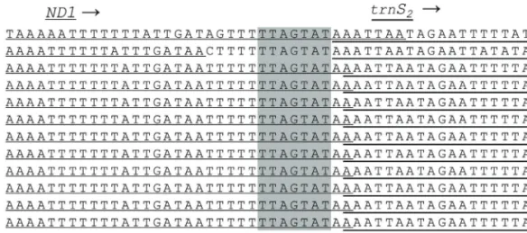
In previous lepidopteran mitogenomic studies, other spacer sequences at thetrnS2andND1junction were
con-sistently reported in lepidopteran lineages (Cameron and Whiting, 2008; Kimet al., 2010; Yanget al., 2013; Kimet al., 2014; Parket al., 2016). The important feature of this spacer is the presence of a short-length TTAGTAT motif within the spacer sequence, which is thought to be a possi-ble binding site for the transcription termination peptide of mtDNA (mtTERM). This characterization is based on the fact that the spacer is located after the final major-strand
coded CytB (Taanman, 1999; Cameron and Whiting,
2008). RegardingK. undans, there is a 7-bp overlap at the ND1andtrnS2junction, butK. undansclearly possesses the
same sequence motif (Figure 4). All other lasiocampid
spe-cies, with the exception ofA. phenax, have a 1-bp gene
overlap in this region, but they also contain the 7-bp motif at theND1andtrnS2junction. On the other hand,A. phenax
has an intergenic spacer sequence at 12 bp, which includes the 7-bp motif. In other macroheteroceran species, the 7-bp motif is found in nearly all species without modification, with the exception of one Noctuoidea species, which has ATAGTAT instead of TTAGTAT. In Macroheterocera, the 7-bp motif is nearly always located at the spacer instead of the coding region at theND1andtrnS2junction (Figure S5).
Thus, the spacing pattern of Lasiocampoidea differs from that of other macroheteroceran superfamilies in this region, so mRNA expression data would be required to clarify the extension ofND1at theND1andtrnS2junction.
In summary, in addition to the typical set of genes, the
15,570-bp complete mitogenome sequence ofK. undans
has an extratrnR. The presence of the additional tRNA is unique in Macroheterocera, including Lasiocampoidea.
The A+T-rich region of K. undanspossesses a few
con-served sequences, which were previously reported in other Macroheterocera (including Lasiocampoidea). Moreover, the intergenic spacing pattern and size forK. undansare largely consistent with those of other Macroheterocera (in-cluding Lasiocampoidea), but instead of an intergenic
spacer, Lasiocampoidea (includingK. undans) exhibit an
overlap at thetrnS2andND1junction.
Acknowledgments
This study was supported by the Basic Science Re-search Program through the National ReRe-search Foundation of Korea (NRF) funded by the Ministry of Education, Sci-ence, and Technology (2015R1D1A3A03018119).
Figure 4- Alignment of the internal spacer sequence located betweenND1andtrnS2of Lasiocampoidea, includingKunugia undans. The shaded
nucleo-tides indicate the conserved heptanucleotide (TTAGTAT) region. Underlined nucleonucleo-tides indicate the adjacent partial sequences ofND1andtrnS2.
References
Burland TG (2000) DNASTAR’s laser gene sequence analysis software. In: Misener S and Krawets SA (eds) Bioinfor-matics Methods and Protocols. Humana Press, Totowa, pp 71-91.
Cameron SL (2014) Insect mitochondrial genomics: implications for evolution and phylogeny. Annu Rev Entomol 59:95-117. Cameron SL and Whiting MF (2008) The complete mitochondrial
genome of the tobacco hornworm,Manduca sexta, (Insecta:
Lepidoptera: Sphingidae) and an examination of
mitochon-drial gene variability within butterflies and moths. Gene
408:112-123.
Cao YQ, Ma C, Chen JY and Yang DR (2012) The complete
mito-chondrial genomes of two ghost moths, Thitarodes
renzhiensisandThitarodes yunnanensis: The ancestral gene arrangement in Lepidoptera. BMC Genomics 13:276. Clary DO and Wolstenholme DR (1987) Drosophila
mitochon-drial DNA: Conserved sequences in the A + T-rich region and supporting evidence for a secondary structure model of the small ribosomal RNA. J Mol Evol 25:116-125. Derks MFL, Smit S, Salis L, Schijlen E, Bossers A, Mateman C,
Piji AS, de Ridder D, Groenen MAM, Visser ME,et al.
(2015) The genome of winter moth (Operophtera brumata) provides a genomic perspective on sexual dimorphism and phenology. Genome Biol Evol 7:2321-2332.
Dowton M, Austin A, Dillon N and Bartowsky E (1997) Molecu-lar phylogeny of the apocritan wasps: The Proctotrupo-morpha and EvanioProctotrupo-morpha. Syst Entomol 22:245-255. Fauron CM and Wolstenholme DR (1980) Intraspecific diversity
of nucleotide sequences within the adenine+ thymine-rich
region of mitochondrial DNA molecules of Drosophila
mauritiana, Drosophila melanogaster and Drosophila simulans. Nucleic Acids Res 8:5391-5410.
Feijao PC, Neiva LS, Azeredo-Espin AML and Lessinger AC (2006) AMiGA: The arthropodan mitochondrial genomes accessible database. Bioinformatics 22:902-903.
Garey JR and Wolstenholme DR (1989) Platyhelminth mitochon-drial DNA: Evidence for early evolutionary origin of a
tRNAserAGN that contains a dihydrouridine arm
replace-ment loop, and of serine-specifying AGA and AGG codons. J Mol Evol 28:374-387.
Hao J, Sun Q, Zhao H, Sun X, Gai Y and Yang Q (2012) The
com-plete mitochondrial genome of Ctenoptilum vasava
(Lepidoptera: Hesperiidae: Pyrginae) and its phylogenetic implication. Comp Funct Genomics 2012:1-13.
Higgs PG, Jameson D, Jow H and Rattray M (2003) The evolution of tRNA-Leu genes in animal mitochondrial genomes. J Mol Evol 57:435-445.
Katoh K, Misawa K, Kuma K and Miyata T (2002) MAFFT: A novel method for rapid multiple sequence alignment based on fast Fourier transform. Nucleic Acids Res 30:3059-3066. Kim I, Lee EM, Seol KY, Yun EY, Lee YB, Hwang JS and Jin BR
(2006) The mitochondrial genome of the Korean hairstreak, Coreana raphaelis(Lepidoptera: Lycaenidae). Insect Mol Biol 15:217-225.
Kim JS, Park JS, Kim MJ, Kang PD, Kim SG, Jin BR, Han YS and Kim I (2012) Complete nucleotide sequence and
organiza-tion of the mitochondrial genome of eri-silkworm, Samia
cynthia ricini(Lepidoptera: Saturniidae). J Asia Pac Ento-mol 15:162-173.
Kim MJ, Wan X, Kim K-G, Hwang JS and Kim I (2010) Com-plete nucleotide sequence and organization of the
mito-genome of endangered Eumenis autonoe (Lepidoptera:
Nymphalidae). Afr J Biotechnol 9:735-754.
Kim MJ, Kang AR, Jeong HC, Kim K-G and Kim I (2011) Recon-structing intraordinal relationships in Lepidoptera using mi-tochondrial genome data with the description of two newly
sequenced lycaenids,Spindasis takanonisandProtantigius
superans(Lepidoptera: Lycaenidae). Mol Phylogenet Evol 61:436-445.
Kim MJ, Wang AR, Park JS and Kim I (2014) Complete mito-chondrial genomes of five skippers (Lepidoptera: Hes-periidae) and phylogenetic reconstruction of Lepidoptera. Gene 549:97-112.
Kim MJ, Kim JS, Kim S-S, Kim SR and Kim I (2016) Complete
mitochondrial genome of the pine moth Dendrolimus
spectabilis (Lepidoptera: Lasiocampidae). Mitochondrial DNA Part B 1:180-181.
Kim SR, Kim MI, Hong MY, Kim KY, Kang PD, Hwang JS, Han YS, Jin BR and Kim I (2009) The complete mitogenome
se-quence of the Japanese oak silkmoth,Antheraea yamamai
(Lepidoptera: Saturniidae). Mol Biol Rep 36:1871-1880. Liao F, Wang L, Wu S, Li Y-P, Zhao L, Huang G-M, Niu C-J, Liu
Y-Q and Li M-G (2010) The complete mitochondrial
ge-nome of the fall webworm,Hyphantria cunea(Lepidoptera:
Arctiidae). Int J Biol Sci 6:172-186.
Lowe TM and Eddy SR (1997) TRNAscan-SE: A program for im-proved detection of transfer RNA genes in genomic se-quence. Nucleic Acids Res 25:955-964.
Lu H-F, Su T-J, Luo AR, Zhu C-D and Wu C-S (2013) Character-ization of the complete mitochondrion genome of diurnal mothAmata emma(Butler) (Lepidoptera: Erebidae) and its phylogenetic implications. PLoS One 8:e72410.
Mao M, Gibson T and Dowton M (2014) Evolutionary dynamics of the mitochondrial genome in the Evaniomorpha (Hymenoptera)-a group with an intermediate rate of gene re-arrangement. Genome Biol Evol 6:1862-1874.
Mao M, Gibson T and Dowton M (2015) Higher-level phylogeny of the Hymenoptera inferred from mitochondrial genomes. Mol Phylogenet Evol 84:34-43.
Margam VM, Coates BS, Hellmich RL, Agunbiade T, Seuf-ferheld MJ and Sun W (2011) Mitochondrial genome se-quence and expression profiling for the legume pod borer Maruca vitrata (Lepidoptera: Crambidae). PLoS One 6:e16444.
Ohtsuki T, Kawai G and Watanabe K (2002) The minimal tRNA:
Unique structure ofAscaris suummitochondrial
tRNASer-UCU having a short T arm and lacking the entire D arm. FEBS Lett 514:37-43.
Ojala D, Montoya J and Attardi G (1981) tRNA punctuation model of RNA processing in human mitochondria. Nature 290:470-474.
Park JS, Kim MJ, Jeong SY, Kim SS and Kim I (2016) Complete
mitochondrial genomes of two gelechioids, Mesophleps
albilinella and Dichomeris ustalella (Lepidoptera: Gelechiidae), with a description of gene rearrangement in Lepidoptera. Curr Genet 62:809-826.
Peng XY, Zhou P, Qiang Y and Qian ZQ (2016) Characterization
of the complete mitochondrial genome ofBombyx huttoni
(Lepidoptera: Bombycidae). Mitochondrial DNA A DNA Mapp Seq Anal 27:4112-4113.
Qin J, Zhang Y, Zhou X, Kong X, Wei S, Ward RD and Zhang A (2015) Mitochondrial phylogenomics and genetic relation-ships of closely related pine moth (Lasiocampidae: Dendrolimus) species in China, using whole mitochondrial genomes. BMC Genomics 16:428-439.
Ramírez-Ríos V, Franco-Sierra ND, Alvarez JC, Saldamando-Benjumea CI and Villanueva-Mejía DF (2016)
Mitochon-drial genome characterization of Tecia solanivora
(Lepidoptera: Gelechiidae) and its phylogenetic relationship with other lepidopteran insects. Gene 581:107-116. Rich A and RajBhandary UL (1976) Transfer RNA: Molecular
structure, sequence, and properties. Annu Rev Biochem 45:805-860.
Saito S, Tamura K and Aotsuka T (2005) Replication origin of mi-tochondrial DNA in insects. Genetics 171:1695-1705. Shin YH (2001) Coloured Illustrations of the Moths of Korea.
Academybook Publishing Co. Ltd., Seoul, 551 p.
Taanman JW (1999) The mitochondrial genome: Structure, tran-scription, translation and replication. Biochim Biophys Acta 1410:103-123.
Tamura K, Stecher G, Peterson D, Filipski A and Kumar S (2013) MEGA6: Molecular Evolutionary Genetics Analysis ver. 6.0. Mol Biol Evol 30:2725-2729.
Tang M, Tan M, Meng G, Yang S, Su X, Liu S, Song W, Li Y, Wu
Q, Zhang A,et al.(2014) Multiplex sequencing of pooled
mitochondrial genomes-a crucial step toward biodiversity analysis using mito-metagenomics. Nucleic Acids Res 42:e166.
Timmermans MJ, Lees DC, Thompson MJ, Sáfián SZ and Brat-tström O (2014) Towards a mitogenomic phylogeny of Lepidoptera. Mol Phylogenet Evol 79:169-178.
van Nieukerken EJ, Kaila L, Kitching IJ, Kristensen NP, Lees DJ,
Minet J, Mitter J, Mutanen M, Regier JC, Simonsen TJ,et al.
(2011) Order Lepidoptera Linnaeus, 1758. Zootaxa 3148:212-221.
Wan X, Kim MJ and Kim I (2013) Description of new
mitochon-drial genomes (Spodoptera litura, Noctuoidea and
Cnaphalocrocis medinalis, Pyraloidea) and phylogenetic re-construction of lepidoptera with the comment on optimiza-tion schemes. Mol Biol Rep 40:6333-6349.
Wolstenholme DR (1992) Animal mitochondrial DNA: Structure and evolution. Int Rev Cytol 141:173-216.
Wu QL, Cui WX and Wei SJ (2015) Characterization of the
com-plete mitochondrial genome of the black cutwormAgrotis
ipsilon (Lepidoptera: Noctuidae). Mitochondrial DNA 26:139-140.
Xuan S, Song F, Cao L, Wang J, Li H and Cao T (2016) The
com-plete mitochondrial genome of the butterfly Euripus
nyctelius(Lepidoptera: Nymphalidae). Mitochondrial DNA A DNA Mapp Seq Anal 27:2563-2565.
Yang L, Wei ZJ, Hong GY, Jiang ST and Wen LP (2009) The complete nucleotide sequence of the mitochondrial genome of Phthonandria atrilineata (Lepidoptera: Geometridae). Mol Biol Rep 36:1441-1449.
Yang X, Xue D and Han H (2013) The complete mitochondrial
genome ofBiston panterinaria(Lepidoptera: Geometridae),
with phylogenetic utility of mitochondrial genome in Lepidoptera. Gene 515:349-358.
Yang X, Cameron SL, Lees DC, Xue D and Han H (2015) A mito-chondrial genome phylogeny of owlet moths (Lepidoptera: Noctuoidea), and examination of the utility of mitochondrial genomes for lepidopteran phylogenetics. Mol Phylogenet Evol 85:230-237.
Zhao J, Sun Y, Xiao L, Tan Y, Dai H and Bai L (2016) Complete
mitochondrial genome of the pink bollwormPectinophora
gossypiella (Lepidoptera: Gelechiidae). Mitochondrial DNA A DNA Mapp Seq Anal 27:1575-1576.
Internet Resources
Basic Local Alignment Search Tool (BLAST),
http://blast.ncbi.nlm.nih.gov/Blast.cgi (November 3, 2016).
Supplementary material
The following online material is available for this article: Table S1 – List of primers used to amplify and sequence the Kunugia undansmitogenome.
Table S2 – Characteristics of Macroheterocera mitoge-nomes.
Table S3 – Genomic summary of Lasiocampoidea. Table S4 – Frequency of the four most frequently used codons in Macroheterocera.
Figure S1 – Predicted secondary cloverleaf structures for the 23 tRNA genes ofKunugia undans, with the duplicated trnR(A and B).
Figure S2 – Alignment of the initiation context of the
MacroheteroceraCOI.
Figure S3 – Schematic illustration of the A+T-rich region of Macroheterocera.
Figure S4 – Alignment of the spacer sequence (located
be-tweentrnQandND2) and neighboring partial
Macrohete-roceraND2.
Figure S5 – Alignment of the internal spacer sequence
lo-cated betweenND1andtrnS2of Macroheterocera.
Associate Editor: Houtan Noushmehr