online | memorias.ioc.fiocruz.br
Whole genome sequence of
Mycobacterium kansasii
isolates of the genotype 1 from Brazilian patients with
pulmonary disease demonstrates considerable heterogeneity
Edson Machado1, Sidra Ezidio Gonçalves Vasconcellos2, Camillo Cerdeira1,3, Lia Lima Gomes2, Ricardo Junqueira3, Luciana Distasio de Carvalho4, Jesus Pais Ramos4, Paulo Redner4,
Carlos Eduardo Dias Campos4, Paulo Cesar de Souza Caldas4, Ana Paula Chaves Sobral Gomes4, Telma Goldenberg4, Fatima Fandinho Montes4, Fernanda Carvalho de Queiroz Mello5,
Vinicius de Oliveira Mussi6, Elena Lasunskaia6, Dick van Soolingen7, Antonio Basílio de Miranda3, Leen Rigouts8, Bouke C de Jong8, Conor J Meehan8, Marcos Catanho1, Philip N Suffys2,8/+
1Fundação Oswaldo Cruz-Fiocruz, Instituto Oswaldo Cruz, Laboratório de Genômica Funcional e Bioinformática, Rio de Janeiro, RJ, Brasil 2Fundação Oswaldo Cruz-Fiocruz, Instituto Oswaldo Cruz, Laboratório de Biologia Molecular Aplicada a Micobactérias, Rio de Janeiro, RJ, Brasil 3Fundação Oswaldo Cruz-Fiocruz, Instituto Oswaldo Cruz, Laboratório de Biologia Computacional e Sistemas, Rio de Janeiro, RJ, Brasil 4Fundação Oswaldo Cruz-Fiocruz, Escola Nacional de Saúde Pública, Centro de Referência Professor Hélio Fraga, Laboratório de Referência
Nacional para Tuberculose, Rio de Janeiro, RJ, Brasil
5Universidade Federal do Rio de Janeiro, Instituto de Doenças do Tórax, Rio de Janeiro, RJ, Brasil
6Universidade Estadual do Norte Fluminense Darcy Ribeiro, Laboratório de Biologia do Reconhecer, Campos dos Goytacazes, RJ, Brasil 7National Institute for Public Health and the Environment, Bilthoven, the Netherlands
8Institute of Tropical Medicine, Unit of Mycobacteriology, Antwerp, Belgium
Mycobacterium kansasii is an opportunistic pathogen and one of the most commonly encountered species in in-dividuals with lung disease. We here report the complete genome sequence of 12 clinical isolates of M. kansasii from patients with pulmonary disease in Brazil.
Key words: Mycobacterium kansasii - genomes - lung disease - genotype I - diversity - drug resistance
doi: 10.1590/0074-02760180085
Financial support: CNPq (scholarships 207422/2014-1, 500769/2014-1, 311554/2013-0; grants 407624/2012-0, 459100/2014-9).
MC and PNS contributed equally to this work. + Corresponding author: psuffys@gmail.com Received 15 February 2018
Accepted 5 June 2018
Nontuberculous Mycobacterium (NTM) species are widespread in the (man-made) environment and some species cause opportunistic infections in humans. Myco-bacterium kansasii, frequently isolated from tap water, is a slow-growing photochromogenic NTM and a pathogen that is commonly isolated from patients with pre-existing lung disease, similar to other NTM clinical species such as M. abscessus, M. avium complex, M. malmoense and M. xenopi.(1) In the USA and South America M. kansasii is the second most isolated NTM after M. avium com-plex(2,3) while in Rio de Janeiro, it is the most frequent NTM to cause pulmonary disease.(4) Besides chronic bronchopulmonary disease, M. kansasii causes other clinical manifestations such as lymphadenitis(5), skin and soft tissue infection(6), tenosynovitis, arthritis, osteomy-elitis and disseminated infection in patients co-infected with human immunodeficiency virus.(7)
Seven major genotypes (I to VII) of M. kansasii have been described and while human isolates are mainly of types I and II (with type II mainly HIV-related)(8,9),
en-vironmental isolates are mostly of the other subtypes.(10) Type I has been described as a heterogeneous group, in-completely characterized on the genomic level.(11,12) Re-cently, the genome sequence of the ATCC strain 12478 Hauduroy isolated in Kansas in 1955 has been compared with that of the M. tuberculosis strain H37Rv(13), while other M. kansasii genomes are available from environ-mental(14), human(15) and simian(16) sources.
Here, we report the genome sequence of 12 clinical M. kansasii isolates belonging to genotype I, as determined by hsp65 sequencing. The strains were isolated from hu-man Brazilian patients with pulmonary disease and pos-sibly earlier tuberculosis. Nine isolates were from sputum, two from bronchoalveolar lavage (7287 and 10742) and one of unknown origin (1580). Isolates were from resi-dents of the state of Rio de Janeiro (n = 7), Pernambuco (n = 3), Rio Grande do Sul (n = 1), and Santa Catarina (n = 1). Genomic DNA libraries were constructed using the Nextera XT DNA library kit and whole-genome shotgun sequencing was performed on the Illumina HiSeq2500 platform, generating paired end reads of 2 × 100 bp. Ge-nomes were de novo assembled using SPAdes software version 3.11.1(17), and annotated with RAST(18) (Table). No evidence for the presence of plasmids was observed.
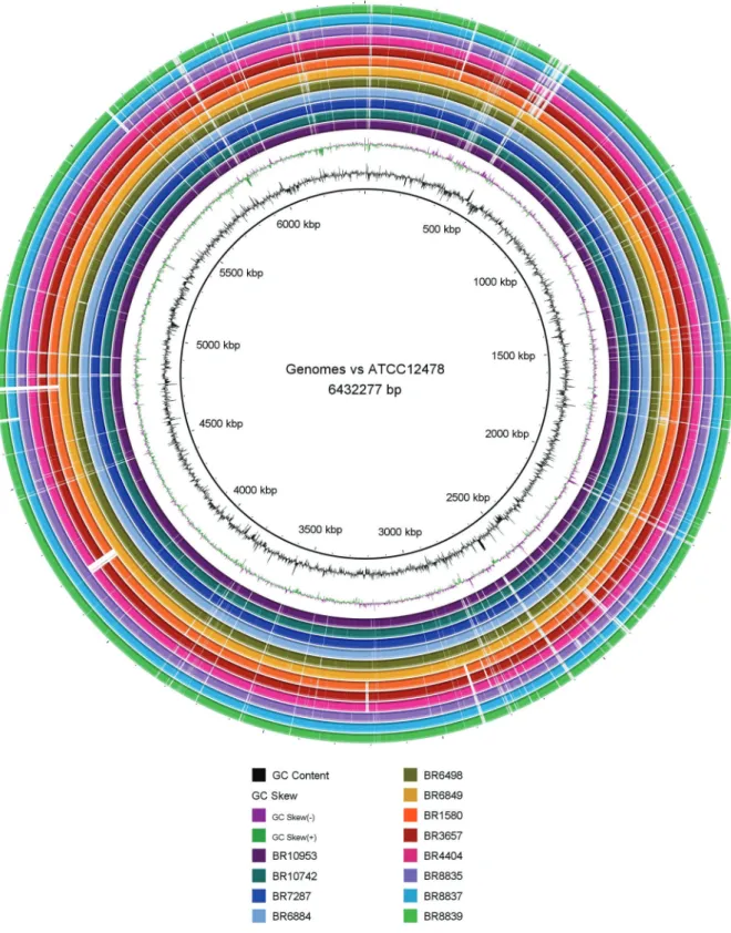
We observed many deletions spread in the genomes that were either shared by all or part of the isolates. Three isolates (1580, 3657 and 4404) presented a region of dif-ference of about 27 kb causing the loss of five helicases, a restriction endonuclease and two hypothetical proteins. We also observed multiple deletions shared by the iso-lates 1580, 3657, 4404, 8835, 8837 and 8839 (Fig. 1).
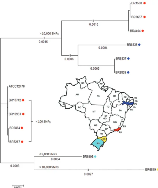
In addition, a reference-based single-nucleotide poly-morphism (SNP) calling was performed against the ge-nome of the M. kansasii ATCC 12478 strain using both Snippy (version 3.2; https://github.com/tseemann/snippy) and the wgSNP module of BioNumerics v7.6 (Applied Maths, Sint-Martens-Latem, Belgium). Most isolates presented homogeneously distributed SNPs over the entire genome, except for isolates 6849 and 6498 which clearly presented regions of higher SNP frequency (data not shown). As demonstrated by a Neighbour-Joining phylogenetic tree based on 5,607,341 sites and 1000 boot-strap replicates (Fig. 2), three groups were observed when compared to the genome of the ATCC strain, one pre-senting less than 100 SNP (n = 4) (middle part), a second with either less than 5,000 SNPs (n = 1) and more than 10,000 SNPs (n = 1) (lower part) and a third group with more than 10,000 SNPs (n = 6) (upper part). We observed an association between SNP-based grouping and phylog-eny on basis of large deletions, that led us suggest that the third group has evolved from the common ancestor.
From two patients, we performed whole genome se-quencing of two isolates each; the pair of isolates from a patient from Rio de Janeiro (10742 and 10953) had 0 SNPs difference; the second pair from a patient from Recife (8837 and 8839) presented 277 mutations (data not shown).
Additionally, two isolates (6498 and 8835) presented a Minimal Inhibitory Concentration of > 1 µg/mL for ri-fampicin in vitro and both presented a non-synonymous SNP, localized respectively at position 4267612 (C to G)
and 4267613 (A to C), causing an amino acid change in codon 411 of rpoB of respectively Gln to Glu and of Gln to Pro. We as such confirm the scarce existing data on the association between resistance against rifampicin and presence of point mutations in M. kansasii rpoB gene. In-deed, point mutations in rpoB gene are the main reason for resistance against rifampicin and this has been con-firmed in several bacteria. Also, molecular determinants of drug resistance in M. kansasii has been recently sug-gested by Bakuła et al.(20). Among the present isolates, we observed considerable differences of in vitro virulence in macrophages as measured by growth and induction of ne-crosis and of cytokine production (data not shown).
In conclusion, these are preliminary data on the variability within the M. kansasii genotype 1 in Brazil and, even on the basis of this small sample number, we evidenced three genetic groups that are separated by the presence of large number of SNPs throughout the ge-nome and by the pattern of large deletions. One grouping of strains from Rio de Janeiro is highly similar to the ATCC strain Hauduroy (< 100 SNPs) that was isolated more than 70 years ago in the US. A second group of hu-man isolates from Rio de Janeiro and Pernambuco pre-sented over 10,000 SNPs when compared to the ATCC strain. We also observed an association between geno-types and geographic origin of isolates, separating those from the states of Rio de Janeiro, Pernambuco and Santa Catarina. Our observation of considerable differences of in vitro and in vivo virulence and their differences on the genome level is under investigation.
ACKNOWLEDGEMENTS
To the platforms at Fiocruz (Plataformas de Bioinformáti-ca RPT04-A/RJ and Sequenciamento de Ácidos Nuclêicos de Nova Geração RPT01-J/RJ). To Applied Maths for offering a one and a half year free access to the Bionumerics software. TABLE
Genome features, predicted genes, and GenBank accession numbers of
Mycobacterium kansasii strains isolated in Brazil from patients with pulmonary disease
Isolate Accession Location Type (hsp65) Reads Genome size (bp) Contigs Coverage (x) Genes
BR3657 PQOL00000000 RJ 1Bd 18,094,408 6,374,282 294 80.90 5,965t
BR6498ª PQOM00000000 RS 1A 9,038,224 6,225,838 317 37.37 5,804
BR7287 PQON00000000 RJ 1A 19,684,890 6,282,326 274 79.59 5,842
BR6884 PQOO00000000 RJ 1A 21,305,254 6,411,468 280 125.91 5,980
BR6849 PQOP00000000 SC 1A 16,399,304 6,464,047 265 78.89 6,023
BR10953b PQOQ00000000 RJ 1A 14,895,938 6,284,879 297 52.02 5,850
BR10742b PQOR00000000 RJ 1A 14,634,490 6,289,619 282 53.58 5,864
BR1580 PQOS00000000 RJ 1B 20,820,372 6,307,487 226 143.20 5,913
BR4404 PQOT00000000 RJ 1B 4,686,134 6,282,923 466 31.43 5,880
BR8835ª PQOU00000000 PE 1B 16,906,236 6,228,381 321 53.45 5,775
BR8837c PQOV00000000 PE 1B 13,311,444 6,213,268 247 75.41 5,773
BR8839c PQOW00000000 PE 1B 9,434,892 6,178,862 270 52.48 5,727
Fig. 2: Neighbor-Joining SNP-based tree of the 12 Brazilian isolates and the ATCC 12478 reference genome.
AUTHORS’ CONTRIBUTION
PNS, EL, CM, LR, BCJ and MC conceived and designed the experiments; SEGV, LLG, RJ, LDC and VOM performed the experiments; EM, CC, SEGV, LLG, ABM and CM ana-lysed the data; LDC, JPR, PR, CEDC, PCSC, APCSG, TG, FFM and FCQM were responsible for samples collection, strain isolation and identification; PNS, EM, EL, DS, SEGV, JPR, LR, BCJ and MC contributed for the manuscript writing.
REFERENCES
1. Wassilew N, Hoffmann H, Andrejak C, Lange C. Pulmonary dis-ease caused by non-tuberculous mycobacteria. Respiration. 2016; 91(5): 386-402.
2. Hoefsloot W, van Ingen J, Andrejak C, Ängeby K, Bauriaud R, Bemer P, et al. The geographic diversity of nontuberculous my-cobacteria isolated from pulmonary samples: An NTM-NET col-laborative study. Eur Respir J. 2013; 42(6): 1604-13.
3. Kaur P, Fishman JA, Misdraji J, Varma MC, Kotton CN.
Dissemi-nated Mycobacterium kansasii infection with hepatic abscesses in
a renal transplant recipient. Transpl Infect Dis. 2011; 13(5): 531-5.
4. de Mello KG, Mello FC, Borga L, Rolla V, Duarte RS, Sampaio EP, et al. Clinical and therapeutic features of pulmonary nontu-berculous mycobacterial disease, Brazil, 1993-2011. Emerg Infect Dis. 2013; 19(3): 393-9.
5. Choi YG, Cho SY, Lee DG, Yim E, Joo H, Ryu S, et al.
Mycobac-terium kansasii pneumonia with mediastinal lymphadenitis in a patient with acute myeloid leukemia : successful treatment to stem cell transplantation. Infect Chemother. 2017; 49(1): 78-83.
6. Han SH, Kim KM, Chin BS, Choi SH, Lee HS, Kim MS, et al.
Disseminated Mycobacterium kansasii infection associated with
skin lesions: a case report and comprehensive review of the litera-ture. J Korean Med Sci. 2010; 25(2): 304-8.
7. Menashe L, Kerr LD, Hermann G. Mycobacterium kansasii
8. Santin M, Alcaide F, Benitez MA, Salazar A, Ardanuy C,
Podzam-czer D, et al. Incidence and molecular typing of Mycobacterium
kansasii in a defined geographical area in Catalonia, Spain. Epi-demiol Infect. 2004; 132(3): 425-32.
9. Taillard C, Greub G, Weber R, Pfyffer GE, Bodmer T, Zimmerli S, et al. Clinical implications of Mycobacterium kansasii species heteroge-neity : Swiss National Survey. J Clin Microbiol. 2003; 41(3): 1240-4.
10. Li Y, Pang Y, Tong X, Zheng H, Zhao Y, Wang C. Mycobacterium
kansasii subtype I is associated with clarithromycin resistance in China. Front Microbiol. 2016; 7: 2097.
11. Alcaide F, Richter I, Bernasconi C, Springer B, Hagenau C, Schul-ze-Röbbecke R, et al. Heterogeneity and clonality among isolates of Mycobacterium kansasii: implications for epidemiological and pathogenicity studies. J Clin Microbiol. 1997; 35(8): 1959-64.
12. Bakuła Z, Safianowska A, Nowacka-Mazurek M, Bielecki J,
Ja-gielski T. Short communication: subtyping of Mycobacterium
kansasii by PCR-restriction enzyme analysis of the hsp65 gene. Biomed Res Int. 2013; 2013: 178725.
13. Wang J, McIntosh F, Radomski N, Dewar K, Simeone R, Enninga
J, et al. Insights on the emergence of Mycobacterium tuberculosis
from the analysis of Mycobacterium kansasii. Genome Biol Evol.
2015; 7(3): 856-70.
14. Strapagiel D, Borówka P, Marciniak B, Bakula Z, van Ingen J,
Safianowska A, et al. Draft genome sequences of Mycobacterium
kansasii strains 1010001454, 1010001458, 1010001468, 1010001493, 1010001495, and 1010001469, isolated from environmental sources. Genome Announc. 2016; 4(3): e00456-16.
15. Borówka P, Lach J, Bakuła Z, van Ingen J, Safianowska A, Br
-zostek A, et al. Draft genome sequences of Mycobacterium
kan-sasii clinical strains. Genome Announc. 2017; 5(22): e00406-17. 16. Panda A, Nagaraj S, Zhao X, Tettelin H, DeTolla LJ. Complete
ge-nome sequences of Mycobacterium kansasii strains isolated from
Rhesus macaques. Genome Announc. 2017; 5(16): e00187-17. 17. Bankevich A, Nurk S, Antipov D, Gurevich AA, Dvorkin M, Kulikov
AS, et al. SPAdes: a new genome assembly algorithm and its applica-tions to single-cell sequencing. J Comput Biol. 2012; 19(5): 455-77.
18. Aziz RK, Bartels D, Best AA, DeJongh M, Disz T, Edwards RA, et al. The RAST server: rapid annotations using subsystems tech-nology. BMC Genomics. 2008; 9: 75.
19. Alikhan NF, Petty NK, Ben Zakour NL, Beatson SA. BLAST Ring Image Generator (BRIG): simple prokaryote genome com-parisons. BMC Genomics. 2011; 12: 402.
20. Bakuła Z, Modrzejewska M, Penningsb L, Proboszczc M, Safi-anowskac A, Bieleckia J, et al. Drug susceptibility profiling and
genetic determinants of drug resistance in Mycobacterium