Human Parvovirus B19 Infection and Hydrops Fetalis in
Rio de Janeiro, Brazil
Rita CN Cubel /**/
+, Aparecida GP Garcia*, Claudia S Pegado
*, Hilda I
Ramos*, Maria EF Fonseca***, Jonhatan P Clewley****, Bernard J
Cohen****, Jussara P Nascimento
Departamento de Virologia, Instituto Oswaldo Cruz, Av. Brasil 4365, 21045-900 Rio de Janeiro, RJ, Brasil *Serviço de Anatomia Patológica, Instituto Fernandes Figueira, Av. Rui Barbosa 716, 22250-020 Rio de Janeiro,
RJ, Brasil **Departamento de Microbiologia e Parasitologia, CCM/UFF, Rua Prof. Ernani Melo 101, 24210-130 Niterói, RJ, Brasil ***Setor de Microscopia Eletrônica, Instituto de Microbiologia, UFRJ, Ilha do Fundão,
21944-790 Rio de Janeiro, RJ, Brasil ****Virus Reference Division, Central Public Health Laboratory, 61 Colindale Av., NW9 5HT, London, UK
Formalin-fixed paraffin embedded lung and liver tissue from 23 cases of non immune hydrops fetalis and five control cases, in which hydrops were due to syphilis (3) and genetic causes (2), were examined for the presence of human parvovirus B19 by DNA hybridisation. Using in situ hybridisation with a biotynilated probe one positive case was detected. Using 32P-labelled probes in a dot blot assay format, five further positives were obtained. These were all confirmed as positive by a nested polymerase chain reaction assay. Electron microscopy revealed virus in all these five positive cases. The six B19 DNA positive cases of hydrops fetalis were from 1974, 1980, 1982, 1987 and 1988, four of which occurred during the second half of the year, confirming the seasonality of the disease.
Key words: human parvovirus B19 - hydrops fetalis - virus detection
This paper is part of the PhD thesis of the first author to be submitted at Instituto de Microbiologia, UFRJ.
+Corresponding author. Fax: 55-21-230.7638
Received 19 May 1995 Accepted 1 September 1995
Human parvovirus B19 was first discovered in blood donors by Cossart et al. (1975) and has been identified as the causative agent of erythema infectiosum, an acute exanthem of childhood (Anderson et al. 1983). In adults, particularly women, the rash illness is less frequent, but there may be involvement of joints leading to acute ar-thritis (Reid et al. 1985). In addition, B19 is also a major cause of aplastic crisis in patients suffering from hemolytic anemias (Pattison et al. 1981).
Infection with the virus was first shown to be associated with fetal death in 1984 (Brown et al. 1984). Since then, several other reports have de-scribed B19 infection in pregnancy (Knott et al. 1984, Bond et al. 1986, Anderson et al. 1988). Fetal infection may cause hydrops fetalis and stillbirth, but normal delivery usually occurs (Kinney et al. 1988). Estimates of the risk of fetal death from intrauterine infection range from 1.66% to 9% (CDC 1989, PHLS 1990, Guidozi et al.1994, Gratacós et al. 1995). More recent reports suggest that the risk of fetal loss in pregnancy may be higher with asymptomatic B19 infections (Smoleniec et al. 1994, Gratacós et al. 1995). The incidence of
congenital malformations after maternal infection is, however, no higher than the expected rate in the general population (Kinney et al. 1988). Thus, parvovirus B19 is considered to be embryocidal rather than teratogenic. The infection is diagnosed by the detection of IgM antibodies in both mater-nal and fetal blood (Knott et al. 1984) but, in some cases, maternal anti-B19 IgM can no longer be detected when hydrops fetalis develops (Bond et al. 1986, Anderson et al. 1988). In most of the reported cases, DNA detection in fetal tissues is the method of choice for diagnosis (Clewley et al. 1987, Salimans et al. 1989). Electron microscopy has also been used (Caul et al. 1988, Knisely et al. 1988, Naides & Weiner 1989, Field et al. 1991).
MATERIALS AND METHODS
Specimens - Formalin-fixed paraffin embed-ded tissues. From a collection of 86 cases of NIHF analyzed during 38 years (1954 to 1992) at the Pathology Department in IFF/FIOCRUZ, 28 fe-tuses were selected because histology suggested intrauterine viral infection. For 23 of these 28 fe-tuses the aetiology of the NIHF was not known (Garcia et al. 1995). In five additional fetuses in-cluded as controls the hydrops was diagnosed as a result of syphilis (three) or genetic causes (two). Lung and liver tissues from all 28 cases were used for screening for B19 DNA. Other tissues, when available, were also examined from the six cases found positive for B19 DNA in lung or liver.
In situ hybridisation (ISH) - ISH was per-formed as described previously (Nascimento et al. 1991). The only modification was that a pBR322 plasmid containing citomegalovirus DNA was used as a negative control probe for each tissue.
Dot blot hybridisation (DBH) - A 10% homogenate of fetal tissue was prepared and DNA extracted as described by Clewley et al. (1987) except that the extracted DNA was applied to ni-trocellulose filters and the blots were hybridised with a 32P- labelled B19 probe. The probe was made from the 5.2 Kb Eco RI fragment excised from the B19/pGem-1 plasmid (Mori et al. 1989). Diluted plasma from a viremic blood donor (Cruz et al. 1989) and normal human serum were used as positive and negative controls.
Nested polymerase chain reaction (PCR) - B19 DNA was recovered from 10% fetal tissue suspen-sions according to the method described by Boom et al. (1990). PCR amplifications were carried out as described by Clewley (1993). Two sucessive sets of amplification were performed using prim-ers derived from the B19 parvovirus non-structural coding sequence. Primers pair H (1417-1424) and C (2160-2141) were used for the first round of re-action, primers pair F (1498-1520) and I (2029-2065) for the second round. The numbers indicate nucleotide positions relative to the clone sequenced by Shade et al. (1986), Genbank accession num-ber, M13178. Ten µl of PCR products were ana-lyzed by electrophoresis on a 4% Nusieve-agarose (3:1) gel. Deionized H2O extracted alongside the specimens was used in other to check for possible contamination. Positive and negative B19 DNA human sera were also used as controls. Amplifica-tions were carried out in a suite of physically sepa-rated PCR rooms.
Electron microscopy - Direct electron micros-copy (DEM) of the 10% fetal tissue suspensions were done as described before (Field et al. 1991). The tissue suspensions were negative stained with 2% PTA pH 7.2 and observed in a Philips 301 EM.
RESULTS
Fifty-six lung and liver tissues from 28 fetuses were tested by ISH. Only one fetus was found to be positive for B19 DNA in both lung and liver tissues. From this case other tissues (placenta, heart, kidney, thymus and adrenal) were available. These were all found to be negative for B19 DNA, when tested by ISH. When DNA was extracted from these tissues and tested by DBH, using nitro-cellulose membranes and a 32P-labelled probe, another two (kidney and adrenal) were found to be positive for B19 DNA. Electron microscopy of ultrathin sections was not able to demonstrate vi-rus particles in any of these tissues (Table I). There was no material remaining for direct electron mi-croscopy (DEM).
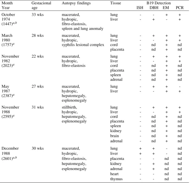
DNA was extracted from the lung and liver tis-sue from the other 27 cases and hybridized against a 32P-labelled probe. Five additional fetuses were found to be positive for B19 DNA for one of each of their tissues tested. A nested PCR was performed to confirm the five positive results found by DBH. Analysis of nested PCR products by agarose gel electrophoresis showed a DNA band of the ex-pected size (591 bp), in at least one of the tissues for each of the samples tested. DEM was also per-formed on these tissues and parvovirus-like par-ticles were observed in all of them. In one of these five positive cases (1757) the NIHF had been first diagnosed as syphilis.
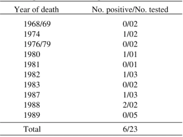
The B19 positive fetuses were found during the years 1974, 1980, 1982, 1987 (one case each year) and 1988 (two cases) as shown in Table II. Of these, only two were not found during the second half of the year.
TABLE II
Cases of NIHF from 1968 to 1989 tested for the presence of human parvovirus B19 Year of death No. positive/No. tested
1968/69 0/02
1974 1/02
1976/79 0/02
1980 1/01
1981 0/01
1982 1/03
1983 0/02
1987 1/03
1988 2/02
1989 0/05
Total 6/23
TABLE I Positive B19 hydrops cases
Month Gestacional Autopsy findings Tissue B19 Detection
Year age ISH DBH EM PCR
October 33 wks macerated, lung - - + +
1974 hydropic, liver - + - +
(1447)a,b fibro-elastosis,
spleen and lung anomaly
March 28 wks macerated, lung - + + +
1980 hydropic, liver - - + +
(1757)a syphilis lesional complex cord - nd + nd placenta - nd + nd November 22 wks macerated, lung - + + +
1982 hydropic, liver - - + +
(2023)a fibro-elastosis cord - nd + nd
placenta - nd + nd spleen - nd + nd adrenal - nd + nd
May 27 wks macerated, lung - + +
-1987 hydropic, liver - - + +
(2387)a hepatomegaly, esplenomegaly
November 31 wks stillbirth, lung - + + +
1988 hydropic, liver - - + +
(2595)a hepatomegaly, cord - nd + nd
esplenomegaly placenta - nd + nd spleen - nd + nd kidney - nd + nd brain - nd + nd adrenal - nd + nd December 30 wks macerated, lung + + - nd
1988 hydropic, liver + + - nd
(2601)a,b fibro-elastosis, placenta - - nd nd
hepatomegaly, kidney - + nd nd esplenomegaly adrenal - + nd nd heart - - nd nd thymus - - nd nd
a: registration number; b: mother with sickle cell disease; nd: not done
lin used in the preservation of the tissues in Brazil is not buffered. This may contribute either to deg-radation of nucleic acids or prevention of probe access to the B19 DNA in the tissue. This would not interfere with DBH since DNA is concentrated by extraction from the tissue and treated with al-kali before binding to the filter and subsequent hybridisation (Clewley 1985).
The difference between the detection of B19 DNA by DBH in different tissues from the same fetus could be explained by postmortem changes associated with the inevitable time lapse between fetal death and formalin fixation of the tissues (Lohr & Neremberg 1990). The site of B19 replication is erythroid progenitors which accumulate in fetal liver between 12 to 30 weeks of gestation (Clewley
DISCUSSION
In the present study using DNA hybridisation we were able to detect six fetuses infected with human parvovirus B19.
forma-et al. 1987, Knisely forma-et al. 1988). Since liver is sub-ject to greater autolysis than lung it is likely that differences in B19 DNA detection in these tissues is due to postmortem changes.
Recent reports have used sensitive PCR assay to detect B19 DNA in clinical specimens (Clewley 1989, Salimans et al. 1989), especially when study-ing B19 infection durstudy-ing pregnancy (Torok et al. 1992, Cassinotti et al. 1993). We used nested PCR to confirm the five positive cases found by DBH. Although a single-step PCR should be suf-ficient for detecting B19 DNA, a nested PCR may be necessary for maximum sensitivity, particularly when investigating fetal tissues in which partially degraded nucleic acids may be recovered (Clewley 1993).
Although a previous study showed that hybridisation is more sensitive than electron mi-croscopy for testing formalin-fixed samples (Field et al. 1991), B19 parvovirus-like particles could be observed in this work, by DEM, in all five fe-tuses positive for B19 DNA by DBH.
A study carried out by the Pathology Depart-ment in IFF/FIOCRUZ showed that of 86 cases of NIHF, 31 were diagnosed as syphilis by identifi-cation of Treponemapallidum. One of these cases (1757) was used as a negative control in our study. B19 DNA was found in this fetus by DBH and PCR. Since congenital syphilis is also considered a cause of hydrops fetalis (Bulova et al. 1972) this may be a case of dual infection.
Four of the parvovirus B19 positive cases were found during the second half of different years. This would be consistent with the seasonality de-scribed in Rio de Janeiro for exanthematic disease (Schatzmayr 1985). Two of the positive cases oc-curred during November and December 1988, co-inciding with the finding by chance of a B19 in-fected blood donor (Cruz et al. 1989). This sup-ports the suggestion that 1988 was an epidemic year for B19 infection in Rio de Janeiro.
Human parvovirus B19 should be considered as a virus to be monitored during pregnancy, since it is clinically very similar to rubella (Shirley et al. 1987) and it has become evident as a cause of fetal infection in countries were rubella is controlled by vaccination (Cohen 1993). It would be par-ticularly important to survey for B19 infection in some Brazilian states, for instance, São Paulo and Paraná which have started rubella vaccination programmes.
REFERENCES
Anderson MJ, Jones SE, Fisher-Hoch SP, Lewis E, Hall SM, Bartlett CRL, Cohen BJ, Mortimer PP, Pereira MS 1983. The human parvovirus, the cause of erythema infectiosum (fifth disease)? Lancet 1:
1378.
Anderson MJ, Khousam MN, Maxwell DJ, Gould SJ, Happerfield LC, Smith WJ 1988. Human parvovirus B19 and hydrops fetalis. Lancet 1: 535. Bond PR, Caul EO, Usher J, Cohen BJ, Clewley JP, Field AM 1986. Intrauterine infection with human parvovirus. Lancet 1: 448-449.
Boom R, Sol CJA, Salimans MMM, Larsen CL, Wertheim-van Dillen PME, Van der Noorda J 1990. Rapid and simple method for purification of nucleic acids. J Clin Microbiol28: 495-503.
Brown T, Anand A, Ritchie LD, Clewley JP, Reid TMS 1984. Intrauterine parvovirus infection associated with hydrops fetalis. Lancet 2: 1033-1034. Bulova SI, Schwartz E, Harrer WV 1972. Hydrops
fetalis and congenital syphilis. Pediat 49: 285-287. Cassinotti P, Weitz M, Siegl G 1993. Human parvovirus B19 infections: routine diagnosis by a new nested polymerase chain reaction assay. J Med Virol 40: 228-234.
Caul EO, Usher MJ, Burton PA 1988. Intrauterine in-fection with human parvovirus B19: a light and electron microscopy study. J Med Virol 24: 55-66. CDC - Centers for Disease Control 1989. Risks associ-ated with human parvovirus B19 infection. MMWR 38: 81-97.
Clewley JP 1985. Detection of human parvovirus B19 using a molecularly cloned probe. JMed Virol 15: 173-181.
Clewley JP 1989. Polymerase chain reaction assay of parvovirus B19 DNA in clinical specimens. J Clin Microbiol 27: 2647-2651.
Clewley JP 1993. PCR detection of parvovirus B19, p.367-373. In DH Persing, TF Smith, FC Tenover, TJ White (eds). Diagnostic Molecular Microbiol-ogy - principles and applications. American Soci-ety for Microbiology, Washington D.C.
Clewley JP, Cohen BJ, Field AM 1987. Detection of parvovirus B19 DNA, antigen, and particles in the human fetus. J Med Virol 23: 367-376.
Cohen BJ 1993. Trends in rubella and parvovirus B19 infections. CDR 3:125.
Cossart YE, Field AM, Cant B, Widdows D 1975. Parvovirus-like particles in human sera Lancet 1: 72-73.
Cruz AS, Andrada Serpa MJ, Barth OM, Nascimento JP 1989. Detection of the human parvovirus B19 in a blood donor plasma in Rio de Janeiro. Mem Inst Oswaldo Cruz 84: 279-280.
Field AM, Cohen BJ, Brown KE, Mori J, Clewley JP, Nascimento JP, Hallam NF 1991. Detection of B19 parvovirus in human fetal tissues by electron mi-croscopy. J Med Virol 35: 85-95.
Garcia AGP, Pegado CS, Ramos HIB, Marques RLS, Cubel RCN, Nascimento JP 1995 Non-immuno-logic hydrops fetalis - study of 86 autopsies. Trop Doc in press.
Guidozzi F, Ballot D, Rothberg AD 1994. Human B19 parvovirus infection in an obstetric population - A prospective study determining fetal outcome. J Reprod Med 39: 36-38.
Kinney JS, Anderson LJ, Farrar J, Strikas RA, Kumar ML, Kliegman RM, Sever JL,Hurwitz ES, Sikes RK 1988. Risk of adverse outcomes of pregnancy after human parvovirus B19 infection. J Infect Dis 157: 663-667.
Knisely AS, O’Shea PA, McMillan P, Singer DB, Magid MS 1988. Electron microscopic identifica-tion of parvovirus virions in erythroid-line cells in fatal hydrops fetalis. Pediatr Pathol 8: 163-170. Knott PD, Welply GAC, Anderson MJ 1984.
Serologi-cally proved intrauterine infection with parvovirus.
BMJ 289: 1660.
Lohr M, Nerenberg MI 1990. Nucleic acid extraction and detection from paraffin embedded tissues, p. 55-62. In MBA Oldstone, Animal Virus Patho-genesis - apratical approach. IRL Press, Oxford. Mori J, Field AM, Clewley JP, Cohen BJ 1989. Dot
blot hybridisation assay of B19 virus DNA in clini-cal specimens. J Clin Microbiol 27: 459-464. Naides SJ, Weiner CP 1989. Antenatal diagnosis and
palliative treatment of nonimmune hydrops fetalis secondary to fetal parvovirus B19 infection. Prenat Diagn 9: 105-114.
Nascimento JP, Buckley MM, Brown KE, Cohen BJ 1990. The prevalence of antibody to human parvovirus B19 in Rio de Janeiro, Brazil. Rev Inst Med Trop São Paulo 32: 41-45.
Nascimento JP, Hallam NF, Mori J, Field AM, Clewley JP, Brown KE, Cohen BJ 1991. Detection of B19 parvovirus in human fetal tissues by in situ
hybridisation. J Med Virol 33: 77-82.
Pattison JR, Jones SE, Hodgson J, Davis LR, White JM, Stroud CE, Murtaza L 1981. Parvovirus infections and hypoplastic crisis in sickle-cell anaemia. Lan-cet 1: 664-665.
PHLS - Public Health Laboratory Service Working Party on Fifth Disease 1990. Prospective study of human parvovirus (B19) infection in pregancy. BMJ 300: 1166-1170.
Reid DM, Reid TMS, Brown T, Rennie JAN, Eastmond CJ 1985. Human parvovirus-associated arthritis: a clinical and laboratory description. Lancet 1: 422-425.
Salimans MMM, van de Rijke FM, Raap AK, van Elsacker-Niele AMW 1989. Detection of parvovirus B19 DNA in fetal tissues by in situ hybridisation and polymerase chain reaction. J Clin Pathol 42: 525-530.
Schatzmayr HG 1985. Aspects of rubella infection in Brazil. Rev Infect Dis (Supl 1): S53-S55.
Shade RO, Blundell MC, Cotmore SF, Tattersall P, Astell CR 1986. Nucleotide sequence and genome orga-nization of human parvovirus B19 isolated from the serum of a child during aplastic crisis. J Virol 58: 921-936.
Shirley JA, Revill S, Cohen BJ, Buckley MM 1987. Serological study of rubella-like illnesses. J Med Virol 21: 369-379.
Smoleniec JS, Pillai M, Caul EO, Usher J 1994. Subclinical transplacental parvovirus B19 infection: an increased fetal risk? Lancet 343: 1100-1101. Torok TJ, Wang QY, Gary GW, Yan CF, Finch TM,