Faculdade de Ciências e Tecnologia
Departamento de Ciências Biomédicas e Medicina
Nano and microparticles as carriers for alveolar
macrophage targeting in pulmonary tuberculosis therapy
Ludmylla Costa Cunha
Faculdade de Ciências e Tecnologia
Departamento de Ciências Biomédicas e Medicina
Nano and microparticles as carriers for alveolar
macrophage targeting in pulmonary tuberculosis therapy
Ludmylla Costa Cunha
PhD in Biomedical Sciences
Thesis supervised by Prof. Ana Grenha, PhD.
ii
Nano and microparticles as carriers for alveolar macrophage targeting in pulmonary tuberculosis therapy
Declaração de autoria de trabalho
Declaro ser a autora deste trabalho, que é original e inédito. Autores e trabalhos consultados estão devidamente citados no texto e constam na listagem de referências incluída.
_____________________________________________________
Copyright – Ludmylla Costa Cunha. Universidade do Algarve. Departamento de Ciências Biomédicas e Medicina.
A Universidade do Algarve tem o direito perpétuo e sem limites geográficos de arquivar e publicitar este trabalho através de exemplares impressos reproduzidos em papel ou de forma digital, ou por qualquer outro meio conhecido ou que venha a ser inventado, de o divulgar através de repositórios científicos e de admitir a sua cópia e distribuição com objetivos educacionais ou de investigação, não comerciais, desde que seja dado crédito ao autor e editor.
iii
Acknowledgements
Agradeço à professora Ana Grenha por me acolher em seu grupo de investigação e pela orientação no desenvolvimento deste trabalho.
Agradeço aos professores que colaboraram diretamente com a realização desta pesquisa, nomeadamente prof. Ana Costa (UAlg), prof. Deborah Power (UAlg), prof. João Lourenço (UAlg), prof. Leonor Faleiro (UAlg), prof. Manuela Gaspar (iMed UL) e prof. Francesca Buttini (Universidade de Parma).
Igualmente, agradeço aos colaboradores dos serviços técnico e administrativo, que com competência e prestimosidade, contribuíram direta ou indiretamente para a realização deste trabalho. Em especial, destaco a colaboração do Sr. Mário Freitas (UAlg) e Sra. Judith Anakayana (Universidade de Parma).
Agradeço o apoio dos meus colegas de laboratório Filipa, Jorge, Susana, e João. E, finalmente, meu eterno agradecimento a meus pais, irmão, Vincent e amigos pelo carinho, incentivo e suporte emocional ao longo dessa jornada.
Muito obrigada!
Esta pesquisa foi financiada pela Fundação para a Ciência e Tecnologia, de Portugal (PTDC/DTP-FTO/0094/2012 and UID/BIM/04773/2013). A bolsa de doutoramento foi financiada pela agência brasileira de fomento à pesquisa CAPES – Coordenação de Aperfeiçoamento de Pessoal de Nível Superior – por meio do programa Ciências sem Fronteiras (BEX 1168-13/4).
iv
v
Contents
1. General Introduction 3
1.1 Tuberculosis 3
1.1.1 Pathogenesis of tuberculosis 5
1.1.2 Diagnosis and treatment of tuberculosis 9
1.2 Pulmonary drug delivery 10
1.2.1 Challenges for pulmonary delivery 13
1.3 Nano and microparticles as drug delivery systems for inhalation purposes 15 1.4 Biopolymers for designing drug delivery systems 19
1.4.1 Fucoidan 21
1.4.2 Chitosan 25
2. Motivation and objectives 31
3. Nanoparticles 37
3.1 Materials and methods 37
3.1.1 Preparation and characterisation of nanoparticles by polyelectrolyte
complexation 37
3.1.2 Preparation and characterisation of nanoparticles by nanoprecipitation 38
3.2 Results and discussion 41
3.2.1 Production of FUC/CS nanoparticles by polyelectrolyte complexation
41
3.2.2 Production of FUC and CS nanoparticles by nanoprecipitation 45
4. Microparticles: Materials and methods 55
4.1 Materials 55
4.2 Preparation of microparticles by spray-drying 55
4.2.1 Fucoidan 55
4.2.2 Chitosan 57
4.2.3 Preparation of fluorescently labelled microparticles 59
4.3 Characterisation of microparticles 60
4.3.1 Morphology 60
4.3.2 Particle size 60
vi 4.3.4 Dry powder analysis using Powder X-Ray Diffraction (PXRD) 61
4.3.5 Drug association efficiency and loading 61
4.4 Evaluation of aerodynamic properties 62
4.5 In vitro drug release profiles 63
4.5.1 Fucoidan microparticles 64
4.5.2 Chitosan microparticles 64
4.6 In vitro biocompatibility studies 65
4.6.1 Cell cultures 65
4.6.2 Assessment of metabolic activity by MTT test 66
4.6.3 Evaluation of cell membrane integrity 67
4.7 Macrophage activation induced by microparticles 68 4.8 Preliminary evaluation of microparticle uptake by macrophages 68
4.9 In vitro antibacterial activity 69
4.9.1 Culture of mycobacteria 69
4.9.2 Determination of minimum inhibitory concentration (MIC) 69
4.10 Preliminary in vivo studies 71
4.10.1 Eosinophil count on blood smears 72
4.10.2 Ouchterlony double immunodiffusion to detect serum
immunoglobulin E (IgE). 73
4.11 Statistical analysis 73
5. Fucoidan microparticles: Results and discussion 77 5.1 Preparation and characterisation of fucoidan microparticles 77 5.1.1 Dry powder analysis using Powder X-Ray Diffraction (PXRD) 82
5.2 Evaluation of aerodynamic properties 85
5.3 In vitro drug release profiles 88
5.4 In vitro biocompatibility studies 91
5.4.1 Assessment of metabolic activity by MTT test 91
5.4.2 Evaluation of cell membrane integrity 96
5.5 Macrophage activation 99
5.6 Preliminary evaluation of microparticle uptake by macrophages 102 5.7 Determination of minimum inhibitory concentration (MIC) 103
5.8 Preliminary in vivo studies 104
vii 6. Chitosan microparticles: Results and discussion 111 6.1 Preparation and characterisation of chitosan microparticles 111 6.1.1 Dry powder analysis using Powder X-Ray Diffraction (PXRD) 115
6.2 Evaluation of aerodynamic properties 117
6.3 In vitro drug release profiles 119
6.4 In vitro biocompatibility studies 122
6.4.1 Assessment of metabolic activity by MTT test 122
6.4.2 Evaluation of cell membrane integrity 127
6.5 Macrophage activation 130
6.6 Preliminary evaluation of microparticle uptake by macrophages 131 6.7 Determination of minimum inhibitory concentration (MIC) 133
6.8 Preliminary in vivo studies 133
6.9 Conclusion 135
7. Final considerations 139
7.1 General discussion 139
viii
ix
Resumo
A tuberculose (TB) é uma das principais causas de morte por infeção no mundo, apesar de uma vacina e vários antibióticos eficazes estarem disponíveis para a prevenção e tratamento da patologia. O controlo global da TB está dificultado por vários fatores, incluindo o diagnóstico tardio e a não adesão do paciente a tratamentos de longo prazo, o que leva a uma alta incidência de resistência aos fármacos tuberculostáticos. Em geral, existem grandes desafios associados à terapêutica convencional, incluindo (i) resistência aos fármacos e toxicidade; (ii) interrupção do tramento pelo paciente, devido à terapêutica prolongada e efeitos secundários graves; (iii) interações medicamentosas, particularmente com antirretrovirais em pacientes co-infectados com TB e HIV. Assim, a gravidade da situação chegou a um ponto em que o desenvolvimento de novas estratégias de intervenção é urgentemente necessário.
Nesse contexto, a administração pulmonar de fármacos tuberculostáticos é uma abordagem promissora no tratamento da TB pulmonar. A doença representa aproximadamente 80% do total de casos e, portanto, o pulmão tem sido explorado como uma via efetiva para a administração de fármacos. Essa estratégia não só permite direcionar instantaneamente os fármacos para o órgão afetado, como também pode reduzir os efeitos adversos sistémicos dos antibióticos, que são as principais razões para a interrupção do tratamento por parte do paciente. No entanto, a administração pulmonar de fármacos enfrenta algumas limitações, como a complexa estrutura das vias aéreas, a degradação local de fármacos e a eliminação mucociliar. A fim de ultrapassar algumas dessas limitações, a microencapsulação de fármacos aparece como uma abordagem com potencial. Nesse sentido, este trabalho teve como objetivo produzir micropartículas inaláveis que associassem eficientemente dois fármacos tuberculostáticos de primeira linha, isoniazida (INH) e/ou rifabutina (RFB), visando uma aplicação na terapia da TB pulmonar. Fucoidan (FUC) e quitosano (CS) foram os biomateriais selecionados para compor a matriz dos transportadores. FUC é um polissacárido composto por unidades de fucose, que foram relatadas como sendo especificamente reconhecidas por recetores de membrana de macrófagos alveolares (a célula hospedeira do agente patogénico da tuberculose - Mycobacterium tuberculosis). Da mesma forma, o CS é também um polissacárido composto por resíduos de
N-x acetilglucosamina e D-glucosamina, sendo os primeiros igualmente reconhecido por macrófagos de acordo com descrições da literatura. Este reconhecimento pelos macrófagos acredita-se que potenciará a fagocitose.
Numa primeira abordagem foram produzidas nanopartículas, tendo-se considerado que o desenvolvimento de uma formulação inalável à base de nanopartículas implicaria uma etapa posterior de microencapsulação para contornar as limitações relacionadas com as propriedades aerodinâmicas dos sistemas nanométricos e as suas dificuldades para alcançar a região alveolar. As nanopartículas foram obtidas por complexação de FUC com CS, tendo-se desenvolvido várias formulações (rácios de massa FUC/CS de 4:1 a 1:4). Os sistemas sem fármaco apresentaram tamanho médio de 159 – 266 nm, PdI entre 0,21 e 0,36 e potencial zeta entre -39 mV e +12 mV, seguindo sensivelmente a alteração das razões de massa. A capacidade das nanopartículas de FUC/CS para associar fármacos tuberculostáticos foi avaliada, tendo-se iniciado estes testes pela incorporação de RFB, associada à razão final polímero/fármaco de 10/1 (m/m). Várias tentativas foram executadas sem sucesso, pelo que o trabalho prosseguiu com uma segunda abordagem metodológica para produção das nanopartículas. Aplicou-se o método de nanoprecipitação, que levou à produção de nanopartículas de FUC e nanopartículas de CS com tamanho médio de 500 nm e 700 nm, respetivamente. No entanto, as nanopartículas obtidas apresentaram uma ampla distribuição de tamanhos, indicada pelos valores de PdI, que variaram entre 0,55 e 0,83. Além disso, um protocolo de nanoprecipitação ideal para obter nanopartículas à base de FUC ou CS que associassem de forma efetiva INH e/ou RFB não foi estabelecido. Tendo em conta a ausência de sucesso da produção de sistemas nanométricos, e considerando as restrições temporais impostas aos trabalhos de doutoramento, foi decidido focar o trabalho no desenvolvimento de micropartículas poliméricas à base de FUC ou CS.
As micropartículas foram produzidas por um processo de atomização, contendo os fármacos modelo (INH e RFB) isoladamente ou de forma combinada. As micropartículas de FUC associaram efetivamente a INH (95%) e a RFB (81%) por separado. Foram igualmente produzidas com sucesso micropartículas de FUC carregadas com os dois fármacos tuberculostáticos simultaneamente, as quais registaram elevada eficácia de associação dos fármacos (97% para INH e 95% para
xi RFB). Todas as formulações de micropartículas de FUC demonstraram ter propriedades aerodinâmicas favoráveis para a distribuição e penetração pulmonar após inalação. Para as micropartículas que associaram os fármacos isoladamente, obteve-se um diâmetro aerodinâmico no intervalo 2,0-3,8 µm. Da mesma forma, a formulação que associa os dois antibióticos apresentou um diâmetro aerodinâmico de 3,6 – 3,9 µm. Em geral, as formulações não evidenciaram efeitos citotóxicos nas células do epitélio alveolar humano (A549), embora tenha sido observada uma ligeira toxicidade nas células THP-1 diferenciadas em macrófagos, na concentração mais elevada que foi testada (1 mg/mL). Contudo, considera-se que esta concentração seja muito mais elevada em relação àquela que seria efetivamente administrada in vivo. As micropartículas de FUC desenvolvidas neste estudo também exibiram propensão para serem capturadas por macrófagos ou células diferenciadas em macrófagos (células-alvo) de forma dose-dependente. Particularmente, as micropartículas que associam os dois fármacos conjuntamente exibiram uma capacidade para ativar as células-alvo e, ainda, inibiram de maneira eficaz o crescimento de micobactéria in vitro, sem alterarem a atividade bactericida dos fármacos. A administração pulmonar in vivo (ratinhos BALB/c) das micropartículas de FUC (sem fármacos) indicou, num ensaio preliminar, que as mesmas não induziram respostas alérgicas.
As micropartículas de CS também associaram eficientemente INH (90%) e RFB (97%) por separado, tendo a formulação com fármacos combinados resultado em 93% de eficácia de associação para INH e 99% para RFB. Todas as formulações apresentaram propriedades adequadas para administração pulmonar, com diâmetro aerodinâmico entre 2,5 e 4 µm. A ausência de toxicidade foi observada no epitélio alveolar humano (células A549), mas, tal como observado para as micropartículas de FUC, a maior concentração de micropartículas testada (1 mg/mL) diminuiu a viabilidade de células THP-1 diferenciadas em macrófagos, após 24 h de exposição, uma dose considerada sobrestimada, como mencionado anteriormente. As micropartículas de CS evidenciaram ainda uma grande capacidade de internalização por macrófagos (percentagem de fagocitose até 99,9%), de forma independente da dose, e as micropartículas contendo ambos os fármacos numa única formulação, induziram a ativação de macrófagos e inibiram eficazmente o crescimento de micobactérias in vitro. Além disso, o biomaterial (CS)
xii foi, aparentemente, bem tolerado por ratinhos BALB/c, após administração pulmonar das micropartículas de CS sem fármaco. No geral, os dados obtidos fornecem indicações positivas sobre o potencial dos sistemas propostos para uma aplicação na terapia inalável da tuberculose pulmonar.
Keywords: atomização, fucoidan, isoniazida, macrófagos alveolares, micropartículas inaláveis, quitosano, rifabutina, terapia pulmonar, tuberculose.
xiii
Abstract
Tuberculosis (TB) is a leading infectious cause of death worldwide, even though a vaccine and several effective antibiotics are available for its prevention and treatment. Global TB control is very difficult due to various factors, including late diagnosis and patient non-compliance to long-term treatments, which leads to a high incidence of extensive resistance to effective anti-TB drugs. Overall, there are significant challenges associated with conventional TB therapy, including (i) drug resistance and toxicity; (ii) patient non-compliance, given the long-term therapy and severe side effects; (iii) and drug-drug interactions, particularly with antiretroviral drugs in patients co-infected with TB and HIV. Thus, the situation has come to a point where the development of novel intervention strategies is urgently needed.
In this context, the pulmonary delivery of anti-TB drugs is a promising approach to treat lung TB. The disease represents approximately 80% of total cases, and thus the lung has been explored as an effective route for the delivery of drugs. This strategy not only allows targeting the infected organ instantly, but it can also reduce the systemic adverse effects of the antibiotics, which are main reasons for patient non-compliance. However, pulmonary drug delivery faces some limitations related with the proper airway structure, local degradation of drugs, and mucociliary clearance. In order to overcome some of these limitations of lung delivery, drug microencapsulation appears as a potential approach. In this sense, this work aimed at producing inhalable microparticles that efficiently associate two anti-TB drugs, isoniazid (INH) and/or rifabutin (RFB), for an application in pulmonary TB therapy. Fucoidan (FUC) and chitosan (CS) were the biomaterials selected to compose the matrix of the carriers. FUC is a polysaccharide composed of fucose units that has been reported to be specifically recognised by surface receptors of alveolar macrophages (the host cells of Mycobacterium tuberculosis). Likewise, CS is a polysaccharide composed of N-acetylglucosamine and D-glucosamine residues, the former being also specifically recognised by macrophages, according to the literature. This recognition by macrophages is believed to potentiate phagocytosis.
The first approach involved the production of nanoparticles and it was considered that subsequent microencapsulation of the nanocarriers would be necessary to overcome aerodynamic limitations of nanosized carriers and their
xiv ability to reach the alveolar zone. Nanoparticles were spontaneously obtained by complexing FUC with CS, resulting from several formulations (polymeric mass ratio varying from 4:1 to 1:4). The produced unloaded FUC/CS nanoparticles presented average size range of 159 – 266 nm, PdI ranging between 0.21 and 0.36, and zeta potential varying from -39 mV to +12 mV, following the alteration of the mass ratios. The ability of FUC/CS nanoparticles to associate anti-TB drugs was assessed, and tests initiated with the incorporation of RFB, which was associated to obtain final polymer/drug mass ratio of 10/1 (w/w). Several attempts were made unsuccessfully, and therefore the work continued using another production method. Nanoprecipitation technique was then used, resulting in FUC nanoparticles and CS nanoparticles with mean size of 500 nm and 700 nm, respectively. However, the obtained nanoparticles showed little uniformity in size, indicated by PdI values, varying between 0.55 and 0.83. Moreover, an optimal protocol to obtain FUC- and CS-based nanoparticles that efficiently encapsulate INH and RFB could not be established. Taking into consideration the unsuccessful nanoparticle production and time restraints to accomplish the aims of the PhD plan, it was decided to focus the study on the development of polymeric microparticles.
Microparticles were produced by spray-drying, associating the model drugs (INH and RFB), either separately or in combination. FUC microparticles effectively associated INH (95%) and RFB (81%), separately. Likewise, FUC microparticles loaded with the two anti-TB drugs simultaneously were also successfully produced, demonstrating high drug association efficiency (97% for INH and 95% for RFB). All FUC-based microparticles evidenced favourable aerodynamic properties for deep lung delivery upon inhalation. Single drug-loaded FUC microparticles showed aerodynamic diameter (MMAD) in the range of 2.0–3.8 µm. Likewise, the dual drug-loaded dry powder presented aerodynamic diameter of 3.6–3.9 µm. Overall, the formulations evidenced no cytotoxic effects on human alveolar epithelium cells (A549), although mild toxicity was observed on macrophage-differentiated THP-1 cells at the highest tested concentration (1 mg/mL). Nonetheless, this dose is considered overestimated compared to that effectively observed in vivo. The produced FUC microparticles also exhibited a propensity to be captured by macrophages or macrophage-like cells (target cells) in a dose-dependent manner. Particularly, dual drug-loaded microparticles displayed ability to activate the target
xv cells and, moreover, effectively inhibited mycobacterial growth in vitro, preserving the bactericidal activity of the drugs. In vivo lung administration (BALB/c mice) of unloaded FUC microparticles indicated, in a preliminary assay, that the carriers induced no allergic responses.
CS microparticles also associated INH (90%) and RFB (97%) efficiently, in separate formulations, whereas dual drug-loaded formulation resulted in 93% association efficiency for INH and 99% for RFB. All formulations presented adequate properties for deep lung delivery, with aerodynamic diameters ranging between 2.5 and 4 µm. Absence of toxicity was observed in human alveolar epithelium (A549 cells) but, as observed for FUC carriers, the highest tested concentration of microparticles (1 mg/mL) decreased the viability of macrophage-differentiated THP-1 cells upon 24 h exposure. This dose is however believed to be overestimated, as aforementioned. CS microparticles further evidenced strong ability to be internalised by macrophage-like cells (percentage of phagocytosis up to 99.9%), regardless of the dose. Yet, dual drug-loaded carriers induced macrophage activation and effectively inhibited the growth of mycobacteria in vitro. Moreover, the biomaterial (CS) was well tolerated by BALB/c mice upon pulmonary administration of unloadead CS microparticles. Overall, the obtained data gave positive indications on the potential of the proposed systems for an application as inhalable tuberculosis therapy.
Keywords: alveolar macrophage, chitosan, fucoidan, inhalable microparticles, isoniazid, lung therapy, rifabutin, spray-drying, tuberculosis.
xvi
List of tables
Table 1.1. The most relevant advantages and limitations of pulmonary drug delivery.
... 12
Table 3.1. Characterisation of unloaded FUC/CS nanoparticles in terms of size,
polydispersity index (PdI) and zeta potential (mean ± SD, n = 3). Different letters represent significant differences in each parameter (p<0.05). ... 42
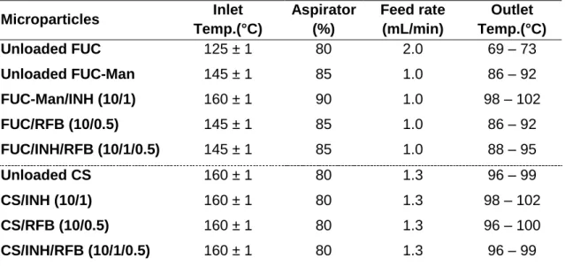
Table 4.1. Spray-drying parameters (inlet temperature, aspirator and feed rate)
used to produce fucoidan- and chitosan-based microparticles and resulting outlet temperature. ... 58
Table 5.1. Spray-drying yield, drug association efficiency, loading capacity, Feret´s
diameter, median volume particle size (Dv50) and density values of fucoidan
(FUC)-based microparticles (mean ± SD, n = 3). INH: isoniazid; Man: mannitol; RFB: rifabutin. ... 78
Table 5.2. Aerodynamic properties of FUC microparticles loaded with INH and/or
RFB. Loaded amount of powder in the capsule was 30 mg, corresponding to approximately 2.6 mg of INH and 1.4 mg of RFB, according to the drug content found in each formulation (n = 3, mean ± SD). FPD: fine particle dose; FPF: fine particle fraction; FUC: fucoidan; INH: isoniazid; Man: mannitol; MMAD: mass median aerodynamic diameter; RFB: rifabutin ... 85
Table 6.1. Spray-drying production yield, drug association efficiency, loading
capacity, Feret´s diameter, median volume particle size (Dv50) and density values
of chitosan (CS)-based microparticles (mean ± SD, n = 3). INH: isoniazid; RFB: rifabutin. ... 113
Table 6.2. Aerodynamic properties of chitosan (CS) microparticles loaded with
isoniazid (INH) and/or rifabutin (RFB). Loaded amount of powder in the capsule was 30 mg, corresponding to approximately 2.8 mg of INH and 1.5 mg of RFB, according to the drug content found in each formulation (n = 3, mean ± SD). FPD: fine particle dose; FPF: fine particle fraction; MMAD: mass median aerodynamic diameter. 117
xvii
List of figures
Figure 1.1. Percentage of new and relapse pulmonary TB cases with bacteriological
confirmation, 2016 (6). ... 4
Figure 1.2. Schematic representation of the granuloma structure. Adapted from
(23). ... 8
Figure 1.3. Number of scientific papers published on the topic “pulmonary drug
delivery” on ISI Web of Science (October/2018), as a function of publication years. ... 11
Figure 1.4. Scheme describing the main mechanisms affecting aerosol transport
and deposition in the human lung. Adapted from (68). ... 13
Figure 1.5. Relationship between aerodynamic diameter and deposition of aerosol
particles in the human respiratory tract. Adapted from (27,76). ... 15
Figure 1.6. Scheme of α‐L-fucose chains observed in fucoidans isolated from
several algae belonging to the taxonomic orders Chordariales and Laminariales (a) and Fucales (b); (a) The chain is only composed of repeating (1→3)-linked fucose residues; (b) The chain consists of alternating (1→3)- and (1→4)-linked α-L-fucose residues. R represents the positions of potential attachment of carbohydrate. ... 23
Figure 1.7. Structure of chitosan showing alternated units of N-acetylglucosamine
and D-glucosamine, linked by β-(1→4) glycosidic bonds. ... 26
Figure 2.1. Illustration of carrier uptake by alveolar macrophages, assuming
targeted drug delivery mediated by the polysaccharides (fucoidan or chitosan). Drug-loaded carriers reach the alveoli upon dry powder aerosolisation. Next, alveolar macrophages, infected with M. tuberculosis, engulf the particles. The polymer is expected to facilitate phagocytosis, because it possesses chemical moieties that are recognisable by the macrophage surface receptors. ... 33
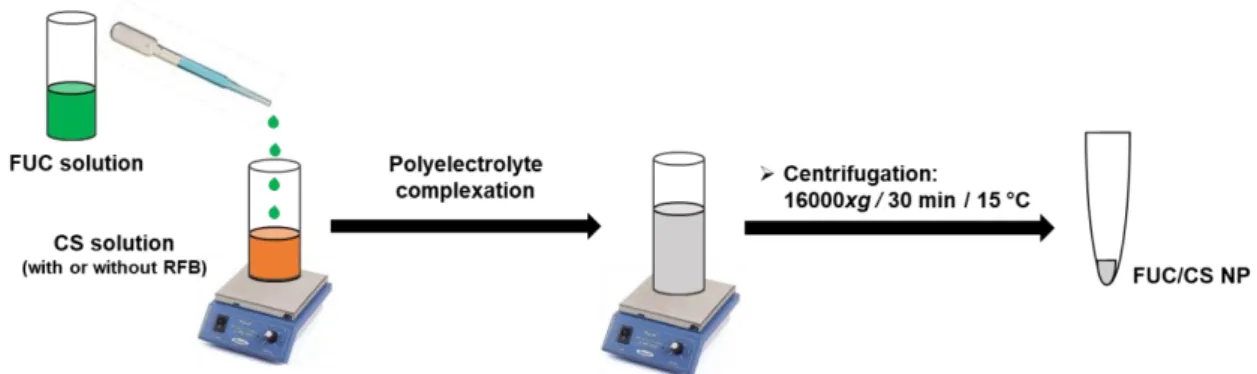
Figure 3.1. Illustration of fucoidan/chitosan nanoparticle (FUC/CS NP) preparation
by polyelectrolyte complexation. ... 38
Figure 3.2. Illustration of the method to produce (a) chitosan and (b) fucoidan
nanoparticles by nanoprecipitation. Drug was solubilised either in the non-solvent (absolute ethanol) or in the polymeric solution. In the latter approach, RFB required solubilisation in HCl 0.01M prior to the addition to the polymeric solution, to address its hydrophobicity. ... 40
Figure 4.1. Scheme of the spray-drying process. ... 57 Figure 4.2. Scheme of the 96-well microplate showing columns 4-11 filled with
solutions of free drugs or microparticles serially diluted with M7H9 broth, containing mycobacteria in triplicate: lines B-C (suspension 1), lines D-E (suspension 2) and lines F-G (suspension 3). Contents of column 2 (only M7H9 medium) and column 3 (bacterial suspensions in broth) were considered negative and positive control, respectively. ... 71
Figure 5.1. Scanning electron microphotographs of fucoidan (FUC)-based
xviii microparticles; (c) FUC/INH/RFB = 10/1/0.5 (w/w) microparticles; (d) unloaded FUC-Man microparticles; (e) FUC-FUC-Man/INH = 10/1 (w/w) microparticles. INH: isoniazid, Man: mannitol, RFB: rifabutin. ... 80
Figure 5.2. Diffractograms of (a) fucoidan (FUC) polymer, unloaded and
drug-loaded FUC microparticles; (b) free isoniazid (INH) before and after spray-drying, (c) free rifabutin (RFB), before and after spray-drying. ... 84
Figure 5.3. In vitro aerodynamic deposition of antitubercular drugs (INH and RFB)
in the Andersen cascade impactor. Drugs associated with fucoidan (FUC) microparticles (MP) either individually (a) or together in a single formulation (b). Values are mean ± SD, n = 3. Cps: capsule; Dev: inhaler device; IP: induction port; F: filter, INH: isoniazid; RFB: rifabutin St: stage. ... 87
Figure 5.4. Drug dissolution and in vitro release profiles of isoniazid (INH) from
FUC-Man/INH (10/1, w/w) microparticles and of rifabutin (RFB) from FUC/RFB microparticles (10/0.5, w/w), in (a) PBS pH 7.4-Tween® 80and (b) citrate buffer pH
5.0-Tween® 80, at 37 °C. Drug dissolution and in vitro release profile of INH and
RFB from FUC/INH/RFB (10/1/0.5, w/w) microparticles in (c) PBS pH 7.4-Tween®
80 and (d) citrate buffer pH 5.0-Tween® 80, at 37 °C. FUC: fucoidan; Man: mannitol;
mean ± SD, n ≥ 3. ... 90
Figure 5.5. A549 cell viabilities upon (a) 3 h and (b) 24 h of exposure to fucoidan
(FUC) polymer, unloaded and drug-loaded FUC microparticles; (c) exposure to INH as a free drug; and (d) exposure to RFB as a free drug. Cell viability was calculated as a percentage of positive control (untreated cells). Data represent mean ± SEM (n = 3, six replicates per experiment at each concentration). Dashed line indicates 70% cell viability. INH: isoniazid; Man: mannitol; MP: microparticles; RFB: rifabutin. ... 93
Figure 5.6. Macrophage-differentiated THP-1 cell viabilities upon (a) 3 h and (b) 24
h of exposure to fucoidan (FUC) polymer, unloaded and drug-loaded FUC microparticles; (c) exposure to INH as a free drug; and (d) exposure to RFB as a free drug. Cell viability was calculated as a percentage of positive control (untreated cells). Data represent mean ± SEM (n = 3, six replicates per experiment at each concentration). Dashed line indicates 70% cell viability. INH: isoniazid; Man: mannitol; MP: microparticles; RFB: rifabutin... 95
Figure 5.7. Release of lactate dehydrogenase (LDH) from (a) A549 cells and (b)
macrophage-differentiated THP-1 cells exposed to fucoidan (FUC) polymer, FUC-based microparticles (1.0 mg/mL), free rifabutin (RFB, 0.05 mg/mL), and free isoniazid (INH, 0.1 mg/mL). Cell culture medium (CCM) and Triton X-100 were used as negative and positive controls, respectively. The released LDH was calculated based on 100% assumed for positive control. Data represent mean ± SEM (n = 3, six replicates per experiment a each concentration) ... 98
Figure 5.8. TNF-α (a) and IL-8 (b) secretion by macrophage-differentiated THP-1
cells upon 24 h exposure to FUC/INH/RFB (10/1/0.5) microparticles (MP) and FUC as raw material. Cell culture medium (CCM) and lipopolysaccharide (LPS) were used as negative and positive controls, respectively. FUC: fucoidan; INH: isoniazid;
xix RFB: rifabutin. Data represent mean ± SEM (n = 3). * p < 0.05 compared to CCM. ... 101
Figure 5.9. Uptake of fluorescently-labelled unloaded fucoidan microparticles by
human macrophage-differentiated THP-1 cells and rat alveolar macrophages (NR8383 cells) upon exposure to 50 µg/cm2 and 200 µg/cm2, for a period of 2 h.
Results are expressed as mean ± SEM (n ≥ 3). ... 103
Figure 5.10. Percentage of white blood cells counted in the blood smears. Mean
value ± SD. ... 105
Figure 6.1. Scanning electron microphotographs of chitosan (CS)-based
microparticles: (a) unloaded CS microparticles, (b) CS/INH = 10/1 (w/w) microparticles; (c) CS/RFB = 10/0.5 (w/w) microparticles; (d) CS/INH/RFB = 10/1/0.5 (w/w) microparticles. INH: isoniazid, RFB: rifabutin. ... 114
Figure 6.2. Diffractograms of (a) chitosan (CS) polymer, unloaded and drug-loaded
CS microparticles; (b) free isoniazid (INH) before and after spray-drying, (c) free rifabutin (RFB), before and after spray-drying. Note to Figure 6.2: Graphics c and d represent the same set of data of Figure 5.2 and were reprinted in this chapter to facilitate reading. ... 116
Figure 6.3. In vitro aerodynamic deposition of antitubercular drugs (isoniazid – INH
and rifabutin – RFB) in the Andersen cascade impactor. Drugs associated with chitosan (CS) microparticles (MP) either individually (a) or together in a single formulation (b). Values are mean ± SD, n = 3. Cps: capsule; Dev: inhaler device; IP: induction port; F: filter, St: stage. ... 119
Figure 6.4. Drug dissolution and in vitro release profiles of isoniazid (INH) from
CS/INH (10/1, w/w) microparticles and of rifabutin (RFB) from CS/RFB microparticles (10/0.5, w/w), in (a) PBS pH 7.4-Tween® 80and (b) citrate buffer pH
5.0-Tween® 80, at 37 °C. Drug dissolution and in vitro release profile of INH and
RFB from CS/INH/RFB (10/1/0.5, w/w) microparticles in (c) PBS pH 7.4-Tween® 80
and (d) citrate buffer pH 5.0-Tween® 80, at 37 °C. CS: chitosan; mean ± SD, n ≥ 3.
... 121
Figure 6.5. A549 cell viabilities upon (a) 3 h and (b) 24 h of exposure to chitosan
(CS) polymer, unloaded and drug-loaded CS microparticles; (c) exposure to INH as a free drug; and (d) exposure to RFB as a free drug. Cell viability was calculated as a percentage of positive control (untreated cells). Data represent mean ± SEM (n = 3, six replicates per experiment at each concentration). Dashed line indicates 70% cell viability. INH: isoniazid; MP: microparticles; RFB: rifabutin. Note to Figure 6.5: Graphics c and d represent the same set of data of Figure 5.5 and were reprinted in this chapter to facilitate reading. ... 125
Figure 6.6. Macrophage-differentiated THP-1 cell viabilities upon (a) 3 h and (b) 24
h of exposure to chitosan (CS) polymer, unloaded and drug-loaded CS microparticles; (c) exposure to INH as a free drug and (d) exposure to RFB as a free drug. Cell viability was calculated as a percentage of positive control (untreated cells). Data represent mean ± SEM (n = 3, six replicates per experiment at each concentration). Dashed line indicates 70% cell viability. INH: isoniazid; MP:
xx microparticles; RFB: rifabutin. Note to Figure 6.6: Graphics c and d represent the same set of data of Figure 5.6 and were reprinted in this chapter to facilitate reading. ... 126
Figure 6.7. Release of lactate dehydrogenase (LDH) from (a) A549 cells and (b)
macrophage-differentiated THP-1 cells exposed to chitosan (CS) polymer, CS-based microparticles (1.0 mg/mL), free rifabutin (RFB, 0.05 mg/mL), and free isoniazid (INH, 0.1 mg/mL). Cell culture medium (CCM) and Triton X-100 were used as negative and positive controls, respectively. The released LDH was calculated based on 100% assumed for positive control. Data represent mean ± SEM (n = 3, six replicates per experiment at each concentration). * p < 0.05 compared to CCM. Note to Figure 6.7: The set of data of free RFB, free INH, CCM and Triton is the same presented in Figure 5.7 – Chapter 5. The data were reprinted in this figure to facilitate reading ... 128
Figure 6.8. TNF-α (a) and IL-8 (b) secretion by macrophage-differentiated THP-1
cells upon 24 h exposure to CS/INH/RFB (10/1/0.5) microparticles (MP) and CS as raw material. Cell culture medium (CCM) and lipopolysaccharide (LPS) were used as negative and positive controls, respectively. CS: chitosan; INH: isoniazid; RFB: rifabutin. Data represent mean ± SEM (n = 3). * p < 0.05 compared to CCM. Note to Figure 6.8: The set of data of CCM and LPS is the same presented in Figure 5.8 – Chapter 5. The data were reprinted in this figure to facilitate reading. ... 131
Figure 6.9. Percentage of human macrophage-differentiated THP-1 cells and rat
alveolar macrophages (NR8383 cells) phagocytosing fluorescently-labelled chitosan microparticles. Cells were exposed (2 h) to 50 and 200 µg/cm2 of
microparticles. Data represent mean ± SEM (n ≥ 3). ... 132
Figure 6.10. Percentage of white blood cells counted in the blood smears. Mean ±
xxi
List of abbreviations, acronyms, and symbols
AE – Association efficiency
ACI – Andersen cascade impactor CCM – Cell culture medium
CD4 – CD4 positive T cells CD8 – CD8 positive T cells CS – Chitosan
Daer – Aerodynamic diameter
ED – Emitted dose
EDAC – N-(3-dimethylaminopropyl)-N′-ethylcarbodiimide hydrochloride ELISA – Enzyme-linked immunosorbent assay
FBS – Fetal bovine serum FUC – Fucoidan
FPD – Fine particle dose FPF – Fine particle fraction
HIV – Human immunodeficiency virus
HPLC – High performance liquid chromatography Ig – Immunoglobulin
IL – Interleukin INH – Isoniazid
ISO – International Organization for Standardization LC – Loading capacity
LDH – Lactate dehydrogenase LPS – Lipopolysaccharide Man – Mannitol
MD – Metered dose
MIC – Minimum inhibitory concentration
MMAD – Mass median aerodynamic diameter MP – Microparticles
MTT – 3-(4,5-dimethylthiazol-2-yl)-2,5-diphenyltetrazolium bromide NP – Nanoparticles
OADC – Oleic acid, albumin, dextrose and catalase OVA – Ovalbumin
PBS – Phosphate-buffered saline PdI – Polydispersity index
PGA – Poly(glycolic acid) PLA – Poly(lactic acid)
PLGA – Poly(lactic acid-co-glycolic acid) copolymer PMA – Phorbol 12-myristate 13-acetate
RFB – Rifabutin
SDS – Sodium dodecyl sulphate
SEM – Scanning electron microphotograph TB – Tuberculosis
TNF – Tumor necrosis factor WHO – World Health Organization
xxii
Chapter 1 – General Introduction
Chapter One
General Introduction
The information presented in this chapter was partially published in the following publication:
Ludmylla Cunha and Ana Grenha. Sulfated seaweed polysaccharides as multifunctional materials in drug delivery applications. Marine Drugs 2016, 14, 42; doi: 10.3390/md1403004
Chapter 1 – General Introduction
2
Nano and microparticles as carriers for alveolar macrophage targeting in pulmonary tuberculosis therapy
3
1. General Introduction
1.1 Tuberculosis
Since ancient times, tuberculosis (TB) or illnesses resembling TB have been described in several parts of the world from many civilisations. Although it was probably described for the first time in Indian scriptures, pulmonary TB is known since the time of Hippocrates as phthisis, which is derived from the Greek for “wasting away”. TB-like diseases have also been documented in ancient Chinese and Arabic literature and have been described under many peculiar names during history, such as consumption (derived from the Latin consumer) and scrofula, a rare manifestation form of TB that affects the lymph nodes, especially of the neck (1,2). The meaning of old names has undergone great alterations with time, mainly due to discoveries about the nature of the disease and its effects on people. The disease was given the name of tuberculosis in 1839 by J.L. Schonlein, derived from the Latin tuberculum (“a small lump”), and came into general use in the last years of the 19th
century (3,4).
The information concerning the actual prevalence and mortality of TB over the centuries is uncertain. It is difficult to obtain accurate data from written documents because the diagnosis was then based exclusively on signs and symptoms. Moreover, the expressions pulmonary consumption and phthisis were often used to designate a variety of unrelated diseases. Therefore, until late in the 19th century, many lung diseases were confused with TB and it was only during
modern times that certain types of non-pulmonary infections have been recognised as being caused by tubercle bacilli, and thus belonging to the class of TB (3). Despite all the uncertainties, TB was the greatest single cause of disease and death by the end of the 19th and beginning of the 20th century in Europe. The lack of
knowledge on TB diagnosis, its nature and treatment certainly contributed to that fact (2,3).
Over the last century, there has been unquestioned scientific and clinical progress on this matter. The understanding of basic concepts related to the pathogenesis, the isolation and cultivation of the aetiological agent of TB by Robert Koch in 1882, have all led to the development of diagnostic techniques, vaccine and effective chemotherapy. Today, the disease can be cured in more than 95% of
Nano and microparticles as carriers for alveolar macrophage targeting in pulmonary tuberculosis therapy
4 cases, when the correct combination of drugs is used (2,5). However, despite the great advances, TB remains depressingly successful as a global epidemic until nowadays.
In 2017, it was estimated that 10 million people (range 9.0–11.1 million) developed TB worldwide, of which 5.8 million were among men, 3.2 million among women and 1 million among children (aged < 15 years old). People living with human immunodeficiency virus (HIV) accounted for 9% of all TB cases. The estimate is that there were 1.3 million TB deaths (range 1.2–1.4 million) in 2017, and an additional 0.3 million deaths resulting from TB diseases among HIV-positive people cases (6).
Figure 1.1. Percentage of new and relapse pulmonary TB cases with bacteriological confirmation, 2016 (6).
It has been 25 years since the World Health Organization (WHO) declared TB a public health emergency, and this rallying call has led to significant investment in research over the last two decades (7). Over the time, great advances have been made: TB mortality has fallen 47% since 1990, with nearly all the improvement taking place since 2000 when the Millennium Development Goals were set (8). Despite the remarkable achievements and efforts, and even though nearly all cases can be cured, TB remains one of the world’s biggest threats, ranking alongside HIV a leading cause of death from an infectious disease (9).
Nano and microparticles as carriers for alveolar macrophage targeting in pulmonary tuberculosis therapy
5 Global TB control is very challenging, essentially due to the emergence of multidrug-resistant TB and extensively drug-resistant TB, which require longer, more aggressive and expensive treatments. In other words, TB cases are becoming increasingly complex and expensive to treat and, to worsen the situation, the spectre of totally drug-resistant TB is now a reality (10).
The severity of the situation requires careful consideration and efforts to control the disease. In this sense, perhaps one effective way to stop the disease spreading and progression would be to directly interrupt the infectious cycle of TB where it starts, exactly in the lungs.
1.1.1 Pathogenesis of tuberculosis
TB is a contagious infectious disease, predominantly affecting the lungs. The commonest causative agent of TB is Mycobacterium tuberculosis, although the disease can be caused by various strains of mycobacteria (4). It is a microscopic, rod-shaped bacterium (0.2-0.6 mm wide and 1-10 mm long), aerobic and non-motile bacillus with a waxy coat that enables retention of the red dye upon treatment with an acidic solution in acid-fast stains (4,11).
There are nearly 60 commonly recognised species of Mycobacterium, but most are saprophytic inhabitants of soil (12). Apart from M. tuberculosis, other mycobacteria are human pathogens such as M. africanum (13), M. marinum (14), M. kansasii (15), M. microti (16), M. leprae (17), M. fortuitum (18), M. bovis (19), M. scrofulaceum (20) and members of the M. avium complex (21).
The clinical interest in mycobacteria started with Robert Koch (1882), who identified, isolated and cultivated the bacilli M. tuberculosis for the first time (2). For clinical purposes, pathogenic mycobacteria are assigned to three main classes: i) Mycobacterium leprae, which causes leprosy; ii) Mycobacterium tuberculosis complex, which can cause TB and include M. tuberculosis, M. bovis, M. africanum, and M. microti; iii) Non-tuberculous mycobacteria or environmental mycobacteria, which are all other strains able to cause lymphadenitis, skin disease or pulmonary diseases resembling TB. These include M. avium complex, M. fortuitum and M. kansasii. The latter can cause both leprosy and TB (4).
As referred before, TB primarily affects the lungs, establishing a condition known as pulmonary TB. However, it can affect any part of the human body (2), in
Nano and microparticles as carriers for alveolar macrophage targeting in pulmonary tuberculosis therapy
6 that case being called extra-pulmonary TB. Nevertheless, only patients with pulmonary TB transmit the infection to new hosts (7). Therefore, transmission of infection within and between species occurs mainly by inhalation. Although M. tuberculosis is the most frequent cause of human TB, M. bovis, the agent responsible for bovine TB, is also known to infect humans by ingestion of infected milk or meat products, and very rarely by inhalation of animal aerosol micro-droplets when humans have contact with infected animals (22).
The infection with M. tuberculosis follows a relatively well-defined sequence of events. Patients with pulmonary TB have tubercle bacilli in their sputum, in a bacterial load that can reach 10 million bacilli/mL (4). When the individual coughs, sneezes or speaks, droplets of saliva containing the pathogen are emitted into the air (12). These droplets can be inhaled by other individuals and the infectious dose is estimated at a single bacterium (23). A cough can generate around 3000 of these droplets, as can talking for 5 minutes, and a single sneeze can produce up to 40000 droplets (24).
In general, the particulate in bioaerosol ranges 0.3 – 100 µm in diameter. However, the infectious aerosols of primary concern are those that are generated as droplets of respirable size (1–10 µm) and have the capability of remaining viable for extended periods in the indoor environment (25,26). The size distribution of infectious aerosols is crucial in the pathogenesis of pulmonary TB. Aerosol particles with an aerodynamic diameter (Daer) over 10 µm will probably be deposited on the
upper airways and cleared away by mucociliary action, whereas those in the range of 1–5 µm will most likely penetrate into lower airways (27). The infection is established when aerosol droplets containing few or even single infectious units bypass the bronchial mucociliary structure to reach the alveolar region of the lungs, where bacilli replicate (24).
Once in the alveoli, alveolar macrophages play an important role in the pathogenesis of TB. These cells are involved in phagocytosis and killing of mycobacteria, as well as in the initiation of adaptive T-cell immunity (28). The defence mechanisms of macrophages include the fusion of the phagosomes containing M. tuberculosis with lysosomes, originating the so-called phagolysosome, which has the bactericidal capacity (29). Other mycobactericidal mechanisms of macrophages include the lysosomal killing of M. tuberculosis mediated by ubiquitin-derived peptides (30) and by the generation of nitric oxide and
Nano and microparticles as carriers for alveolar macrophage targeting in pulmonary tuberculosis therapy
7 other reactive nitrogen intermediates, which have a toxic effect on the bacilli (31). The ubiquitination destroys tubercle bacilli by autophagy as a ubiquitin-derived peptide impairs the integrity of mycobacterial membrane, allowing nitric oxide to act more efficiently (32).
Few microorganisms can survive inside macrophages, due to the abundance of acidic phagocytic vacuoles and hydrolytic enzymes. Despite that, M. tuberculosis has evolved mechanisms that potentiate survival and replication inside the host (33). Viable and virulent M. tuberculosis displays the capacity of blocking the fusion of phagosomes containing mycobacteria with lysosomes. Furthermore, the mycobacteria appear to have the ability to disrupt the normal functioning of phagosomes, preventing them from developing into acidic hydrolase-rich compartments (34,35). Therefore, antimicrobial activity may require a more aggressive response from the phagocyte, and thus macrophages are equipped with a full range of Toll-like receptors and other pattern-recognition receptors capable of recognising and inducing a preliminary, inflammatory response against a microbial presence (29). Among these pattern-recognition receptors are the complement receptor (the mannose receptor) and scavenger receptors, differing in the pathogen recognition motif. For instance, C-type lectin receptors recognise conserved carbohydrate structures, including mannose and galactose, found on the surface of many respiratory pathogens, as M. tuberculosis (36). Additionally, the mannose receptor, like other C-type lectin receptors, is important in the phagocytic and pinocytic uptake of sugar-containing molecules (37). In fact, the mannose receptor is reported as capable of recognising mannose, fucose, N-acetylglucosamine units and sulphated sugars (36). Likewise, macrophage scavenger receptors can bind to a wide range of negatively charged macromolecules, including carbohydrates (e.g. fucoidan) (38,39).
All these receptors induce rearrangements in the actin cytoskeleton that lead to the internalisation of the pathogen. Phagocytosis of pathogens by macrophages initiates the innate immune response (40) while inducing the activation of macrophages (32). Activated phagocytes, then, release cytokines that limit the growth of ingested organisms, and recruit additional leukocytes from the peripheral circulation (41).
It should be stressed that the infection with M. tuberculosis does not necessarily lead to active disease. In most cases, the individual is asymptomatic
Nano and microparticles as carriers for alveolar macrophage targeting in pulmonary tuberculosis therapy
8 and non-infectious owing to a successful immune response that restrains the pathogen, although not eliminating it (42). In these cases, a condition known as latent TB infection is established (43), which often extends for the lifetime of the individual. In this state, the host does not transmit the infection to others, but reactivation of the latent infection can occur in response to perturbations of the immune response, leading to active TB (23,42).
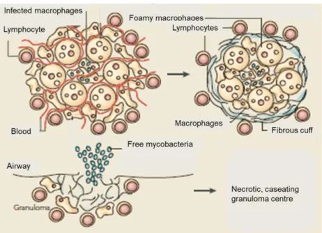
Briefly, alveolar macrophages trigger an inflammatory response immediately after taking up M. tuberculosis. The bacilli that manage to escape the initial intracellular destruction can multiply and cause the apoptosis of macrophage cells (28). The apoptotic cells participate in host defence by inducing the activation of CD4 and CD8 T-cells, which migrate to the site of inflammation, i.e. the lung (32,44). The presence of activated T cells at the infection site attracts other cells of the immune system that are organised in a highly specific way to form the so-called granuloma or tubercle (32,45). The granuloma basically consists of a cluster of infected macrophages surrounded by foamy macrophages, and other defence cells, covered by lymphocytes in association with a fibrous cuff of collagen and extracellular matrix components (23). A schematic representation of the granuloma structure is depicted in Figure 1.2.
Figure 1.2. Schematic representation of the granuloma structure. Adapted from (23).
In some cases, with granuloma formation, most tubercle bacilli are killed and disease progression is halted (32). In others, the bacilli resist the hostile
Nano and microparticles as carriers for alveolar macrophage targeting in pulmonary tuberculosis therapy
9 environment of the granuloma (e.g. hypoxia, nutrient deficiency, acidic pH) and induce a state of bacteriostasis, allowing them to survive during extended periods of latency (46). In patients with latent TB, reactivation occurs when the granuloma wall shatters and releases thousands of viable/infectious mycobacteria into the host airways, a process known as caseation (23).
The probability of developing active clinical TB (or primary infection) after being infected with M. tuberculosis is small. Less than 10% of infected individuals develop symptoms and signs of active disease over a lifetime. However, containment usually fails when the person is immunocompromised (primary TB cases) or when the immune status of the host changes (reactivation of latent TB), which is usually a consequence of ageing, malnutrition, treatment with corticosteroids, alcohol or drug abuse, co-infection with HIV or basically any condition that reduces the number or impairs the function of CD4 T cells, mainly (23,42). Not surprisingly, infection with HIV is the most potent of these risk factors, with the risk of people infected with HIV developing TB being more than 20-times greater than that of people not infected with HIV (47).
1.1.2 Diagnosis and treatment of tuberculosis
The definitive test for TB disease is the detection of M. tuberculosis bacilli in clinical specimens from symptomatic patients. Therefore, sputum microscopy and culture, with subsequent drug-susceptibility testing are currently recommended as standard methods to diagnose pulmonary TB (48).
Although much work has been done to develop modern diagnostic tools (e.g. nucleic acid amplification tests), direct sputum smear microscopy remains necessary and it is recommended to evaluate the response of the disease to treatment (49). Overall, detection of TB relies heavily on direct smear microscopy, solid culture and chest radiography, tools that often perform poorly, and require infrastructure frequently unavailable in the periphery of the health system where patients first seek care (50). Moreover, multiple investigations may be necessary over a period of weeks or months before a diagnosis is made. Delay in diagnosis is crucial to both disease prognosis at the individual level and transmission within the community. Most transmissions occur between the onset of a cough and initiation
Nano and microparticles as carriers for alveolar macrophage targeting in pulmonary tuberculosis therapy
10 of treatment. Therefore, early diagnosis and immediate initiation of treatment are essential for an effective TB control program (51,52).
Currently, the recommended treatment for new cases of drug-susceptible TB is a 6-month regimen of four first-line drugs: isoniazid (INH), rifampicin, pyrazinamide, and ethambutol. It involves an initial phase of a four-drug regimen for the first 2 months followed by a continuation phase of two drugs: INH and rifampicin for the next 4 months. Up to 95% of people with drug-susceptible TB reach the cure with this four-drug regimen (53). In cases in which rifampicin resistance is suspected, other rifamycin derivatives, such as rifapentine and rifabutin (RFB) may be used (54). Moreover, RFB is the first-line anti-TB drug recommended for patients who are taking medications incompatible with rifampicin, such as antiretroviral drugs (55).
In summary, TB is a curable disease as long as a rapid diagnosis is performed, and a proper antibiotic therapy is established and followed. Although advances have been made in TB therapy, standard TB regimens are limited and cause severe side effects that include ototoxicity, hepatotoxicity and nephrotoxicity and hyperuricemia, especially in the treatment of drug-resistant TB (56,57). For this reason, new TB drugs have started to emerge from the pipeline, and combination regimens that include new compounds are being tested in clinical trials. However, the evolution of resistance will inevitably follow the introduction of new drugs, making it unlikely that existing agents are removed from clinical use.
The situation has, thus, come to a point where the development of novel therapeutic strategies is urgently needed. In this regard, the advent of drug delivery systems may hold the key to the prevention and treatment of TB disease. The delivery of anti-TB drugs directly to the site of infection would potentially enable the reduction of dosing frequency, possibly shortening treatment duration, thereby avoiding or reducing systemic side effects and improving patient compliance (12).
1.2 Pulmonary drug delivery
Site-specific delivery of drugs permits delivering drugs to a patient in a very specific manner that allows concentrating the drugs in the site of interest while reducing their concentration in the remaining tissues (58). This not only improves the inherent efficacy of drugs, but also potentially reduces side effects. The selective
Nano and microparticles as carriers for alveolar macrophage targeting in pulmonary tuberculosis therapy
11 delivery of drugs is, therefore, very attractive and a real need, as it provides one of the most potent ways to improve the therapeutic effect of drugs.
Since it is very difficult for drug molecules to reach with high efficiency the intended destination in the complex cellular network of an organism, the assistance of a drug carrier may strongly benefit the process. Drug delivery systems offer an intelligent approach for carrying and protecting drugs, while in many cases providing simultaneous modulation of their release and absorption. However, their success is frequently limited by short residence times at the site of absorption or action. For this reason, it is deemed advantageous to potentiate the intimate contact of drug delivery systems with the referred sites.
In light of this, particulate carriers are one of the most used classes of drug delivery systems. They offer many advantages that include the possibility of tailoring particle sizes and surface characteristics, improvement of drug pharmacokinetics and pharmacodynamics, and the possibility for delivery through various routes of administration, including the respiratory route (59,60). As it is well demonstrated in Figure 1.3, the pulmonary delivery of drugs has been increasingly studied over the last two decades, given the advantages over traditional routes.
Figure 1.3. Number of scientific papers published on the topic “pulmonary drug delivery” on ISI Web of Science (October/2018), as a function of publication years.
In general, the main reasons why the lung is an attractive route for drug delivery are non-invasiveness, the possibility of direct delivery to the site of action when treating pulmonary diseases, avoidance of the first-pass metabolism, and the
0 200 400 600 800 1000 1200 1400 1600 1800 1990-1994 1995-1999 2000-2004 2005-2009 2010-2014 2015-to date Num be r of pu bl is he d pa pe rs
Nano and microparticles as carriers for alveolar macrophage targeting in pulmonary tuberculosis therapy
12 availability of a large surface area for systemic delivery of drugs, along with highly vascularised epithelium (61). Besides, drug efflux transporters and metabolising enzymes are present in the lung at much lower levels comparing with the gastrointestinal tract, which is the most common route of delivery (62). Table 1.1 summarises the most relevant advantages and limitations of drug delivery by inhalation.
Table 1.1. The most relevant advantages and limitations of pulmonary drug delivery.
Advantages Limitations
Non-invasive route Respiratory tract is structurally complex Avoidance of the first-pass metabolism
and limitation of side effects Lung defence mechanisms
Extensive vascularisation Specific aerodynamic requirements needed to reach each zone of respiratory tree
Possibility of administering lower drug doses
Inhalation devices and special dosage form are required for drug delivery
Despite marked advantages of pulmonary administration, delivery of free drugs is not easy and their efficiency has been potentiated by means of the use of drug carriers, which need to be specifically designed to meet aerodynamic requirements (63). Nevertheless, lung drug delivery is not only affected by characteristics of the inhalable formulation, further having great contribution from biophysical and physiological factors, e.g. aerosol particle size and breathing manoeuvre (inspired volume, inspiratory flow, and end-inspiratory breath holding time). Importantly, the physical and biochemical stability of pharmaceutical formulations designed for aerosolisation (dry powder, suspensions or solutions) are preconditions for the administration of adequate and reproducible drug doses via the pulmonary route (64). In parallel, inhalation devices are required to be designed to overcome all the obstacles and potentiate the performance of formulations.
Nano and microparticles as carriers for alveolar macrophage targeting in pulmonary tuberculosis therapy
13
1.2.1 Challenges for pulmonary delivery
The lung presents several defence mechanisms that comprise relevant limitations to drug delivery, including cough, mucociliary apparatus and airway anatomic barriers. Understanding the complex architecture of the respiratory tract is important because it is a biophysical factor affecting the deposition of particulate drug delivery systems in the airways (64). As depicted in Figure 1.4, there are several mechanisms involved in aerosol transport and deposition in the human lung, including inertial impaction, gravitational sedimentation and Brownian diffusion, which are the most prevalent (65). Inertial impaction refers to the inability of particles (larger than 5 µm) to adjust its course according to the sudden change in air flow direction at airway bifurcations (65,66). Gravitational sedimentation is the settling of particles under the action of gravity and occurs mainly in small airways and alveolar region. Deposition by Brownian diffusion results from the random motion of particles of size lower than 0.5 µm, as a consequence of their collisions with gas molecules. It increases with decreasing particle size and is the dominant mechanism of deposition for particles in this size range (65,67).
Figure 1.4. Scheme describing the main mechanisms affecting aerosol transport and deposition in the human lung. Adapted from (68).
As a consequence of the physical forces acting on aerosol particles, their deposition in the lungs is highly dependent on diameter (66). The size of particles aimed at inhalation is conventionally defined as the Daer, which is the diameter of
Nano and microparticles as carriers for alveolar macrophage targeting in pulmonary tuberculosis therapy
14 a spherical particle with unit density that has the same settling velocity as the particle in consideration (69). Some equations have been proposed to be used in the theoretical calculation of Daer of aerosol particles. One of the most used is displayed below (70,71):
Daer = d
( )
where d is the particle geometric diameter, ρ is the particle density, ρ0 = 1 g/cm3
and λ is the dynamic shape factor of the particle, which is 1 in the case of spherical particles (72). Nevertheless, accurate determination of Daer is only possible experimentally, which is routinely performed using techniques based on inertial impaction (e.g. cascade impactors, twin-stage impinger, etc.) as indicated in the European Pharmacopeia (73). Although several terminologies are used to characterise this parameter, the mass median aerodynamic diameter (MMAD) is that commonly used after characterisation by cascade impaction. MMAD represents the median of the distribution of airborne particle mass with respect to the aerodynamic diameter. This is usually accompanied by the geometric standard deviation, which characterises the variability of the particle size distribution, referring to mono- or polydisperse aerosols (66,74). Additionally, the fine particle fraction (FPF) is defined as the percentage relative to the total quantity of drug collected in the impactor that has the size equal or lower than 5 µm (75).
In other words, aerosols with larger MMADs will deposit higher in the respiratory tract and a polydisperse aerosol is also more likely to show greater deposition in the tracheobronchial region than a monodisperse aerosol of the same MMAD (66). The MMAD of aerosols is, therefore, critical factors in determining the deposition patterns within the lung, although other conditions will also affect this behaviour, as detailed above. Figure 1.5 represents the relationship between Daer and deposition of aerosol particles in the human respiratory tract.
λ ρ0
Nano and microparticles as carriers for alveolar macrophage targeting in pulmonary tuberculosis therapy
15
Figure 1.5. Relationship between aerodynamic diameter and deposition of aerosol particles in the human respiratory tract. Adapted from (27,76).
In brief, particles greater than 5 μm in diameter will mostly impact in the upper airways and are rapidly removed by coughing and mucociliary processes. In turn, smaller particles in the size range of 1–5 μm may escape impaction in the upper airways and will deposit in the lower tracheobronchial and alveolar regions. On the other hand, particles <1 μm may not be deposited at all, since many will be removed from the lung on the exhaled air stream before sedimentation can occur (66,77). Therefore, to reach the lower respiratory tract, particularly the alveoli, and optimise pulmonary drug deposition, aerosols should have aerodynamic diameters <5 µm. (65,78).
1.3 Nano and microparticles as drug delivery systems for inhalation purposes
Nanoparticles (NP) and microparticles (MP) are the most cited of the particulate carriers, in most cases presenting a matrix composed of polymeric materials. The International Organization for Standardization (ISO) defines nanoparticles as particles having at least one dimension below 100 nm (79). The definition is, however, not consensual in the area of drug delivery and the term is frequently used for spherical particles with submicron size (<1000 nm) (80,81). Microparticles present diameters in the micrometre range (from 1 μm to 1000 μm). Structurally, these systems are typically divided into two categories, according to a classification that is now widely accepted: nanocapsules/microcapsules, when the
Nano and microparticles as carriers for alveolar macrophage targeting in pulmonary tuberculosis therapy
16 drug is mainly confined to a cavity surrounded by a polymer membrane (shell); and nanospheres/microspheres when the drug is dispersed within the polymeric matrix (82). The drug release mechanism from carriers primarily involves drug diffusion through the carrier material. Additional mechanisms depend on the considered drug carrier. For instance, the drug may be released through pores present in the carrier or by water penetration into the systems or by hydrolysis/erosion of polymeric matrix (83).
In general, carrier systems aim to minimise drug degradation and loss, enhance the solubility of poorly soluble drugs, prevent adverse effects (targeted therapy) and increase the availability of the drug at the target site. To reach these goals, carriers can be designed to slowly degrade, react to stimuli or to be site-specific. Targeted therapy generally requires lower total doses to achieve clinically effective results, comparing with conventional approaches (62). In this context, the use of polymeric systems offers potential advantages over free drug formulations or even other carriers (such as liposomes), considering their higher stability and the possibility to modulate the systems for targeted delivery (84). For instance, inhalable PLGA particles containing rapamycin were more effective in clearing intracellular mycobacteria and presented lower cytotoxicity compared to the free drug (85). Nonetheless, the development of polymeric drug carriers presents some limitations, including lower reproducibility compared to conventional formulations, higher cost of materials and processing, and even the environmental impact of degradation products (86).
There are many methods available to prepare nano and microparticles and it is not uncommon that the same method can be used to prepare both types of particles. Obtaining one system or the other naturally depends on specific conditions of the process, such as the used concentrations, intensity of stirring, etc. (87). Methods such as polyelectrolyte complexation, ionic gelation, solvent evaporation, coacervation, supercritical fluids technology and spray-drying, have been frequently reported (88,89). Polyelectrolyte complexation is a widely used technique to obtain polymeric carrier systems, because it generally offers simple and mild preparation processes that do not involve harsh conditions. The absence of additives results in low toxicity and low cost. This technique is based on electrostatic interactions between oppositely charged polymers, which allow spontaneous formation of complexes (90,91). Another method commonly used is coacervation, that also