Antige nic characte rizatio n o f Brazilian
bo vine viral diarrhe a virus iso late s
by m o no clo nal antibo die s and
cro ss-ne utralizatio n
Departamento de Medicina Veterinária Preventiva, Microbiologia e Parasitologia, Universidade Federal de Santa Maria, Santa Maria, RS, Brasil
S.A. Botton, A.M. da-Silva, M.C.S. Brum, R. Weiblen and E.F. Flores
Abstract
Nineteen Brazilian isolates of bovine viral diarrhea virus (BVDV) were characterized antigenically with a panel of 19 monoclonal anti-bodies (mAbs) (Corapi WV, Donis RO and Dubovi EJ (1990) Ameri-can Journal of Veterinary Research, 55: 1388-1394). Eight isolates were further characterized by cross-neutralization using sheep mono-specific antisera. Analysis of mAb binding to viral antigens by indirect immunofluorescence revealed distinct patterns of reactivity among the native viruses. Local isolates differed from the prototype Singer strain in recognition by up to 14 mAbs. Only two mAbs - one to the non-structural protein NS23/p125 and another to the envelope glyco-protein E0/gp48 - recognized 100% of the isolates. No isolate was recognized by more than 14 mAbs and twelve viruses reacted with 10 or less mAbs. mAbs to the major envelope glycoprotein E2/gp53 revealed a particularly high degree of antigenic variability in this glycoprotein. Nine isolates (47.3%) reacted with three or less of 10 E2/ gp53 mAbs, and one isolate was not recognized by any of these mAbs. Virus-specific antisera to eight isolates plus three standard BVDV strains raised in lambs had virus-neutralizing titers ranging from 400 to 3200 against the homologous virus. Nonetheless, many antisera showed significantly reduced neutralizing activity when tested against heterologous viruses. Up to 128-fold differences in cross-neutraliza-tion titers were observed for some pairs of viruses. When the coeffi-cient of antigenic similarity (R) was calculated, 49 of 66 comparisons (74.24%) between viruses resulted in R values that antigenically distinguish strains. Moreover, one isolate had R values suggesting that it belongs to a distinct serologic group. The marked antigenic diversity observed among Brazilian BVDV isolates should be considered when planning diagnostic and immunization strategies.
Co rre spo nde nce E.F. Flores
Departamento de Medicina Veterinária Preventiva, UFSM
97015-900 Santa Maria, RS Brasil
Fax: + 55-55-220-8257 E-mail: flores@ ccr.ufsm.br
Research supported by MCT, CNPq, CAPES and FINEP (PRO NEX em
Virologia Veterinária, No. 215/96) and FAPERGS (No. 96/1471.6). S.A. Botton and A.M. Silva are recipients of CAPES and CNPq scholarships, respectively. E.F. Flores and R. Weiblen are recipients of CNPq fellowships
(Nos. 352386/96 and 520011/95, respectively).
Received March 20, 1998 Accepted August 13, 1998
Ke y wo rds
•Bovine viral diarrhea virus
•BVDV
•Monoclonal antibodies
•Cross-neutralization
Intro ductio n
Bovine viral diarrhea virus (BVDV) is an important cattle pathogen which causes sig-nificant losses to the livestock industry around the world (1,2). BVDV is currently classi-fied within the Flaviviridae, genus Pestivirus, along with the classical swine fever virus (CSFV) and border disease virus (BDV) of sheep (3). Pestiviruses are small enveloped, positive-sense RNA viruses (4,5) which natu-rally infect swine and domestic and wild ruminants (6). Pestiviruses display consider-able genetic and antigenic diversity within the genus, although all members cross-react serologically to some extent (7).
BVDV infections in cattle are associated with a variety of clinical manifestations in-cluding inapparent infections, gastroenteric, respiratory, and hemorrhagic syndromes and the deadly mucosal disease (MD) (8-10). Infection of pregnant cows by the noncyto-pathic BVDV biotype may result in a variety of outcomes: early or late embryonic death, abortion or mummification, malformations, stillbirth and birth of weak and non-thriving calves (10). Infection of fetuses between 40 and 120 days of gestation often leads to fetal immunotolerance, resulting in the birth of immunotolerant persistently infected (PI) calves (9,10). Most persistently infected ani-mals develop and die of mucosal disease within the first 6 to 24 months of life (2,9). Cytopathic and noncytopathic BVDV bio-types are usually isolated from animals af-fected by MD (9). Cytopathic BVD viruses originate from their noncytopathic counter-part through mutations, recombinations or rearrangements in the viral RNA genome (11,12).
Serological studies have shown that al-though BVDV field isolates are antigeni-cally related to each other, antigenic differ-ences can be readily detected among viruses (7,13-15). These antigenic differences have been demonstrated by the use of monoclonal antibodies (mAbs) and by in vitro and in vivo
cross-neutralization studies (7,13-16). How-ever, in spite of the antigenic variability, no clear definition of serotypes has been pos-sible to date (7,16). The marked antigenic variability of BVD viruses allows differen-tiation between strains, but also represents a potential problem for diagnosis and immuni-zation strategies (7,16).
Very little is known about BVDV infec-tion and disease in Brazil. The presence of BVDV infection in the country was first indicated by clinical reports and serological studies conducted in the 60’s and 70’s (17-19). Thereafter, serologic and virologic data have confirmed the widespread distribution of BVDV infection among Brazilian cattle (20,21). To date, approximately 20 BVD isolates have been isolated in Brazil (Botton SA and Flores EF, unpublished results). The present article reports the antigenic charac-terization of 19 of these isolates. Analysis of reactivity with a panel of mAbs and cross-neutralization studies revealed a marked an-tigenic diversity among these viruses. This antigenic diversity may have important im-plications for epidemiologic studies and for the diagnosis and control of BVDV infection in Brazil.
Mate rial and Me tho ds
Virus iso latio n, viruse s and ce lls
Col-lection. Rockville, MD) and submitted to three blind passages at 48-h intervals. At the end of the third passage, the inoculated cells were submitted to an indirect immunofluo-rescence assay (IFA) for viral antigens. IFA was performed in acetone-fixed cells, using a pool of anti-BVDV Singer mAbs (22) as the primary antibody, and an FITC-conju-gated anti-mouse IgG (Sigma, St. Louis, MO, USA) as the secondary antibody. The slides were counterstained with Evans blue, mounted with PBS:glycerol (1:1) and ob-served in an epifluorescence microscope. Samples positive for BVDV antigens were identified and the respective viruses were further cultured in MDBK cells.
Re activity with mo no clo nal antibo die s
A panel of 19 mAbs to the prototype BVDV Singer strain (22) was used to char-acterize the Brazilian isolates. The mAbs were provided by Dr. Ruben Donis, Depart-ment of Veterinary and Biomedical Sciences, University of Nebraska at Lincoln, Lincoln, NE, USA. The ability of each individual mAb to recognize and bind to viral antigens was assayed by IFA. Cells infected with each isolate were incubated with individual mAbs as the primary antibody, followed by incubation with the secondary antibody as described above. Either ascites fluid (1:500 in phosphate buffered saline; PBS) or super-natants of hybridoma cultures were used. Mock-infected cells, cells infected with the prototype Singer strain and stained with each individual mAb, and cells infected with each isolate and stained with a pool of mAbs were used as controls.
Pro ductio n o f virus-mo no spe cific antise ra
Virus-specific antisera against three ref-erence BVDV strains (Singer, NADL and Oregon/C24v) and eight Brazilian isolates were produced in lambs. Eleven BVDV-seronegative, 6- to 8-month-old lambs were
inoculated intranasally and intramuscularly with approximately 107 TCID50 (tissue cul-ture median infectious dose) of each virus. The animals were housed individually until the second serum collection. Blood was col-lected at 15 and 30 days post-inoculation (pi) to obtain serum which was heat inactivated at 56oC for 30 min prior to virus neutraliza-tion (VN) assays.
Virus-ne utralizatio n assays
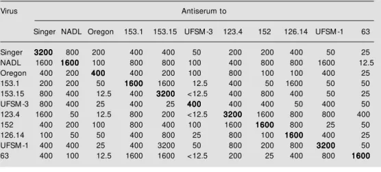
Individual serum samples collected at 15 and 30 days pi were initially titrated against their homologous viruses in a standard VN assay (15). The serum samples obtained at 30 days pi (which had the highest VN titer) were titrated against each heterologous virus and the neutralization end points were deter-mined for each virus-serum combination. VN assays were performed in polystyrene 96-well plates, using doubling two-fold dilu-tions of each serum (starting at 1:12.5) against a fixed dose of virus (approximately 100 TCID50/well) and MDBK cells as indicators. Readings were performed after 96 h of incu-bation. Virus growth or neutralization was monitored by microscopic examination of the cytopathic effect for the cytopathic strains (Singer, NADL and Oregon/C24v) or by staining the indicator cells for viral antigens by IFA for the noncytopathic viruses. When-ever a certain serum was titrated with differ-ent viruses, the VN tests were performed at the same time on the same plate, using the same preparation of MDBK cells. The cross-neutralization tests yielded 121 values, cor-responding to the VN titer of each virus-serum combination. These values are re-ported as the reciprocal of the highest dilu-tion of serum capable of preventing viral replication (Table 2).
Calculatio n o f antige nic similarity
calculate the antigenic similarity among the isolates. The neutralization end points for each virus-serum combination were com-bined as described by Howard et al. (15) to obtain a percent relatedness value for each pair of viruses. The coefficient of antigenic similarity (R) was calculated according to Archetti and Horsfall (23) using the follow-ing formula:
Re sults
BVD V iso late s
BVD viruses were isolated and identified in 0.75% (11/1396) of the blood samples collected from bovine fetuses. One addi-tional virus was isolated from a clinical case
of gastroenteric disease and another from the serum of an apparently healthy calf from a dairy herd. All these isolates were of the noncytopathic biotype. The origin and pre-liminary characterization of these and other 6 viruses isolated in other laboratories will be described elsewhere (Botton SA and Flores EF, unpublished data).
Re activity with mo no clo nal antibo die s
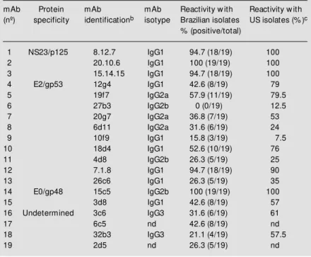
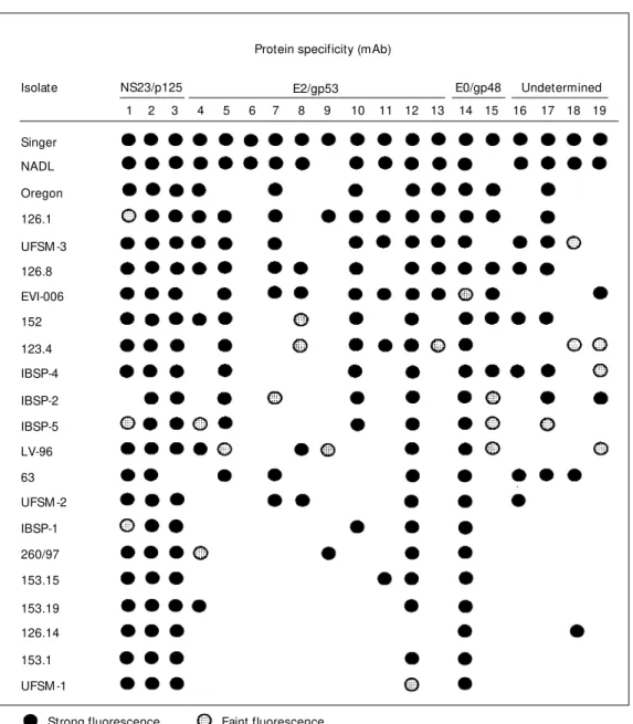
The protein specificity of the mAbs used in this study and a summary of their reactiv-ity with Brazilian BVDV isolates are pre-sented in Table 1. The spectrum of reactivity of these mAbs with 81 North-American (US) BVDV isolates is also presented (22,24). mAbs to the non-structural protein NS23/ p125 (and its cleavage product NS3/p80) showed a broad spectrum of reactivity, rec-ognizing 94.7 to 100% of the isolates both in the US and Brazilian groups of viruses. One mAb to the envelope glycoprotein E0/gp48 (15c5) also reacted with 100% of the iso-lates. mAbs to the major envelope glycopro-tein E2/gp53 showed a high variation in the spectrum of reactivity with native viruses. Except for a mAb that recognized 18 (94.7%) native viruses (and 90% of the US’s), all other nine mAbs reacted with zero to 11 isolates (57.9%). Most of the E2/gp53 mAbs recognized a higher number of US than Bra-zilian isolates, although equivalent and lower recognition rates of US viruses were also observed (Table 1). Four mAbs of undeter-mined protein specificity displayed a vari-able spectrum of reactivity recognizing from 21.1 to 47.4% of the native isolates (Table 1). The profile of reactivity of the 19 mAbs with each Brazilian isolate plus the standard US BVDV strains is presented in Figure 1.
Virus-ne utralizatio n
Virus-specific antisera produced in lambs against three BVDV standard strains and 8
Table 1 - Reactivity of a panel of monoclonal antibodies (mAb) w ith Brazilian isolates of bovine viral diarrhea virus (BVDV)a.
aAssayed by an indirect immunofluorescence (IFA) assay. bPanel of monoclonal
anti-bodies to the standard BVDV Singer produced and characterized by Corapi et al. (22).
cReported by Corapi et al. (22) and Carman et al. (24). nd, Not determined.
mAb Protein mAb mAb Reactivity w ith Reactivity w ith
(nº) specificity identificationb isotype Brazilian isolates US isolates (% )c
% (positive/total)
1 NS23/p125 8.12.7 IgG1 94.7 (18/19) 100
2 20.10.6 IgG1 100 (19/19) 100
3 15.14.15 IgG1 94.7 (18/19) 100
4 E2/gp53 12g4 IgG1 42.6 (8/19) 79
5 19f7 IgG2a 57.9 (11/19) 79.5
6 27b3 IgG2b 0 (0/19) 12.5
7 20g7 IgG2a 36.8 (7/19) 53
8 6d11 IgG2a 31.6 (6/19) 24
9 10f9 IgG1 15.8 (3/19) 7.5
10 18d4 IgG1 52.6 (10/19) 76
11 4d8 IgG2b 26.3 (5/19) 25
12 7.1.8 IgG1 94.7 (18/19) 90
13 26c6 IgG1 26.3 (5/19) 35
14 E0/gp48 15c5 IgG2b 100 (19/19) 100
15 3d8 IgG1 42.6 (8/19) 57
16 Undetermined 3c6 IgG3 31.6 (6/19) 61
17 6c5 nd 42.6 (8/19) nd
18 32b3 IgG3 21.1 (4/19) 57.5
19 2d5 nd 26.3 (5/19) nd
R = 100 x titer strain I with antiserum II x titer strain II with antiserum I
native isolates had moderate to high levels of neutralizing antibodies. The VN titers against the homologous virus in sera col-lected at 30 days pi ranged from 400 to 3200 (Table 2). Testing these sera against heterol-ogous viruses, however, yielded variable and often significantly lower VN titers. Sixty-four to 128-fold reductions in titers were observed for some pairs of heterologous se-rum-virus combinations. Antisera UFSM-1 and 153-1 showed a reduction from 3200 to 25 when tested against isolates 152 and Singer/UFSM-3, respectively. Antiserum 63
also showed a significant reduction of neu-tralizing activity when tested against most of the viruses examined. Eight-, 16- and 32-fold differences in VN titers were common among the different combinations of heter-ologous viruses (Table 2).
The VN values for each pair of heterolo-gous viruses were used in order to calculate a coefficient of antigenic similarity (R) be-tween viruses (Table 3). An R value of ≤25 represents four-fold differences in titers be-tween homologous and heterologous anti-sera, indicating significant antigenic
differ-Protein specificity (mAb)
Isolate NS23/p125 E0/gp48 Undetermined
1 2 3 4 5 6 7 8 9 10 11 12 13 14 15 16 17 18 19
Singer
NADL
Oregon
126.1
UFSM -3
126.8
EVI-006
152
123.4
IBSP-4
IBSP-2
IBSP-5
LV-96
63
UFSM -2
IBSP-1
260/97
153.15
153.19
126.14
153.1
UFSM -1
Strong fluorescence Faint fluorescence
Figure 1 - Reactivity of a panel of monoclonal antibodies (mAb) to t he prot ot ype BVDV Singer strain w ith Brazilian BVDV iso-lates in an indirect immunofluo-rescence assay.
ences that are greater than would be ex-pected from the variation in the assay (15,25). R values ≤5 are obtained when >20-fold differences in titers occur between homolo-gous and heterolohomolo-gous antisera and have been suggested as the threshold to differenti-ate serotypes. Forty-nine (74.24%) of the 66 R values calculated in this study gave values
≤25. These results corroborate those from the monoclonal antibody analysis, which re-vealed a considerable antigenic variation within this group of viruses. Ten (15.1%) of
the calculated R values were <5. Five of them (50%) involved isolate 63, and 3 in-volved isolate UFSM-3. Three other viruses (UFSM-1, 152 and 153.15) yielded two R values below 5. These results suggest that within an antigenically variable group of viruses isolate 63 is more distantly related and may belong to a distinct serologic group.
D iscussio n
Much of the early work on BVDV
fo-Table 2 - Virus-neutralizing (VN) antibody titers of monospecific sheep antisera against homologous and heterologous BVDVa.
aReciprocal of the highest dilution of serum capable of neutralizing 100 TCID50 of the respective virus.
Virus Antiserum to
Singer NADL Oregon 153.1 153.15 UFSM -3 123.4 152 126.14 UFSM -1 63
Singer 32003200320032003200 800 200 400 400 50 200 200 400 50 25
NADL 1600 16001600160016001600 100 800 800 100 400 800 800 1600 12.5
Oregon 400 200 400400400400400 400 200 100 800 100 100 400 25
153.1 200 200 50 16001600160016001600 1600 12.5 400 50 1600 50 50
153.15 800 400 12.5 400 32003200320032003200 <12.5 400 800 400 50 25
UFSM -3 800 400 25 400 25 400400400400400 400 400 50 400 50
123.4 1600 50 12.5 800 200 <12.5 32003200320032003200 1600 800 800 400
152 400 200 100 800 400 100 1600 16001600160016001600 800 25 50
126.14 100 50 50 400 800 25 800 100 16001600160016001600 400 25
UFSM -1 400 400 25 400 3200 50 800 200 800 32003200320032003200 50
63 400 100 12.5 1600 1600 <12.5 200 25 400 800 16001600160016001600
Table 3 - Coefficient of antigenic similarity (R) for standard strains and Brazilian BVDV isolates, calculated w ith the VN values for homologous and heterologous pairs of virusesa.
aAccording to Archetti and Horsfall (23). bR values <5 are underlined.
Virus Antiserum to
Singer NADL Oregon 153.1 153.15 UFSM -3 123.4 152 126.14 UFSM -1 63
Singer 100
NADL 50 100
Oregon 25 17.68 100
153.1 12.5 25 17.68 100
153.15 17.68 25 4.42b 35.3 100
UFSM -3 17.68 25 12.5 8.84 15.6 100
123.4 17.68 6.25 8.84 25 8.84 6.25 100
152 12.5 25 12.5 12.5 25 25 70.71 100
126.14 8.84 12.5 8.84 50 25 4.42 35.35 17.6 100
UFSM -1 4.42 3535 8.84 6.25 12.5 12.5 25 3.12 25 100
cused on the antigenic similarities rather than on the differences among isolates. The first in vivo cross-protection studies showed that two isolates from different regions of the US were antigenically related to each other, suggesting that immunization with one isolate would protect against heterologous BVDV (25). Likewise, in vitro VN tests demonstrated that BVDV isolated from dif-ferent clinical syndromes were antigenically related, suggesting that distinct serotypes did not exist (14,26). Thereafter, observa-tions of antigenic relaobserva-tionships were extended to the other Pestiviruses - classical swine fever virus (CSFV) and border disease virus (BDV) (7,27). The concepts of antigenic similarity and of the existence of a single serologic type for BVDV persisted until re-cently, so that most BVDV vaccines pro-duced up to the 90’s were based on a single BVDV isolate.
It was only with the identification of cytopathic strains that the antigenic diversity of BVDV could be more thoroughly exam-ined. The availability of cytopathic viruses greatly facilitated and improved the in vitro virus-neutralization studies (7,16). Nonethe-less, more definitive and precise character-ization of the antigenic variability was only possible with the development of BVDV-mAbs in the late 1980’s (22,27) and more recently, as more BVDV gene sequences became available (28-30).
Production of BVDV-specific mAbs with standard laboratory strains has resulted in two major groups of mAbs: 1) group-specif-ic, which recognize most - if not all - isolates and even other Pestiviruses, and 2) type or strain-specific, which allow differentiation and discrimination among isolates. The first group of mAbs is mainly directed against highly conserved epitopes, mostly in the soluble, non-structural polypeptide NS23/ p125 (7). NS23/p125 is a multifunctional protein whose amino-terminal half (NS3/ p80) is the most conserved polypeptide a-mong Pestiviruses (12). As many epitopes
across their sequence are conserved, mAbs to NS3/p80 have a wide application in diag-nostic tests by enhancing the accuracy of identification of these viruses in clinical material (16,22).
Type or strain-specific mAbs are mainly directed against the two major envelope gly-coproteins (E0/gp48 and E2/gp53). E2/gp53 is thought to be involved in the binding of virions to cellular receptors in such a way that most mAbs directed against it are neu-tralizing (12,31,32). The E2/gp53 gene con-tains one of the three hypervariable regions of the viral genome (12,31). Consequently, many E2/gp53 mAbs are directed against epitopes that are differently expressed a-mong isolates, failing to recognize the equiva-lent epitopes in field isolates. The pattern of reactivity of 10 E2/gp53 mAbs, however, demonstrated that the variability is not uni-formly distributed throughout the protein. mAbs reacting with most isolates, such as mAb 7.1.8, are probably directed against conserved epitopes which are under strong structural constraints or simply not subjected to immunologic pressure, or both. In con-trast, mAbs reacting with few viruses (10f 9, 27b3) are probably directed to epitopes that are highly variable due to immunological pressure by neutralizing antibodies. These strain-specific or discriminatory mAbs have been widely used and have proven to be extremely useful in several areas of research (7,16,22,27,30-32).
viruses examined. Nonetheless, it was pos-sible to identify discrete clusters of viruses whose antigenic profile may provide the ba-sis for a tentative group allocation. Patterns of reactivity revealed a group of viruses including UFSM-1, 153.1, 153.15, 153.19 and 126.14 which are very distinct from the reference strains. The main variability of these viruses is related to the lack of recogni-tion by most E2/gp53 mAbs. These isolates also showed an almost identical mAb reac-tivity profile within the group. The reacreac-tivity profile of the other isolates was highly di-verse, not allowing an easy identification of distinct groups of antigenically related vi-ruses.
Neutralization studies using monospe-cific bovine or sheep antisera have detected up to 100-fold differences in cross-neutral-ization titers among BVDV field isolates (15,30,33). In the present study, a few 68-and 128-fold differences were observed for some pairs of viruses, whereas differences of 16- and 32-fold were common throughout the comparisons. Interestingly, many of the 16-, 32- and 64-fold differences in cross-neutralization titers involved the Singer strain (virus/serum) against the isolates (or serum) mentioned above. The low cross-reactivity observed among many of the pairs examined reflects the marked antigenic diversity de-tected by mAb analysis, suggesting that the immune response to a given virus may not be protective against an antigenically distinct isolate.
Recently, genotypically different BVDV strains have been associated with high mor-tality in calves in the US and Canada (8,28,29). These viruses, tentatively termed BVDV type II (28,29), display a low sero-logic cross-reactivity with the classical BVDV strains, now termed type I BVDV (29). In our study, VN titers involving isolate 63 showed significant variations all across the comparisons, with its antiserum showing a poor neutralizing activity against most of the viruses examined. The calculation of
antigenic similarity reinforced the marked antigenic diversity and indicated the exist-ence of rather distinct viruses among the Brazilian isolates. R values that antigeni-cally distinguish strains (<25) were observed for several pairs of viruses, most consis-tently for isolates 63 (all its R values were below 25), UFSM-1 and 3, 126.14 and 123.4. Furthermore, R values that suggest alloca-tion of viruses to distinct serologic groups were observed for isolate 63 (five R values below 5) but also for UFSM-1, UFSM-3 and 123.4. Noteworthy are the low R values obtained for isolate 63 when compared to the Singer, NADL and Oregon/C24v strains. These laboratory reference strains have been unequivocally classified as BVDV type I (28,29) and have shown a low serological cross-reactivity with North-American type II BVD viruses (29). The quite distinct anti-genic profile displayed by isolate 63 and its distant antigenic relatedness to reference type I BVDV strongly suggest that this virus be-longs to a distinct serologic group within BVDV. Genomic sequencing and analysis of these isolates are currently underway and will better define a possible group allocation for Brazilian BVDV isolates.
isolates that are poorly neutralized by anti-sera to reference vaccine strains raises the question about the degree of protection con-ferred by such vaccines. Since most BVDV vaccines available in Brazil are based on a single US or European strain, it is pertinent to question whether these vaccines will pro-tect against native viruses.
The antigenic characterization of an ad-ditional number of isolates is needed to pro-vide a more comprehensive understanding of the antigenic characteristics of BVDV present in Brazilian cattle. Nevertheless, the antigenic variability demonstrated in this study indicates that a reevaluation of diag-nostic procedures, vaccine manufacturing and immunization strategies may be neces-sary to efficiently diagnose and control
BVDV infection in Brazil.
Ackno wle dgm e nts
We thank Dr. Ruben Donis (University of Nebraska at Lincoln, Lincoln, NE, USA) for providing the mAbs and the reference BVDV strains. Isolates IBSP-1, 2, 4 and 5 were isolated and provided by Dr. E. Maristela Pituco (Instituto Biológico de São Paulo, SP) and isolates LV96 and EVI-006 were provided by Dr. Valéria Moojen (Fa-culdade de Veterinária, Universidade Fed-eral do Rio Grande do Sul, Porto Alegre, RS) and Dr. Paulo M. Roehe (Centro de Pesquisas Veterinárias Desidério Finamor, Eldorado do Sul, RS), respectively.
Re fe re nce s
1. Horzinek M (1991). Pestiviruses - taxo-nomic perspectives. Archives of Virology, Suppl 3: 1-5.
2. Bolin SR (1990). The current understand-ing about the pathogenesis and clinical forms of BVD. Veterinary M edicine, 85: 1123-1132.
3. Francki RIB, Fauquet CM , Knudson DL & Brow n F (1991). Classification and nomen-clature of viruses. Fifth Report of the In-ternational Committee on the Taxonomy of Viruses. Archives of Virology, Suppl 2: 223-233.
4. Collett M S, M oenning V & Horzinek M C (1989). Recent advances in pestivirus re-search. Journal of General Virology, 70: 253-266.
5. Collett M S, Larson RE & Gold C (1988). M olecular cloning and nucleotide se-quence of the pestivirus bovine viral diar-rhea virus. Virology, 70: 253-266. 6. Nettleton PF (1990). Pestivirus infections
in other ruminants than cattle. Revue Scientifique et Technique, Office Interna-tional des Epizoties, 9: 131-145. 7. Edw ards S & Paton D (1995). Antigenic
differences among pestiviruses. Veteri-nary Clinics of North America, 11: 563-578.
8. Corapi W V, French TW & Dubovi EJ (1989). Severe t hrom bocyt openia in young calves experimentally infected w ith
noncytopathic bovine viral diarrhea virus.
Journal of Virology, 62: 2823-2827. 9. Brow nlie J (1991). The pathw ays for
bo-vine viral diarrhea virus biotypes in the pathogenesis of disease. Archives of Vi-rology, Suppl 3: 79-86.
10. Baker JC (1995). The clinical manifesta-tions of bovine viral diarrhea infecmanifesta-tions.
Veterinary Clinics of North America,11: 427-444.
11. M eyers G, Tautz N & Dubovi EJ (1991). Viral cytopathogenicity correlated w ith in-tegration of ubiquitin-coding sequences.
Virology, 180: 602-610.
12. Donis R (1995). M olecular biology of bo-vine viral diarrhea virus and its interac-tions w ith the host. Veterinary Clinics of North America,11: 393-421.
13. Fernelius AL, Lambert G & Boot GD (1971). Bovine viral diarrhea virus-host cell interactions: serotypes and their relation-ship to biotypes by cross-neutralization.
American Journal of Veterinary Research, 32: 229-236.
14. Hafez SM & Liess B (1972). Studies on bovine viral diarrhea-mucosal disease vi-rus. I. Cultural behaviour and antigenic relationship of some strains. Acta Virolo-gica, 16: 388-398.
15. How ard CJ, Brow nlie J & Clarke M C (1987). Comparison by the neutralization assay of pairs of non-cytopathogenic and
cytopathogenic strains of bovine virus di-arrhoea virus isolated from cases of mu-cosal disease. Veterinary M icrobiology, 13: 361-369.
16. Dubovi EJ (1992). Genetic diversity and BVD virus. Comparative Immunology and M icrobiology of Infectious Diseases, 15: 155-162.
17. Correa W M , Netto ZC & Barros HM (1968). Nota clínico-patológica de uma enfermidade das mucosas em São Paulo.
Arquivos do Instituto Biológico de São Paulo, 35: 141-151.
18. Correa WM , Gottschalk AF & Correa C (1972). Observações na diarréia a vírus dos bovinos no Estado de São Paulo.
Arquivos do Instituto Biológico de São Paulo, 38: 145-147.
19. Soares LA & Carvalho Pereira OA (1974). Neutralizing antibodies against bovine vi-ral diarrhea virus-mucosal disease virus in cattle sera from São Paulo, Brazil. Revista de M icrobiologia, 5: 1-5.
20. Roehe PM , Oliveira EAS & Oliveira LG (1995). Characterization of field and cell culture isolates of bovine viral diarrhea virus (BVDV) w ith monoclonal antibodies.
Anais da 5ªVirológica, Ribeirão Preto, SP, 26-29 Novembro 1995, B-6.
Presença de pestivírus e anticorpos con-t ra pescon-t ivírus em soros e culcon-t ivos celulares. Arquivo Brasileiro de M edicina Veterinária e Zootecnia, 48: 513-521. 22. Corapi WV, Donis RO & Dubovi EJ (1990).
Characterization of a panel of monoclonal antibodies and their use in the study of the antigenic diversity of bovine viral diar-rhea virus. American Journal of Veterinary Research, 51: 1388-1394.
23. Archetti I & Horsfall FL (1950). Persistent antigenic variation of influenza A viruses after incomplete neutralization in ovo w ith heterologous immune serum. Journal of Experimental M edicine, 92: 441-462. 24. Carman S, Von Dreumel T, Hazlett M ,
Alves D, Dubovi EJ, Ridpath J, Bolin SR & Tremblay R (1996). A diagnostic labora-tory in the middle of an acute outbreak of BVDV. Proceedings of the International Symposium on BVDV: A 50 year review. Ithaca, NY, June 22-24, 1996, 79-85. 25. Baker JA, York CJ, Gillespie JH & M itchell
GB (1954). Virus diarrhea in cattle. Ameri-can Journal of Veterinary Research, 15: 525-531.
26. Taylor DON, Gustaffson DP & Claflin RM (1963). Properties of some viruses of the mucosal disease-virus diarrhea complex.
American Journal of Veterinary Research, 24: 143-149.
27. Edw ards S, M oenning V & Wensvoort G (1991). The development of an interna-tional reference panel of monoclonal anti-bodies for the differentiation of hog chol-era virus from other pestiviruses. Veteri-nary M icrobiology, 29: 101-108. 28. Ridpath J, Bolin S & Dubovi E (1994).
Segregation of bovine viral diarrhea virus into genotypes. Virology, 205: 66-74. 29. Pellerin C, van den Hurk J & Lecomte J
(1994). Identification of a new group of bovine viral diarrhea virus strains associ-ated w ith severe outbreaks and high mor-talities. Virology, 203: 260-267.
30. Paton D, Sands JJ, Low ings JP, Smith JE, Ibata G & Edw ards S (1995). A proposed division of the pestivirus genus using m onoclonal ant ibodies, support ed by cross-neutralization assays and genetic sequencing. Veterinary Research, 26: 92-109.
31. Weiland E, Stark R, Haas B, Rumenapf T, M eyers G & Thiel HJ (1990). Pestivirus glycoprotein w hich induces neutralizing antibodies forms part of a disulfide-linked heterodim er. Journal of Virology, 64: 3563-3569.
32. Bolin S, M oennig V, Gourley NEK & Ridpath J (1988). M onoclonal antibodies w ith neutralizing activity segregate iso-lates of bovine viral diarrhea virus into groups. Archives of Virology, 99: 117-118. 33. Wensvoort G, Terpstra C & de Kluyver EP (1989). Characterization of porcine and some ruminant pestiviruses by cross-neu-tralization. Veterinary M icrobiology, 20: 291-306.
34. Flores EF & Donis R (1995). Isolation and characterization of a mutant M DBK cell line resistant to infection w ith bovine viral diarrhea virus due to a block in viral entry.
Virology, 208: 565-575.