RESUMO.- [Aspectos patológicos e microbiológicos de Psittaciformes de companhia infectados por Escherichia coli e Salmonella Typhimurium.] A participação de Esche-richia coli na microbiota saudável de Psicittaciformes e a de Salmonella spp. já indica possível doença. O objetivo deste estudo foi pesquisar a presença de E. coli e Salmonella spp. em psittaciformes de companhia na cidade de Fortaleza/ Ceará, traçando os aspectos de patogenicidade destas cepas através das lesões e da sensibilidade antimicrobiana. Foram necropsiados os psittaciformes de companhia
encaminha-dos ao Laboratório de Estuencaminha-dos Ornitológicos da Universi-dade Estadual do Ceará durante o período de 2014 a 2015. No momento da necropsia foram coletados fragmentos de
fígado, rins, intestino, pulmão, coração, baço e encéfalo para
posterior processamento microbiológico e histopatológico. As lesões foram graduadas e as cepas isoladas submetidas a antibiograma. Das setenta aves necropsiadas, dezenove foram positivas para E. coli e apenas uma para Salmonella
Typhimurium. As lesões de congestão e infiltrado inflamató -rio linfoplasmocitá-rio variaram de leve a moderado, e foram as principais lesões encontradas. Nas cepas analisadas foi constatada multiresistência a diferentes grupos de antibió-ticos testados. De acordo com os achados, pode-se concluir que os isolados de E. coli e Salmonella Typhimurium
produ-ziram lesões significativas em psittaciformes em Fortaleza, Brasil, e a multirresistência pode dificultar o tratamento com
antibióticos usados na clínica de aves de companhia.
Pathologic and microbiologic aspects of pet psittacine infected
by
Escherichia coli
and
Salmonella
Typhimurium
1Raul A.S. Siqueira2*, William C. Maciel2, Ruben H. Vasconcelos2, Windleyanne G.A. Bezerra2, Elisângela S.Lopes2, Débora N. Machado2, Marcel F. de Lucena2
and Ricardo B. de Lucena3
ABSTRACT.- Siqueira R.A.S., Maciel W.C., Vasconcelos R.H., Bezerra W.G.A., Lopes E.S., Ma-chado D.N., Lucena M.F. & Lucena R.B. 2017. Pathologic and microbiologic aspects of pet psittacine infected by Escherichia coli and Salmonella Typhimurium. Pesquisa Veteriná-ria Brasileira 37(4):379-384. Laboratório de Estudos Ornitológicos, Universidade Estadual do Ceará, Itaperi, Fortaleza, CE 60740-000, Brazil. Email: [email protected]
The role of Escherichia coli in healthy microbiota of psittacine is controversial, and the presence of Salmonella sp. indicates possible disease. Therefore, this study aimed to iden-tify the presence of E. coli and Salmonella spp. in a psittacine pet that died in Fortaleza, Brazil, correlating pathogenicity aspects of the isolates through the evaluation of lesions and antimicrobial susceptibility. Psittacine pets sent to the Laboratory of Ornithological Studies, State University of Ceará, that died in 2014 and 2015 were necropsied. Fragments of liver, kidneys, intestine, lung, heart, spleen and brain were collected for microbiologi-cal and histopathologimicrobiologi-cal analyses. Scores were attributed to lesions and isolated strains submitted to antimicrobial susceptibility test. From the seventy necropsied birds, ninete-en were positive for E. coli and one for Salmonella Typhimurium. Congestive lesions and
lymphoplasmocitic inflammatory infiltrate were observed varying from light to moderate and were the main findings. In the analyzed strains, multidrug resistance against different
groups of antibiotics was observed. In conclusion, according to the results, E. coli strains and the Salmonella Typhimurium isolate produced significant lesions in the psittacine pets, and multidrug resistance may hinder treatments with antibiotics used in avian pet medicine.
INDEX TERMS: Escherichia coli, Salmonella Typhimurium, colibacillosis, salmonellosis, Psittacidae, anatomopathological features, antimicrobial susceptibility test.
1 Received on November 6, 2015.
Accepted for publication on July 13, 2016.
2Laboratório de Estudos Ornitopatlógicos, Universidade Estadual do
Ceará (UECE), Av. Silas Munguba 1700, Fortaleza, CE 60740-000, Brazil. *Corresponding author: [email protected]
3 Laboratório de Patologia Animal, Universidade Federal da Paraíba,
TERMOS DE INDEXAÇÃO: Escherichia coli, Salmonella Typhimu-rium, colibacilose, Salmonelose, Psittacidae, características ana-tomopatológicas.
INTRODUCTION
Escherichia coli is an anaerobic Gram negative bacillary bacterium, which is a potential pathogen of birds, named Avian Pathogenic E. coli (APEC) when are responsible for avian colibacillosis. This disease may develop in different forms, such as: colisepticemia, coligranuloma, cellulitis, ae-rossacullitis, sinusitis, pericarditis, peritonitis, salpingitis, hepatitis, panoftalmitis and osteomyelitis; however res-piratory disease followed by septicemia and death is the most frequent process (Janben et al. 2001). The genus Sal-monella has two species, S. bongori and S. enterica, which presents six subspecies and 2759 serotypes which cause three distinct diseases: pullorum disease, caused by Salmo-nella enterica serotype Pullorum; Fowl typhoid, caused by Salmonella enterica serotype Gallinarum; and paratyphoid infection, caused by any of the remaining serotypes (Seo et al. 2000). Both Salmonella sp. and E. coli are not regu-lar members of the intestinal microbiota of psittacine and, therefore isolating these bacteria from asymptomatic or immunosupressed individuals indicates a possibility of di-sease (Marietto-Gonçalves & Almeida 2010, Marietto-Gon-çalves et al. 2010).
Antimicrobial resistance is the mechanism by which bacteria may overcome the activity of antibiotics, which ac-cording to WHO (World Health Organization) mainly occurs due to the indiscriminate use of these drugs important for human and veterinary medicine (Tortora et al. 2012). The-re is scarce information about antimicrobial The-resistance and diseases in pet birds, however there are reports involving free-living birds as potential disseminators of E. coli and Salmonella sp. resistant to cefalosporins, ampicillin, strep-tomycin, sulfoxazole and tetracycline isolated from passe-rines and columbids (Andrés et al. 2013, Poirel et al. 2012, Lopes et al. 2014). This study aimed to perform a survey of E. coli and Salmonella spp. from pet psittacine that died during one year and to describe the mains lesions in the affected organs attributing scores, but also to identify the
antimicrobial susceptibility profiles of the isolates.
MATERIALS AND METHODS
Seventy five psittacine pets from Fortaleza, Brazil delivered dead to or that died in the Laboratory of Ornithological Studies from August 2014 to July 2015 were submitted to necropsy to identify the presence of E. coli and Salmonella sp. This study was approved by the Ethics Committee of the Use of Animals of the State Univer-sity of Ceará under protocol number 1586195/2015 and with the consent from all the owners. The species are shown in Table 1.
Fragments were collected from liver, kidneys, intestine, lung, heart, spleen and brain in formaldehyde and submitted to histo-pathological analysis. In addition, from each organ, samples were collected with aseptic conditions and material close to the Bun-sen burner and placed in tubes containing 5mL of buffered pep-tone water, which were incubated. Then, 0.5mL of this broth was transferred to tubes containing brain heart infusion broth and selenite-cystine broth, which were incubated and aliquots were streaked in Petri dishes with MacConkey agar and brilliant green
agar. After incubation, a single colony with morphological char-acteristics compatible with E. coli was selected and submitted to the following biochemical tests: triple-sugar-iron (TSI) agar, sul-fite-indole-motility (SIM) agar, lysine-iron-agar (LIA), citrate, Vog -es-Proskauer, methyl red, urease broth and malonate. In addition, a single colony from each plate with morphological aspects sim-ilar to Salmonella sp. was selected and submitted to TSI, urease and LIA, followed by rapid slide agglutination test, whenever the biochemical results indicated the presence of this pathogen. If the serological test was positive, the isolate was submitted to the En-terobacteria Laboratory of the Oswaldo Cruz Institute Foundation (Fiocruz) to be confirmed and serotyped.
All the isolates were maintained in nutrient agar in refriger-ator until the antimicrobial susceptibility test, which was per-formed with the isolates being diluted until a turbidity equivalent to 1.0 in McFarland scale was achieved. Then, the inoculum was streaked in plate with Mueller-Hinton agar, to which discs con-taining the following antibiotics were added: amoxicillin (10µg), ampicillin (10µg), azithromycin (15µg), ciprofloxacin (5µg), dox -ycycline (30µg), enrofloxacin (5µg), fosfomycin (200µg), genta -mycin (10µg), tetracycline (30µg) and sulfazotrim (25µg). After incubation, inhibition zone diameters were measured and com-pared to standards previously established (CLSI 2012). Multidrug resistance was considered whenever an isolate was resistant to at least two antibiotics of different groups and in all incubation steps afore mentioned, the conditions in bacteriological incubator were temperature of 37°C and duration 24h.
Histopathological procedure was performed at the Animal Pathology Laboratory of the Federal University of Paraíba, where slides of 5μm were prepared and stained with hematoxylin-eosin to be evaluated for the presence of hemosiderosis (HEMO), inflamma -tory infiltrate (INFINT), congestion (CNGT) and necrosis (NECRO). Lesions were observed and to each a mild (+), moderate (++) or marked (+++) score was assigned according to the intensity.
RESULTS
From the seventy five investigated psittacine, nineteen
were positive for Escherichia coli (nine Melopsittacus undu-latus, four Nymphicus hollandicus, three Amazona aestiva, two Eupsittula cactorum and one Psephotus haematono-tus), from which kidneys and liver were the organs with the highest isolation frequency. In addition, from a single Amazona aestiva, a strain of Salmonella enterica serotype Typhimurium was isolated and the results as well as le-sions scores are displayed in Table 2. These histopatholo-Table 1. Absolute Frequency and Relative Frequency from
species of seventy five psittacine birds necropsied Species Frequency of birds
AF RF
AA 13 17,33
AC 1 1,33
AR 2 2,67
AZ 1 1,33
ER 1 1,33
ET 11 14,67
MT 25 33,33
NH 19 25,33
PH 2 2,67
gic lesions were in most cases congestion and inflamma
-tory infiltrates, most frequently in liver and kidneys. The
case of salmonellosis presented a multifocal hepatocytes necrosis, accompanied by lymphoplasmocitary and
hetero-philic inflammatory infiltrates (Fig.1). Hemosiderosis was
present only in E. coli cases most frequently in liver, follo-wed by spleen and kidneys; however, lymphoplasmocitary
infiltrate was also observed varying from light to moderate
(Fig.2). Other simultaneous lesions and/or diseases were observed in six birds (one Amazona aestiva, two Aratinga cactorum and three Melopsittacus undulatus) all positive for E. coli (Table 3), which consisted of traumas, secondary infections and neoplasms.
The antimicrobial susceptibility tests revealed that sul-fonamide was the antibiotic to which the isolates presen-ted the most frequent resistance, which was 75%, followed by sulfazotrim (67%), both from sulfa group, ampicillin
(67%), nalidixic acid (58%), tetracycline (50%), norfloxa -cin (42%) and azithromy-cin (33%). In three colibacillosis cases, birds were treated with tetracycline, sulfonamide or
enrofloxacin, and the respective isolates presented resis -tance to the same used antibiotics and multidrug resistan-ce. Unfortunately, 66.7% of the cases did not have
informa-tion about antibiotics use; however, from five of these, the
isolated strains presented resistance to at least one of the tested drugs.
DISCUSSION
The role of Esherichia coli in as a commensal member of the regular microbiota of healthy psittacine with regular diet (grains, fruits, vegetables and sprouts) is still contro-versial, however Salmonella sp. is not consider as a part of the intestinal microbiota of these birds. Therefore, he-althy budgerigars (Melopsittacus undulatus) and cockatiels (Nymphicus hollandicus) should present an intestinal mi-crobiota composed mostly by Gram positive bacteria Lamb
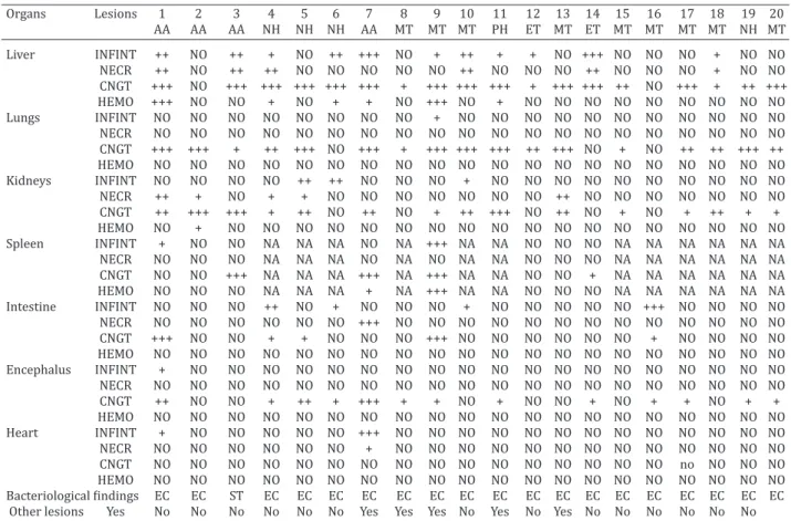
Table 2. Histopathological and bacteriological findings in twenty pet psittacine from Fortaleza, Brazil, with colibacillosis and salmonellosis
Organs Lesions 1 2 3 4 5 6 7 8 9 10 11 12 13 14 15 16 17 18 19 20 AA AA AA NH NH NH AA MT MT MT PH ET MT ET MT MT MT MT NH MT Liver INFINT ++ NO ++ + NO ++ +++ NO + ++ + + NO +++ NO NO NO + NO NO NECR ++ NO ++ ++ NO NO NO NO NO ++ NO NO NO ++ NO NO NO + NO NO CNGT +++ NO +++ +++ +++ +++ +++ + +++ +++ +++ + +++ +++ ++ NO +++ + ++ +++ HEMO +++ NO NO + NO + + NO +++ NO + NO NO NO NO NO NO NO NO NO Lungs INFINT NO NO NO NO NO NO NO NO + NO NO NO NO NO NO NO NO NO NO NO NECR NO NO NO NO NO NO NO NO NO NO NO NO NO NO NO NO NO NO NO NO CNGT +++ +++ + ++ +++ NO +++ + +++ +++ +++ ++ +++ NO + NO ++ ++ +++ ++ HEMO NO NO NO NO NO NO NO NO NO NO NO NO NO NO NO NO NO NO NO NO Kidneys INFINT NO NO NO NO ++ ++ NO NO NO + NO NO NO NO NO NO NO NO NO NO NECR ++ + NO + + NO NO NO NO NO NO NO ++ NO NO NO NO NO NO NO CNGT ++ +++ +++ + ++ NO ++ NO + ++ +++ NO ++ NO + NO + ++ + + HEMO NO + NO NO NO NO NO NO NO NO NO NO NO NO NO NO NO NO NO NO Spleen INFINT + NO NO NA NA NA NO NA +++ NA NA NO NO NO NA NA NA NA NA NA NECR NO NO NO NA NA NA NO NA NO NA NA NO NO NO NA NA NA NA NA NA CNGT NO NO +++ NA NA NA +++ NA +++ NA NA NO NO + NA NA NA NA NA NA HEMO NO NO NO NA NA NA + NA +++ NA NA NO NO NO NA NA NA NA NA NA Intestine INFINT NO NO NO ++ NO + NO NO NO + NO NO NO NO NO +++ NO NO NO NO NECR NO NO NO NO NO NO +++ NO NO NO NO NO NO NO NO NO NO NO NO NO CNGT +++ NO NO + + NO NO NO +++ NO NO NO NO NO NO + NO NO NO NO HEMO NO NO NO NO NO NO NO NO NO NO NO NO NO NO NO NO NO NO NO NO Encephalus INFINT + NO NO NO NO NO NO NO NO NO NO NO NO NO NO NO NO NO NO NO NECR NO NO NO NO NO NO NO NO NO NO NO NO NO NO NO NO NO NO NO NO CNGT ++ NO NO + ++ + +++ + + NO + NO NO + NO + + NO + + HEMO NO NO NO NO NO NO NO NO NO NO NO NO NO NO NO NO NO NO NO NO Heart INFINT + NO NO NO NO NO +++ NO NO NO NO NO NO NO NO NO NO NO NO NO NECR NO NO NO NO NO NO + NO NO NO NO NO NO NO NO NO NO NO NO NO CNGT NO NO NO NO NO NO NO NO NO NO NO NO NO NO NO NO no NO NO NO HEMO NO NO NO NO NO NO NO NO NO NO NO NO NO NO NO NO NO NO NO NO
Bacteriological findings EC EC ST EC EC EC EC EC EC EC EC EC EC EC EC EC EC EC EC EC
Other lesions Yes No No No No No No Yes Yes Yes No Yes No Yes No No No No No No AA = Amazona aestiva, NH = Nymphicus hollandicus, MT = Melopsittacus undulatus, ET = Eupsittula cactorum, PH = Psephotus haematonotus,
INFINT = Inflammatory infiltrate, NECRO = Necrosis, CNGT = Congestion, HEMO = Hemosiderosis, NO = Not observed, NA = Not avaliable, EC = Escherichia coli, ST = Salmonella Typhimurium. + mild, ++ moderate, +++ severe.
Table 3. Other findings from non-Escherichia coli lesions in six pet psittacine from Fortaleza, Brazil, with colibacillosis Case Other lesions or disease Isolated
1 Granulomatous fungic celomitis EC AA
8 Fusiform sarcoma EC
MT
9 Egg retention EC
MT
10 Caseous sinusitis EC
MT
12 Skin laceration EC
ET
14 Limb edema EC
ET
et al. (2014). However, as observed in a study with healthy hispaniolan amazon parrots (Amazona ventralis) in captivi-ty, which had the intestinal microbiota assessed with Gram staining and bacterial culture, E. coli was the most frequent Gram negative rod (23.8%) (Evans et al. 2014). In addition, a study with three species of Amazona (A. farinosa, A. aesti-va and A. amazônica) and other psittacine with a conside-rable health risk due to management conditions of illegal wildlife trade, E. coli was isolated from 57.3% individuals (Hidasi et al. 2013).
In birds that died with suggestive colibacillosis lesions (aerossaculitis, cachexy, pericarditis, perihepatitis, hemor-rhage in the intestinal mucosa and accumulation of feces around the cloacae) or symptoms, as observed in this study and in a study with red-spectacled amazon (A. pretrei) par-rots, E. coli may be isolated frequently from several distinct organs (Corrêa et al. 2013). Enteropathogenic E. coli has been detected from asymptomatic psittacine from several species, among which Amazona spp. and Eupsittula spp. in captivity (Saidenberg et al. 2012). The frequency of E. coli isolation (19%) from psittacine of illegal wildlife trade or in rehabilitation previous to reintroduction in the wild is more frequent than Salmonella sp. (1.12%), which was isolated from an A. aestiva as previously described,
simi-lar to the findings from this study (Marietto-Gonçalves et
al. 2010). There are some reports of Salmonella sp. being isolated from psittacine of conservationist, breeding and rehabilitation facilities, even in Fortaleza, Brazil from the species Ara chloroptera, Amazona aestiva and Melopsit-tacus undulatus, however this is the first report in Brazil with a pet psittacine (Marietto-Gonçalves & Almeida 2010, Bezerra et al. 2013, Hidasi et al. 2013, Lopes et al. 2014, Almeida et al. 2015).
Avian colibacillosis aspects were described in a case re-port with a pet A. aestiva (Marietto-Gonçalves et al. 2007),
in which E. coli was isolated from several organs and le-sions, which consisted of multiple granulomas and
hete-rophilic infiltrate in lungs, congestion in kidneys, liver and brain, which are similar to the findings in this study. The
most frequent lesions of colibacillosis are coagulative liver necrosis, aerossaculitis, granulomas, acute lung hemorrha-ge and conhemorrha-gestion, enteritis with fusion of vili, epithelium hypertrophy and exfoliation, cellulitis with intense
hetero-philic infiltrate, multinuclear giant cells and fibrin-necrotic
plaques; salpingitis with intense follicle necrosis and mixed
infiltrate (Crespo et al. 2001, Andrade et al. 2006, Seeley et
al. 2014).
The most frequent characteristics of avian salmonello-sis are poor body condition, muscular atrophy, granulomas varying from multifocal to coalescent, transmural ulcerati-ve necrosis of the gastrointestinal tract with clear presen-ce of bacterial aggregation inside and outside the lesions, necrotizing hepatitis, interstitial pneumonia, myocarditis, epicarditis and necrotizing encephalitis (Madadgar et al.
2009, Giovaninni et al. 2012). In Australia, a flock of budge -rigars with mortality caused by E. coli presented congestion
and hemorrhage (Seeley et al. 2014) as the main findings
observed, similar to the results from this study, except the encephalitis present in different species of psittacine. The lesions caused by Salmonella Typhimurium in passerines in Switzerland had similar intensity to the ones caused by the strain in this study isolated from the A. aestiva, which was also sever necrotizing hepatitis (Giovaninni et al. 2012). The occurrence colibacillosis in birds with other simulta-neous diseases or lesions are frequent, which may serve as an entry port for the infection by E. coli or even Salmonella spp. (Crespo et al. 2001, Seeley et al. 2014).
Elevated rates of resistance to tetracycline or other an-tibiotics from the same group may occur via transference between microorganisms in the microbiota of birds, which
Table 4. Antimicrobial susceptibility profiles of Escherichia coli and Salmonella Typhimurium strains isolated from different organs of dead pet psittacine
Case and Organs AMP AZI CLO CTF FOS GEN NAL NOR POL SUL SUT TET Previous treatment Isolate R S R S R S R S R S R S R S R S R S R S R S R S R
3 AA ST L + - + - - + - + - + - + + - + - - + + - + - + - Tetracycline and S + - + - - + - + - + - + + - + - - + + - + - + - Prednizolone 4 NH EC K + - + - - + - + - + - + - + - + - + + - + - - + Tetracycline and
Sulfonamide
5 NH EC S - + - + - + - + - + - + + - - + - + - + - + - + Not informed
I - + - + - + - + - + - + + - - + - + + - - + - +
6 NH EC L - + - + - + - + - + - + - + - + - + - + - + - + Not informed 9 MT EC L + - - + - + - + - + - + + - - + + - + - - + + - Ketoprophen
K - + - + - + - + - + - + - + - + - + - + - + - +
10 MT EC L + - - + + - + - + - - + + - + - - + + - + - + - Enrofloxacin
12 ET EC L - + - + - + - + - + - + - + - + - + + - + - + - Not informed 14 ET EC K + - + - - + - + - + - + - + + - - + + - + - + - Not informed 15 MT EC L + - - + - + - + - + - + + - + - - + - + + - - + Not informed
I + - - + - + - + - + - + - + - + - + + - + - - +
16 MT EC L - + - + - + - + - + - + - + - + - + - + - + - + Not informed 18 MT EC I - + - + - + - + - + - + - + - + - + - + - + - + Not informed 20 MT EC L - + - + - + - + - + - + - + - + - + - + - + - + Not informed RF of strains 66,7 33,3 8,3 8,3 8,3 0,0 58,3 41,7 8,3 75,0 66,7 50,0
may be a direct or indirect risk to the human health (Hu et al. 2013). Resistance to tetracycline in E. coli strains isola-ted from pet birds have been reporisola-ted in Australia (13.9%), from a total of 594 analyzed samples and multidrug resis-tance was also reported (Blynton et al. 2015). In another study, multidrug resistance to other groups, such as amino-glycosides, quinolones, sulfas and others has been
identi-fied in E. coli strains isolated from psittacine.
Similar to other studies involving E. coli and Salmonella sp. isolated from psittacine (Corrêa et al. 2013), antibiotic resistance to tetracycline, sulfonamide and ampicillin pre-sented different results in organs and intestine samples (Case 9 and 14, Table 4). The Salmonella Typhimurium strain isolated in this study presented multidrug resistan-ce to six different antibiotic groups, similar to the strain isolated from parrots in rehabilitation prior to
reintroduc-tion in the wild that presented resistance to enrofloxacin, ceflacor, ciprofloxacin and sulfonamide (Marietto-Gonçal
-ves et al. 2010). These findings indicate an indiscriminate
use of antibiotics in avian medicine and the occurrence of multidrug resistance involved with failure in antimicrobial therapy.
CONCLUSIONS
Pet psittacine that died in Fortaleza, Brazil positive for Escherichia coli and Salmonella Typhimurium had coliba-cillosis and salmonellosis showed mild to moderate lesions and liver was the most affected organ.
Isolated strains presented multidrug resistance and the most frequent was to tetracycline, followed by ampicillin, sulfonamide, sulfazotrim and nalidixic acid; which suggests an ill use of antimicrobials in pet birds.
REFERENCES
Almeida P.M., Otutumi L.K., Gerônimo E., Messa V., Suenaga S.S., Amaral P.F.G.P., Lima E.T., Vendrame A., Gonçalves D.D. & Cestari E.D. 2015. Study of the presence of Salmonella spp.and gastrointestinal parasites in ex-creta from ornamental birds from breeders in the city of Umuruama, Paraná. Afr. J. Microbiol. Res. 9(4):253-257.
Andrade C.L., Ferreira G.F., Franco R.M., Nascimento E.R. & Tortelly R.
2006. Alterações patológicas e identificação da Escherichia coli como
agente causal da celulite aviária em frangos de corte inspecionados em um matadouro em São Paulo. Revta Bras. Clín. Vet. 13(3):139-143. Andrés S., Vico J.P., Garrido V., Grilló M.J., Samper S., Gavin P., Herrera-León
S. & Mainar-Jaime R.C. 2013. Epidemiology of subclinical salmonelosis in wild birds from a area of high prevalence of pig salmonelosis:
pheno-typic and genetic profiles of Salmonella isolates. Zoonoses Publ. Health
60:355-365.
Bezerra W.G.A., Cardoso W.M., Teixeira R.S.C., Vasconcelos R.H., Machado D.N., Lopes E.S., Albunquerque A.H. & Rocha-e-Silva R.C. 2013. Survey of Salmonella sp. in budgerigars (Melopsittacus undulatus) in Fortaleza, Brazil. Acta Scient. Vet. 41:1714-1723.
Blynton M.D.J., Pi H., Vangcchia B., Abraham S., Trott D.J., JohnsonJ.R. & Gordon D.M. 2015. The genetic structure and antimicrobial resistence of Escherichia coli and cryptic clades in birds with diverse human asso-ciations. Appl. Environ. Microbiol. 81(15):5123-5133.
CLSI 2012. Perfomance standards for antimicrobial disk susceptibility tests: Approved Standards-Eleventh Edition. CLSI document M02-A11. Clinical and Laboratory Standards Institute, Wayne, PA.
Corrêa I.M.O., Flores F., Schneiders G.H., Pereira L.Q., Brito B.G. & Lovato M. 2013. Detecção de fatores de virulência de Eschericia coli e análise de
Salmonella spp. em psitacídeos. Pesq. Vet. Bras. 33(2):241-246.
Crespo R., Walker R.L., Nordhausen R., Sawyer S.J. & Manalc R.B. 2001. Sal-pingitis in Pekin ducks associated with concurrent infection with Treta-trichomonas spp. and Escherichia coli. J. Vet. Diagn. Invest. 13:240-245. Evans E.E., Mitchell M.A., Whittington J.K. & Tully Jr T.N. 2014. Measuring
the level of agrément between cloacal gram´s stains and bacterial cul-tures in Hispaniolan Amazon parrots (Amazona ventralis). J. Avian Med. Surg. 28(4):290-296.
Giovaninni S., Pewsner M., Hüssy D., Hächler H., Degiorgis M.P.R., Hirschheydt J. von & Origgi F.C. 2012. Epidemic of Salmonellosis in pas-serine birds in switzerland with spillover to domestic cats. Vet. Pathol. 50(4):597-606.
Hidasi H.W., Hidasi Neto J., Moraes D.M.C., Linhares G.F.C., Jayme V.S. & An-drade M.A. 2013. Enterobacterial detection and Escherichia coli antimi-crobial resistance in parrots seized from the illegal wildlife trade. J. Zoo Wildl. Med. 44(1):1-7.
Hu G.Z., Pan Y.S., Wu H., Han H., Xu R., Yuan L., Liu J.H. & Feng J.K. 2013. Prevalence of tetracycline resistence genes and identification of tet(M) in clinical isolates of Escherichia coli from sick ducks in China. J. Med. Microbiol. 62:851-858.
Janben D.W., Schwarz C., Preikschat P., Voss M., Philipp H.C. & Wieler L.H. 2001. Virulence-associated genes in avian pathogenic Escherichia coli
(APEC) isolated from intestinal organs of poultry having died from coli-bacillosis. Int. J. Med. Microbiol. 291(5):371-378.
Lamb S., Sobczynski A., Starks D. & Sitinas N. 2014. Bacteria isolated from the skin of congo African grey parrots (Psittacus erithacus), budgerig-ans (Melopsittacus undulatus), and cockatiels (Nymphicus hollandicus). J. Avian Med. Sug. 28(4):275-279.
Lopes E.S., Cardoso W.M., Albuquerque A.H., Teixeira R.S.C., Salles R.P.R., Bezerra W.G.A., Rocha-e-Silva R.C., Lima S.V.G., Sales R.J.P.F. & Vasconce-los R.H. 2014. Isolation of Salmonella spp. em Psitaciformes from zoos and a comercial establishment of Fortaleza, Brazil. Arq. Bras. Med. Vet. Zootec. 66(3):965-968.
Madadgar O., Zharaei-Salehi T., Ghafari M.M., Tamai A., Madani A.S. & Yahyareyat R. 2009. Study of an unusal paratyphoid epornitic in canar-ies (Serinus canarius). Avian Pathol. 38(6):437-441.
Marietto-Gonçalves G.A. & Almeida S.M. 2010a. Isolation of Salmonella entérica Serovar Enteritidis in Blue-fronted amazona parrot (Amazona aestiva). Avian Dis. 54:151-155.
Marietto-Gonçalves G.A., Almeida S.M., Lima E.T. & Andreatti-Filho R.L. 2010b. Detecção de Escherichia coli e Salmonella spp. em microbiota in-testinal de Psitaciformes em fase de reabilitação para soltura. Brazilian J. Vet. Res. Anim. Sci. 47(3):185-189.
Marietto-Gonçalves G.A., Lima E.T., Siqueira J.L. & Andreatti-Fiho R.L. 2007. Colisepticemia em papagaio-verdadeiro (Amazona aestiva). Revta Bras. Saúde Prod. Anim. 8(1):56-60.
Poirel L., Potron A., de La Cuesta C., Cleary T., Nordmann P. & Munoz-Price
S. 2012. Wild coast birds as reservoirs of Broad-Spectrum-β-Lact
-amase-producing Enterobacteriaceae in Miami beach, Florida. Antimi-crob. Agents Chemoterapy 56(5):2756-2758.
Saidenberg A.B., Teixeira R.H.F., Guedes N.M.R., Allgayer M.C., Melville P.N. & Benites N.R. 2012. Molecular detection of Enteropathogenic
Escherichia coli in asymptomatic captive psittacines. Pesq. Vet. Bras. 32(9):922-926.
Seeley K.E., Baitchman E., Bartlett S., Debroy C. & Garner M.M. 2014. In-vestigation and control of an attaching and effacing Escherichia coli
outbreak in a colony of captive budgerigars (Melopsittacus undulatus). J. Zoo Wildl. Med. 45(4):875-882.
Seo K.H., Holt T.P.S., Gast R.K. & Hofacre C.L. 2000. Elimination of early
Salmonella Enteritidis infection after treatment with competitive-exclu-sion culture and enrofloxacin in experimentally infected chicks. Poult. Sci. 79(10):1408-1413.
Tortora G.J., Funke B.R. & Case C.L. 2012. Microbiologia. 10ª ed. ArtMed, Porto Alegre. 964p.