A study o f the inte ractio n be twe e n
Helicob acter p ylori
and co mpo ne nts
o f the human fibrino lytic syste m
1Laboratorio de Biología y Medicina Experimental, Facultad de Ciencias, and 2Laboratorio de Microbiología, Facultad de Medicina, Universidad de Los Andes,
Mérida, Venezuela A. Yarzábal1, L. Avilán1,
K. Hoelzl2, M. de Muñoz2,
J. Puig1 and I. Kansau2
Abstract
The interaction of plasminogen, tissue plasminogen activator (t-PA) and urokinase with a clinical strain of Helicobacter pylori was stud-ied. Plasminogen bound to the surface of H. pylori cells in a concen-tration-dependent manner and could be activated to the enzymatic form, plasmin, by t-PA. Affinity chromatography assays revealed a plasminogen-binding protein of 58.9 kDa in water extracts of surface proteins. Surface-associated plasmin activity, detected with the chro-mogenic substrate CBS 00.65, was observed only when plasminogen and an exogenous activator were added to the cell suspension. The two physiologic plasminogen activators, t-PA and urokinase, were also shown to bind to and remain active on the surface of bacterial cells. e -Aminocaproic acid caused partial inhibition of t-PA binding, suggest-ing that the krsuggest-ingle 2 structure of this activator is involved in the interaction with surface receptors. The activation of plasminogen by t-PA, but not urokinase, strongly depended on the presence of cells and a 25-fold enhancer effect on the initial velocity of activation by t-PA compared to urokinase was established. Furthermore, a relationship between cell concentration and the initial velocity of activation was demonstrated. These findings support the concept that plasminogen activation by t-PA on the bacterial surface is a surface-dependent reaction which offers catalytic advantages.
Co rre spo nde nce
L. Avilan
LABIO MEX, Facultad de Ciencias Universidad de Los Andes Apartado Postal 281 Mérida 5101 Venezuela Fax: + 58-74-63-4587 E-mail: avilan@ ciens.ula.ve
Research supported by CO NICIT (No. S1-97001300) and CDCHT-ULA (No. C-925-98-03-B).
Received December 1, 1999 Accepted June 12, 2000
Ke y words
·Helicobacter pylori
·Plasminogen
·t-PA
·Urokinase
·Plasminogen activation
Intro ductio n
Plasminogen, a 92-kDa glycoprotein pre-sent in plasma and extracellular fluids, is the main component of the fibrinolytic system (1). It circulates in plasma as an inactive zymogen which, under different conditions, could be activated to the active form, plas-min, by plasminogen activators (PA) such as the tissue-type PA (t-PA) and urokinase. The activation reaction involves the hydrolysis of
a single Arg-Val peptide bond (2), convert-ing the zymogen to active two-chain plas-min, a broad spectrum serine protease, which degrades fibrin clots, as well as other pro-teins (3).
nu-merous cell types and fibrin clots facilitates plasmin generation, resulting in an enhance-ment of the catalytic efficiency (7,8); fur-thermore, surface plasmin cannot be regu-lated efficiently by host inhibitors such as
a2-antiplasmin (9).
It has been recently established that bind-ing and activation of human plasminogen on the surface of bacterial cells may be a com-mon mechanism used by invasive bacteria to facilitate movement through normal tissue barriers (10,11). Several species generate their own plasminogen activators: this is the case of group A streptococci and Staphylo-coccus aureus, which secrete streptokinase and staphylokinase, respectively, two bacte-rial PAs widely used in the therapy of acute myocardial infarction (12). Other pathogens, not known to produce a PA, may be capable of using host PAs to generate surface plas-min activity, a mechanism which is pre-dicted to be less efficient (11). These organ-isms can possibly express surface receptors for eukaryotic activators which enable them to acquire plasmin-like enzymatic activity in the human host (10).
Helicobacter pylori is a gram-negative human pathogen that colonizes the gastric mucosa and is associated with various stom-ach and duodenal disorders such as active chronic gastritis, peptic ulceration and pos-sibly gastric carcinoma (13). Like other bac-terial pathogens, H. pylori expresses surface proteins with affinity for several human pro-teins, components of the mammalian extra-cellular matrix such as laminin, vitronectin, collagen types I and IV and plasminogen (14-18). Plasminogen binding to the surface of H. pylori has been shown to be inhibited by lysine, lysine analogues and miniplas-minogen (fifth kringle and catalytic domain (1)), suggesting an important role of the fifth kringle structure of the zymogen which pos-sibly interacts with two surface proteins of 42 and 57 kDa (19).
In the present study we have examined the binding of plasminogen and its
activa-tion by t-PA and urokinase on the surface of an H. pylori clinical isolate. The effect of bacterial cells on plasminogen activation was studied. In addition, the capability of H. pylori to bind other components of the plas-minogen system such as t-PA and urokinase was also explored.
Mate rial and Me thods
Bacte rial isolate s
A clinical H. pylori isolate was obtained from a patient with gastric ulcer at the Hos-pital Universitario de Los Andes (HULA), University of Los Andes, Mérida, Venezu-ela. Bacteria were recovered from gastric biopsies collected by endoscopy. The isolate represents a single colony obtained from a biopsy, which was processed for Gram stain-ing, urease test and histology as well as H. pylori culture, as previously described (20). In some experiments, comparisons were made among different clinical isolates ob-tained as previously described.
In order to prepare cell suspensions, H. pylori cells incubated for 6-7 days under microaerophilic conditions on blood-agar plates were collected in sterile PBS (0.14 M NaCl, 0.06 M sodium phosphate, pH 7.2), washed twice and resuspended in the same buffer.
Re age nts
eluted with 50 mM e-aminocaproic acid (EACA) and plasminogen-containing frac-tions were further purified by filtration chro-matography. t-PA was from Biopool (Umeå, Sweden). Urokinase was purchased from Sanofi Winthrop (Gentilly, France).
Plasminoge n binding and activation assays
H. pylori (108 cells) were incubated with plasminogen (0-0.4 µM) in 0.1 ml PBS con-taining 1% BSA for 1 h at 25oC under con-tinuous mixing. At the end of the incubation period, the cells were pelleted. Bacteria were washed with 10 volumes of PBS containing 0.1% Tween 20 followed by washing in 50 mM Tris HCl, pH 7.4 (assay buffer), and finally resuspended in assay buffer. Bound plasminogen was activated with t-PA and the resultant plasmin activity monitored in the mixture at 405 nm with the chromogenic substrate CBS 00.65 (0.5 mM) after 30 min incubation at 37oC.
Plasminogen activation by t-PA was stud-ied in the presence or absence of H. pylori cells. For this purpose, 1.5 µM plasminogen, 2.1 nM t-PA, 0.5 mM CBS 00.65 and in-creasing cell concentrations from 107 to 109/ ml were mixed in 0.1 ml assay buffer. The A405 of the mixture was monitored continu-ously at 37oC. Initial velocities of activation were calculated from plots of A405 versus t2, according to Wohl et al. (22).
De te ction of ce ll-associate d t-PA and urokinase activity
Urokinase and t-PA were tested for their ability to bind to the surface of H. pylori cells. For this, increasing concentrations of both activators (0-14 nM t-PA; 0-50 nM urokinase) were incubated with 108 H. py-lori cells as described above. Washed cells were incubated with plasminogen (1.5 µM) in the presence of CBS 00.65 (0.5 mM). The A405 of the mixture was recorded at different time intervals, and plotted against the PA
concentration.
Affinity chromatography assays
Human plasminogen was covalently coupled to NHS-activated Sepharose (HiTrap; Pharmacia) according to the manu-facturer’s instructions. H. pylori protein frac-tions were batch adsorbed to the plasmino-gen-Sepharose resine in PBS for 1 h at room temperature under continuous shaking. Fol-lowing extensive washing with PBS, bound proteins were eluted with either 50 mM EACA or 50 mM glycine, 0.1 M NaCl, pH 2.7, and further analyzed by SDS-PAGE.
Ele ctro pho re sis
SDS-PAGE was performed according to Laemmli (23). H. pylori total cell extracts, surface proteins extracted with water, both obtained in the presence of 0.2 mM phenyl-methylsulfonyl fluoride and 1 mM benzami-dine, as well as proteins eluted from plas-minogen-Sepharose assays, were separated on 10% acrylamide gels under reducing con-ditions. The proteins were not boiled prior to electrophoresis and were silver stained.
Re sults
Plasminoge n binding and activation on the surface of H. pylori
H. pylori proteins involved in the interac-tion with human plasminogen were isolated by affinity chromatography. This assay re-vealed the presence of one polypeptide of 58.9 kDa in water extracts of surface pro-teins (Figure 2). This protein was eluted with
50 mM EACA; however, the same result was obtained when glycine buffer, pH 2.7, was used in the elution step.
Enhance me nt of t-PA-catalyze d plasminoge n activation by H. pylori ce lls
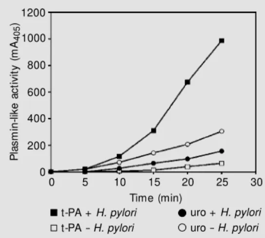
The effect of H. pylori cells on plasmin-ogen activation rate by t-PA and urokinase was explored. When t-PA was the activator used, as shown in Figure 3, the addition of cells induced a »25-fold higher amidolytic activity (expressed in mA405/min2) compared with the activity in the absence of cells. In contrast to t-PA, when bacteria were incu-bated with plasminogen and urokinase un-der conditions identical to those used in the t-PA experiment, only a two-fold increase in plasminogen activation was observed (Fig-ure 3). It thus appears that H. pylori enhance-ment of plasminogen activation is more spe-cific with t-PA.
It was also observed that the initial rate of plasminogen activation (expressed as mA405/ min2) increased significantly with cell con-centration (Figure 4). A definite effect was seen at bacterial concentrations as low as 5 x 10⁷ cells/ml.
Binding of t-PA and urokinase to H. pylori To assess whether H. pylori cells may express surface receptors for physiological plasminogen activators, a binding assay was developed. In both cases, an enzymatic ac-tivity proportional to the initial concentra-tion of the activator was observed (Figure 5A and B), showing that both activators bind to the surface of H. pylori cells in a concen-tration-dependent manner and are able to activate plasminogen. These results suggest that the activators remain active following immobilization on the surface of bacterial cells. The interaction between t-PA and H. pylori cells was inhibited by addition of the lysine analogue EACA, as shown in Figure 5A (inset).
94.0
14.4 67.0
43.0
30.0
20.1
1 2 3
Figure 2 - Plasminogen-Sepha-rose affinity purification of
plas-minogen-binding protein. H.
py-lori w ater extracts of surface
pro-teins w ere allow ed to bind to plasminogen-Sepharose for 1 h. After extensive w ashing, bound proteins w ere eluted w ith 50 mM EACA and further analyzed by SDS-PAGE under reducing
conditions. Lane 1, M olecular
mass standards; lane 2, w ater
extract of surface proteins; lane
3, purified proteins after elution
from plasminogen-Sepharose af-finity matrix. The arrow indicates the position of the 58.9-kDa plas-minogen-binding protein.
Figure 1 - Activation of plasmin-ogen to plasmin on the surface
of H. pylori. Cells (108) w ere
in-cubated w ith different amounts of human plasminogen. After ex-tensive w ashing, surface-bound plasminogen w as activated w ith 2.1 nM t-PA. Plasmin activity w as detected by digestion of the chrom ogenic subst rat e CBS 00.65 after 30-min incubation at
37oC (circles). Data are reported
as the mean change in milliunits of absorbance at 405 nm ob-tained from at least three deter-minations. Standard errors are show n.
kDa
P
la
s
m
in
-l
ik
e
a
c
ti
v
it
y
(
m
A4
0
5
/3
0
m
in
)
1200
1000
800
600
400
200
0
0 100 200 300 400 500
D iscussio n
Plasminogen binding and activation on the surface of pathogenic bacteria have been implied in the invasion of tissues, hydrolysis of host immunoglobulins as well as availa-bility of peptides from proteins for bacterial growth (11,24,25). Several mechanisms are used by these organisms to activate host plasminogen. These include the expression of membrane-anchored activators, the secre-tion of soluble activators and/or the presence of receptors for both plasminogen and its physiological activators on the surface of bacteria (10). H. pylori has been recently added to the list of such organisms due to its ability to bind plasminogen, a zymogen which may be subsequently activated by host acti-vators (19).
In the present study, we demonstrated that a clinical isolate of H. pylori is able to bind not only human plasminogen but also the two physiological activators, t-PA and urokinase. Binding of t-PA to H. pylori was inhibited by the lysine analogue EACA, sug-gesting an important role of the lysine-bind-ing sites located in the activator krlysine-bind-ingle 2 structure (12). We also demonstrated that, once bound, these proteins remain active and may be used by the cells to express a surface plasmin activity. This strategy ap-pears to be a common and efficient mechan-ism shared by invasive pathogens (11). It has already been observed that Borrelia burg-dorferi expresses a receptor for urokinase in addition to binding plasminogen and that Escherichia coli and Salmonella enteritidis bind both t-PA and plasminogen, suggesting that organisms which bind plasminogen but do not produce their own plasminogen acti-vators may acquire surface plasmin activity through host activators (10). H. pylori can be now included in this class of pathogens which share an alternative strategy to efficiently acquire cell-surface, unregulatable, plasmin activity under physiological conditions. Al-though we believe it is likely that specific
P la s m in -l ik e a c ti v it y ( m A4 0 5 )1200 1000 800 600 400 200 0
0 5 10 15 20 25
Time (min) 30 6 5 4 3 2 1 0
0 40 80 120
Cells/ml (x 107)
1200 1000 800 600 400 200 0
0 4 8 12 16
0.5 1.0 1.5 2.0 0 0 40 80 120 1000 800 600 400 200 0
0 10 20 30 40 50 60
Urokinase (nM )
EACA (mM ) A
B
Figure 3 - Effect of H. pylori cells
on t-PA- and urokinase-catalyzed plasmin formation. Human plas-minogen (0.4 µM ) w as activated
by 2.1 nMt-PA (squares) or 0.2
nM urokinase (circles) in the presence (filled symbols) and
ab-sence (open symbols) of 108 H.
pylori cells. The formation of
plasmin activity w as measured using a chromogenic substrate.
Figure 4 - Effect of cell density on t-PA-catalyzed plasminogen activation. Plasminogen (1.5 µM ) w as activated w ith 2.1 nM t-PA in the presence of increasing
concentrations of H. pylori cells.
The resultant plasmin activity w as measured w ith the chromo-genic substrate CBS 00.65 at dif-ferent time intervals and the ini-tial rates of plasmin generation determined. Each point repre-sents the mean value of at least three determinations.
Figure 5 - t-PA and urokinase
binding to the surface of H.
py-lori. Different amounts of t-PA
(A) and urokinase (B) w ere
al-low ed to interact w ith 108 H.
pylori cells. Follow ing extensive
w ashing, plasminogen (1.5 µM ) w as added and the resultant plasmin activity recorded w ith CBS 00.65 after 5-min (uroki-nase) or 15-min (t-PA) incubation
at 37oC. Each point represents
the mean value of at least three determinations. Standard errors
are show n. Inset, Inhibition by e
-aminocaproic acid (EACA) of the
binding of t-PA (14 nM ) to H.
pylori cells.
P la s m in o g e n a c ti v a ti o n (m A4 0 5 /m in 2) P la s m in -l ik e a c ti v it y ( m A4 0 5 )
t-PA (nM )
% M a x im a l a c ti v it y
t-PA + H. pylori
t-PA - H. pylori
uro + H. pylori
receptors for both t-PA and urokinase may be present on the surface of H. pylori cells, their identity remains obscure and needs further experiments to be elucidated.
The receptors for human plasminogen in several pathogenic bacteria have been char-acterized (10). In the case of group A strep-tococci, such proteins appear to play a sig-nificant biological role since almost four different proteins are produced by these spe-cies (26). In the particular case of H. pylori, two proteins of 42 and 57 kDa have been recently demonstrated to be involved in plas-minogen binding, using immunoblot proce-dures (19). In the present study, with a dif-ferent experimental approach, affinity bind-ing assays of water extracts of surface pro-teins allowed the detection of a plasmino-gen-binding polypeptide of 58.9 kDa, a pro-tein of a molecular mass similar to that of one of the proteins found in the other strain of H. pylori already studied (19). This pro-tein was eluted with EACA, indicating spe-cific binding through lysine-binding sites in the plasminogen molecule. The identifica-tion of this protein will allow to determine whether it is structurally related to other proteins which have been identified as plas-minogen receptors (27,28).
A significant enhancement of plasmino-gen activation by t-PA in the presence of H. pylori cells has been clearly established; when urokinase was used, only small differ-ences were seen. These results are not sur-prising since others have observed the same behavior in the presence of platelets (7). It is well known that plasminogen activation in vivo is a surface-dependent reaction that of-fers a catalytic advantage for enzyme com-plex formation and that assembly of plas-minogen and plasplas-minogen activators on many cell types facilitates plasmin generation (re-viewed in 29). Molecular assembly of t-PA and plasminogen on the surface of fibrin or
cells results in a ternary complex that leads to a profound enhancement of the reaction rate (29). In the case of platelets, this en-hancement appears to be particularly effi-cient when t-PA is the activator, resulting in a 5- to 8-fold increase in catalytic efficiency; however, when either urokinase or strep-tokinase is used, no enhancement is observed (7). Furthermore, enhancement of plasmino-gen activation by a number of bacteria ex-pressing receptors for t-PA has been reported (10,30). These observations correlate with the requirement of a third macromolecular component for the formation of a complex between plasminogen and t-PA. Fibrin, casein, denatured proteins, aggregated IgG and the eukaryotic cell surface are among the factors which enhance the activation of plasminogen (31,32). In agreement with this, it is tempting to speculate that the cell sur-face of H. pylori may also promote complex formation between plasminogen and t-PA through specific receptors, leading to the pronounced rate of activation observed. From a biological perspective, our data suggest that t-PA, instead of urokinase, might be the physiological activator of plasminogen on the H. pylori surface. However, a decrease in t-PA and an increase in urokinase levels in H. pylori-associated gastritis have been pre-viously reported (33). The activator respon-sible for the in vivo plasmin formation on the H. pylori surface is still to be elucidated.
Re fe re nce s
1. Ponting CP, M arshall JM & Cederholm-Williams SA (1992). Plasminogen: a
struc-tural review . Blood Coagulation and
Fi-brinolysis, 3: 605-614.
2. Robbins RC (1987). The plasminogen-plasmin enzyme system. In: Coleman RW, Hirsh J, M arder VJ & Salzman EW
(Editors), Hemostasis and Thrombosis.
Lippincott, Philadelphia.
3. Castellino FJ & Pow ell JR (1981). Human
plasminogen. M ethods in Enzymology,
80: 365-378.
4. Strickland S, Reich E & Sherman M I (1976). Plasminogen activator in early em-bryogenesis: Enzyme production by
tro-phoblast and parietal endoderm. Cell, 9:
231-240.
5. M cNeill H & Jensen PJ (1990). A high-affinity receptor for urokinase plasmino-gen activator on human keratinocytes: characterization and potential modulation
during migration. Cell Regulation, 1:
843-852.
6. Danø K, Andreasen PA, Grøndahl-Hansen J, Kristensen P, Nielsen LS & Skriver L (1985). Plasminogen activators, tissue
degradation and cancer. Advances in
Can-cer Research, 44: 139-266.
7. Gao S, M orser J, M cLean K & Shuman M (1990). Differential effect of platelets on plasminogen activation by tissue plasmin-ogen activator, urokinase and
streptoki-nase. Thrombosis Research, 58: 421-433.
8. Lijnen HR, Bachmann F, Collen D, Ellis V, Pannekoek H, Rijken DC & Thorsen S (1994). M echanisms of plasminogen
acti-vation. Journal of Internal M edicine, 236:
415-424.
9. M iles LA & Plow EF (1988). Plasminogen receptors: ubiquitous sites for cellular
regulation of fibrinolysis. Fibrinolysis, 2:
61-71.
10. Boyle M & Lottenbergh R (1997). Plas-minogen activation by invasive human
pathogens. Thrombosis and Haemostasis,
77: 1-10.
11. Lottenbergh R, M inning-Wenz D & Boyle M D (1994). Capturing host plasmin(ogen): a common mechanism for invasive
patho-gens? Trends in M icrobiology, 22: 20-24.
12. Bachmann F (1995). Fibrinolytic agents.
Fibrinolysis, 9: 1-15.
13. Vandenplas Y (1999). Helicobacter pylori
infection. Clinical M icrobiology and
Infec-tion, 5: 1-11.
14. Khin M M , Ringnér M , Aleljung P, Wadström T & Ho B (1996). Binding of human plasminogen and lactoferrin by
Helicobacter pylori coccoid forms.
Jour-nal of M edical M icrobiology, 45: 433-439.
15. Ringnér M , Paulsson M & Wadström T
(1992). Vitronectin binding by
Helico-bacter pylori. FEM S M icrobiology and
Im-munology, 5: 219-224.
16. Ringnér M , Valkonen KH & Wadström T (1994). Binding of vitronectin and
plas-minogen to Helicobacter pylori. FEM S
Im-munology and M edical M icrobiology, 9:
29-34.
17. Trust TJ, Doig P, Emödy L, Kienle Z, Wadström T & O’Toole P (1991). High-affinity binding of the basement mem-brane proteins collagen type IV and
lami-nin to the gastric pathogen Helicobacter
pylori. Infection and Immunity, 59:
4398-4404.
18. Valkonen KH, Ringnér M , Ljungh A & Wadström T (1993). High affinity binding
of laminin by Helicobacter pylori: evidence
for a lectin-like interaction. FEM S
Immu-nology and M edical M icrobiology, 7:
29-37.
19. Pantzar M , Ljungh A & W adström T (1998). Plasminogen binding and
activa-tion at the surface of Helicobacter pylori
CCUG 17874. Infection and Immunity, 66:
4976-4980.
20. Raymond J, Begeret M , Benhamou PH, M ensah K & Dupont C (1994). A 2-year
study of H. pylori in children. Journal of
Clinical M icrobiology, 32: 461-463.
21. Deutsch DG & M ertz ET (1970). Plasmin-ogen: purification from human plasma by
affinity chromatography. Science, 170:
1095-1096.
22. Wohl RC, Summaria L & Robbins KC (1980). Kinetics of activation of human plasminogen by different activator
spe-cies at pH 7.4 and 37 degrees C. Journal
of Biological Chemistry, 255: 2005-2013.
23. Laemmli UK (1970). Cleavage of struc-tural proteins during the assembly of the
head of bacteriophage T4. Nature, 227:
680-685.
24. Chuba JV (1994). Susceptibility of mono-clonal IgG paraproteins to plasmin cleav-age using glycerol stabilised human
plas-min. Biochemical and Biophysical
Re-search Communications, 202: 367-373.
25. Leigh JA (1993). Activation of bovine
plas-minogen by Streptococcus uberis. FEM S
M icrobiology Letters, 114: 67-72.
26. Poon-King R, Bannan J, Viteri A, Cu G & Zabriskie JB (1993). Identification of an extracellular plasmin binding protein from
nephritogenic streptococci. Journal of
Ex-perimental M edicine, 1178: 759-763.
27. Redlitz A & Plow EF (1995). Receptors for
plasminogen and t-PA: an update.
Bail-lieres Clinical Haematology, 8: 313-327.
28. Pancholi V & Fischetti VA (1998). Alpha-enolase, a novel strong plasmin(ogen) binding protein on the surface of
patho-genic streptococci. Journal of Biological
Chemistry, 273: 14503-14515.
29. Anglés-Cano E (1994). Overview on fibrin-olysis: plasminogen activation pathw ays
on fibrin and cell surfaces. Chemistry and
Physics of Lipids, 67/68: 353-362.
30. Lähteenmäki K, Westerlund B, Kuusela P & Korhonen TK (1993). Immobilization of
plasminogen on Escherichia coli flagella.
FEM S M icrobiology Letters, 106:
309-314.
31. Kuusela P & Saksela O (1990). Binding and activation of plasminogen at the
sur-face of Staphylococcus aureus. Increase
in affinity after conversion to the Lys form
of the ligand. European Journal of
Bio-chemistry, 193: 759-765.
32. M achovich R & Ow en WG (1997). Dena-tured proteins as cofactors for
plasmino-gen activation. Archives of Biochemistry
and Biophysics, 15: 343-349.
33. Gotz JM , Vergouw e Y, Verspaget HW, Biem ond I, Sier CF, Lam ers CB & Veenendaal RA (1996). Gastric mucosal
plasminogen activators in Helicobacter
pylori infection. Digestive Diseases and
Sciences, 41: 1577-1582.
34. Scott D, Weeks D, M elchers K & Sachs G
(1998). The life and death of Helicobacter
pylori. Gut, 43: S56-S60.
35. M ullins DE & Rohrlich ST (1983). The role
of proteases in cellular invasiveness.