R E S E A R C H
Open Access
Antitumor potential of the myotoxin
BthTX-I from
Bothrops jararacussu
snake
venom: evaluation of cell cycle alterations
and death mechanisms induced in tumor
cell lines
Cássio Prinholato da Silva, Tássia R. Costa, Raquel M. Alves Paiva, Adélia C. O. Cintra, Danilo L. Menaldo,
Lusânia M. Greggi Antunes and Suely V. Sampaio
*Abstract
Background:Phospholipases A2(PLA2s) are abundant components of snake venoms that have been extensively
studied due to their pharmacological and pathophysiological effects on living organisms. This study aimed to assess the antitumor potential of BthTX-I, a basic myotoxic PLA2isolated fromBothrops jararacussuvenom, by evaluating in vitroprocesses of cytotoxicity, modulation of the cell cycle and induction of apoptosis in human (HL-60 and HepG2) and murine (PC-12 and B16F10) tumor cell lines.
Methods:The cytotoxic effects of BthTX-I were evaluated on the tumor cell lines HL-60 (promyelocytic leukemia), HepG2 (human hepatocellular carcinoma), PC-12 (murine pheochromocytoma) and B16F10 (murine melanoma) using the MTT method. Flow cytometry technique was used for the analysis of cell cycle alterations and death mechanisms (apoptosis and/or necrosis) induced in tumor cells after treatment with BthTX-I.
Results:It was observed that BthTX-I was cytotoxic to all evaluated tumor cell lines, reducing their viability in 40 to 50 %. The myotoxin showed modulating effects on the cell cycle of PC-12 and B16F10 cells, promoting delay in the G0/G1 phase. Additionally, flow cytometry analysis indicated cell death mainly by apoptosis. B16F10 was more susceptible to the effects of BthTX-I, with ~40 % of the cells analyzed in apoptosis, followed by HepG2 (~35 %), PC-12 (~25 %) and HL-60 (~4 %).
Conclusions:These results suggest that BthTX-I presents antitumor properties that may be useful for developing new therapeutic strategies against cancer.
Keywords:Bothrops jararacussu, BthTX-I, Antitumor potential, Apoptosis, Cell cycle alterations
Background
According to the World Health Organization, cancer is one of the leading causes of morbidity and mortality worldwide. Cancer is a generic term for a large group of diseases that may affect any part of the body [1]. These diseases are char-acterized by uncontrolled growth and spread of abnormal cells, and may be caused by external factors (smoking,
infectious organisms, radiation and chemical products) and internal factors (inherited or metabolic mutations, hormones and immune conditions) [2].
A correct diagnosis of cancer is essential for appropriate and effective treatment, because each cancer type requires specific treatments that may include surgery, radiotherapy and/or chemotherapy. Nonetheless, chemotherapy is flawed especially considering that no chemotherapeutic agent available acts exclusively on tumor cells, affecting also many types of normal body cells and leading to several undesirable effects [2, 3].
* Correspondence:suvilela@usp.br
Department of Clinical Analyses, Toxicology and Food Sciences, School of Pharmaceutical Sciences of Ribeirão Preto, University of São Paulo (USP), Avenida do Café, s/n, Ribeirão Preto, SP CEP 14040-903, Brazil
Currently there is great medical and scientific interest in the investigation of natural products for therapeutic purposes, in search of new molecules that could be used as antitumor agents with fewer side effects than the usual chemotherapy, or serve as molecular models for the development of more effective drugs against malignant tumors [4, 5].
In recent years, studies on snake venoms and their components have demonstrated the antitumor effects of different toxins [6–8]. The focus of these researches has been the understanding of the mechanisms of action of toxins on different types of tumors. It is known that the cytotoxicity induced by animal venoms is mainly related to changes in the cellular metabolism of cells, especially tumorous ones, however, the mechanisms of action of these toxins are still to be clarified, which makes them target of many studies with interest in their therapeutic potential [8]. Some phospholipases A2(PLA2s) have been described to possess antitumor and antiangiogenic properties [9–11]. PLA2s are enzymes of great scientific interest due to their involvement in several human inflammatory diseases and in envenomations by snakes and bees. In addition, PLA2s present important effects on lipid membranes and are related to the formation of various substances involved in numerous biological activities, such as prostaglandins, prostacyclins, thromboxanes and leukotrienes [12, 13].
Myotoxins are PLA2homologues that present a substi-tution of the aspartic acid residue at position 49 (Asp49) by a lysine (Lys49), with changes in their calcium binding site. These variations are related to the low or no catalytic activity presented by these molecules [14]. Nevertheless, myotoxic PLA2s have diverse biological effects, such as myotoxicity, edema, anticoagulation, neurotoxicity, indir-ect hemolysis, cytotoxicity, and also antibacterial, antiviral and antiparasitic activities [15].
Bothropstoxin-I (BthTX-I) is a single chain protein of 13.7 kDa and pI 8.2, with 121 amino acid residues and 7 disulfide bonds, isolated fromBothrops jararacussusnake venom [16]. This basic myotoxin showed action on the gastrocnemius muscle of mice and cytotoxicity in murine muscle cells (C2C12) and also had its cytotoxic effects evaluated bothin vitroandin vivoon different tumor cell lines such as Jurkat, SKBR3, B16F10 and S180, showing promising antitumor properties [17, 18].
Considering these previous findings on BthTX-I and the wide variety of cytotoxic effects presented by snake toxins, additional studies using different tumor cell lines are necessary in order to increase the knowledge of the antitumor and biotechnological potential of thisB. jarara-cussumyotoxin. Thereby, this study aimed to evaluate the in vitroeffects of BthTX-I on human (HL-60 and HepG2) and murine (PC-12 and B16F10) tumor cell lines by assessing its induced cytotoxicity, cell cycle alterations and death mechanisms.
Methods
Materials
Toxin
BthTX-I was isolated from Bothrops jararacussu venom according to the methodology described by Cintraet al. [19]. The lyophilized protein was stored at −20 °C and solubilized in phosphate buffered saline (PBS) immedi-ately before its use in the tests.
Cell lines
HL-60 (CCL-240, promyelocytic leukemia), HepG2 (HB-8065, human hepatocellular carcinoma), PC-12 (CRL-1721, murine pheochromocytoma) and B16F10 (CRL-6475, murine melanoma) tumor cell lines were obtained from ATCC (American Type Culture Collection, USA).
Methods
Cell culture
Cells were grown in monolayer in 25 cm2flasks. HL-60 cells were cultured in 5 mL of RPMI culture medium (Gibco 31800–022, USA) supplemented with 10 % fetal bovine serum (FBS, Gibco 12657, USA) and 1 % antibiotic (streptomycin and penicillin, P4333 Sigma, USA). PC-12 cells were cultured in 5 mL of RPMI supplemented with 15 % fetal equine serum (Gibco 26050–088, New Zealand), 5 % FBS and 1 % antibiotic. B16F10 and HepG2 cells were grown in DMEM culture medium (Gibco 31600–034, USA) also supplemented with 10 % FBS and 1 % antibiotic. The vials containing the cells were incu-bated at 37 °C in a humidified incubator containing 5 % CO2, until reaching a state of confluence (~5 × 10⁶cells) when they require subculture. The tests with BthTX-I were performed with cells between the 3rd and 6th day of subculture. Cell viability tests using the Trypan blue dye were performed before any experimentation to ensure the accuracy of the results.
Cytotoxic assays using MTT
For the cytotoxicity assay, tumor cells (HL-60, PC-12, HepG2 or B16F10) were seeded into 96-well plates, followed by incubation for 24 h at 37 °C in a humidified incubator containing 5 % CO2. After this period, the cells were treated with 50 μL of PBS (negative control)
or 50 μL of BthTX-I samples at different concentrations
(5; 10; 25; 50 or 100μg/mL). Experimental positive
con-trol received 50 μL of a cisplatin solution at 1 mg/mL
(Incel, Darrow®), which is an antineoplastic agent that binds to DNA, inducing structural changes and, conse-quently, apoptosis. After treatment, the wells received 20μL of MTT
[3-(4,5-dimethylthiazol-2-yl)-2,5-diphenyl-tetrazolium bromide] (Sigma M2128, USA) (500 μg/mL,
followed by addition of 100 μL of DMSO (Sigma D2650,
USA) to each well. The plates were kept under stirring until complete dissolution of crystals (~20 min) and then the absorbance at 570 nm was determined in a Powerwave XS2 microreader (Biotek) [20].
Cell cycle analysis
Tumor cells (HL-60, PC-12, HepG2 or B16F10) were plated into 24-well plates (1 × 10⁶cells/well), followed by incubation for 24 h at 37 °C in a humidified incubator containing 5 % CO2. After this period, the cells were treated with 50μL of PBS (negative control) or 50μL of
BthTX-I samples at different concentrations (5; 10; 25; 50 or 100 μg/mL). After 24 h of treatment, cells were
transferred to flow cytometry tubes and centrifuged at 900gfor 5 min, supernatant was discarded and pellet re-suspended in 1 mL of PBS. Then, cells were centrifuged again, the supernatant was discarded, the pellet resus-pended in 100 μL of cold PBS and 2 mL of cold 70 %
ethanol and the samples were stored at−20 °C for 24 h. At the end of this period, the tubes were centrifuged, the supernatant discarded and the pellet was resuspended in 300 μL of cold PBS with 10 μL of RNase (10 mg/mL)
and incubated for 30 min at 37 °C. Immediately after the incubation, samples were placed in an ice bath with 164μL
of HFS (hypotonic fluorescence solution, a reagent that measures the amount of DNA in the cell and defines its cell cycle position) and incubated on ice for 1 h, followed by analysis of 10,000 events in a FACSCanto flow cytometer (Becton Dickinson, USA). The files generated by the equip-ment were read in the FlowJo 7.6.1 software for the analysis of cell division phases: G0/G1, S and G2/M. The results were expressed as the percentage of cells in each phase of the cycle [6].
Analysis of apoptosis/necrosis by flow cytometry
The assessment of the apoptotic and necrotic effects in-duced by BthTX-I on different tumor cell lines (HL-60, PC-12, HepG2 or B16F10) was determined by flow cy-tometry, using a kit containing annexin V FITC and propidium iodide (PI) [21]. The population of necrotic cells is marked with PI (PI+), while the cells in apop-tosis are marked with annexin V FITC (AV+) and the population of both necrotic and apoptotic cells is marked as PI+/AV+.
Tumor cells were plated into 24-well plates (5 × 10⁵ cells/well), followed by incubation for 24 h at 37 °C in a humidified incubator containing 5 % CO2. After this period, the cells were treated with 50μL of PBS
(nega-tive control) or 50μL of BthTX-I samples at different
concentrations (5; 10; 25; 50 or 100μg/mL). Experimental
positive control received 50 μL of a cisplatin solution at
1 mg/mL. After 24 h of treatment, cells were transferred to flow cytometry tubes, and centrifuged at 900g for
5 min, supernatant was discarded and pellet resuspended in 1 mL of PBS. The same process was repeated, this time with addition of 300 μL of 1X annexin V to the
resus-pended pellet and 5 μL of FITC-annexin V to each tube.
The samples were incubated for 15 min in ice, adding 1μL
of propidium iodide (PI) immediately after incubation, followed by analysis of 10,000 events in a FACSCanto flow cytometer (Becton Dickinson, USA) using Diva software.
Statistical analysis
Results were analyzed by the software Graph Pad Prism 5 using the method one-way ANOVA and Tukey’s post-test, considering values ofp< 0.05 as significant. All treatments were compared to the negative control (PBS).
Results
Evaluation of the cytotoxicity induced by BthTX-I
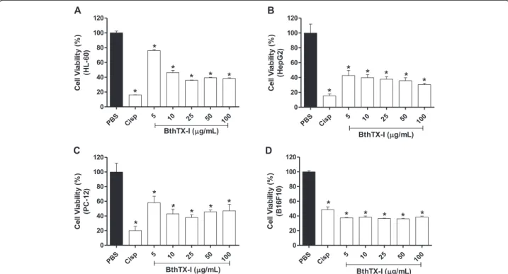
The antitumor activity of BthTX-I was assessed by treat-ing tumor cell lines with different concentrations of the myotoxin. For HL-60, it was observed that the cell via-bility after treatment with BthTX-I was of approximately 80 % for the lowest concentration evaluated (5 μg/mL),
while at the concentrations of 10, 25, 50 and 100μg/mL,
cell viabilities of 46; 36; 40 and 38 %, respectively, were observed (Fig. 1–a). For HepG2, BthTX-I concentrations from 5 to 25μg/mL led to cell viabilities of approximately
40 %, whereas at the concentrations of 50 and 100μg/mL,
the values of viability observed were of 35 % and 30 %, re-spectively (Fig. 1–b). For PC-12, cell viability ranged from 40 to 60 %, with lowest values observed after treatment with BthTX-I at the concentration of 25μg/mL (Fig. 1–c).
B16F10 cells showed around 40 % cell viability at all concentrations tested (Fig. 1–d).
Cell cycle kinetics analysis
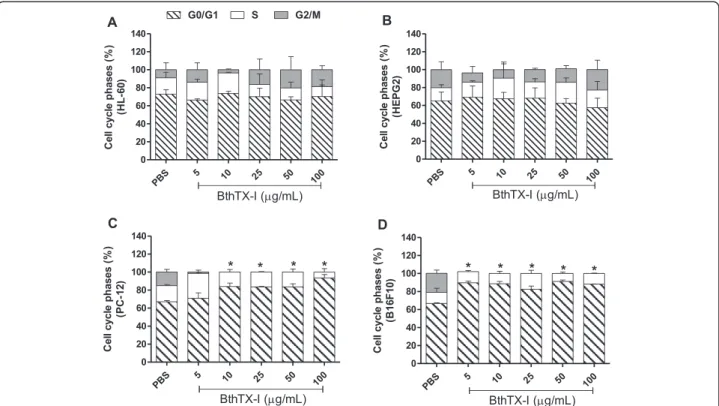
The cell cycle kinetics analysis was performed in order to detect the role of BthTX-I in the cell division process of tumor cells. HL-60 and HepG2 cells treated with dif-ferent concentrations of the toxin showed distribution in all three phases of the cell cycle (G0/G1, S, G2/M), with no significant differences (p< 0.05) in comparison with the negative control (PBS). Around 70 % of the HL-60 cells remained in G0/G1, 15 % in S and 15 % in G2/M phases (Fig. 2–a), while 70 % of HepG2 cells remained in G0/G1, 20 % in S and 10 % in G2/M phases (Fig. 2–b).
PC-12 and B16F10 cells treated with different concen-trations of BthTX-I showed delayed cell cycles, with an increased number of cells in the G0/G1 phase, followed by a reduction in the percentage of cells in the S phase and an absence of cells in the G2/M phase. The increase in the percentage of PC-12 cells in the G0/G1 phase, compared with the negative control, was of ~16 % for the treatments with 10; 25 and 50μg/mL of BtTX-I, and
increase for B16F10 cells was of approximately 24 % after treatment with BthTX-I at all the concentrations evaluated (Fig. 2–d).
Analysis of the apoptotic/necrotic effects of BthTX-I
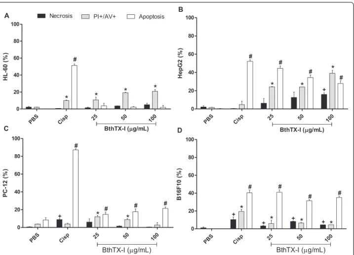
All concentrations of BthTX-I induced apoptosis/necro-sis (PI+/AV+) in leukemic cells (HL-60), with the highest percentage of PI+/AV+ cells (~20 %) observed at the concentrations of 50 and 100 μg/mL. There was also a
small percentage of cells that were only in necrotic process (1-5 %) at all the concentrations evaluated (Fig. 3–a).
The treatment of HepG2 cells with BthTX-I at the concentrations of 25 and 50 μg/mL induced apoptosis/
necrosis (PI+/AV+) in 24 % of the cell population, and apoptosis (AV+) in 44 and 34 % of cells, respectively. At the concentration of 100 μg/mL, 16 % of cells were in
the process of necrosis (PI+), 40 % in apoptosis/necrosis (PI+/AV+) and 28 % in apoptosis (AV+) (Fig. 3–b).
BthTX-I at the concentrations of 25 and 50 μg/mL
induced about 10 % of PC-12 cell death by apoptosis/ necrosis and 15 % by apoptosis. The concentration of 100μg/mL promoted apoptosis in approximately 20 %
of cells (Fig. 3–c).
The treatment of B16F10 cells with different concen-trations of BthTX-I (25, 50 and 100μg/mL) promoted a
high percentage of cell death by apoptosis (AV+) (30 to 40 %), with lower percentages of cells exclusively in
the processes of necrosis (3, 8 and 4 %, respectively) or apoptosis (~5 %) (Fig. 3–d).
Discussion
Many toxins isolated from snake venoms have been used as tools for understanding different pathophysiological events, since these proteins present several biological ef-fects such as antiparasitic, antimicrobial and antitumor activities [22–24]. In that context, snake venom PLA2s of both acidic and basic character, and also synthetic peptides derived from homologous PLA2s, have been extensively studied due to their wide variety of pharmaco-logical effects, including their antitumor and antiangiogenic properties [25–27].
In recent years, PLA2s have been investigated as tools for better understanding the mechanisms related to cancer. This class of toxins seems to act directly on the metabolism of phospholipid membranes, promoting changes in the lipid biosynthesis and deregulation of lipogenesis in dif-ferent cell lines, including tumorous ones [28].
In the present study, BthTX-I induced significant cytotox-icity in all tested tumor cell lines. In general, all concentra-tions of the myotoxin inhibited the viability of HL-60, HepG2, PC-12 and B16F10 cells in 50 to 60 %. These re-sults are consistent with many others which have shown that these toxins have the ability to induce death of various tumor and non-tumor cell lines [11, 29, 30].
Fig. 1Cell viability of the tumor cell lines HL-60 (a), HepG2 (b), PC-12 (c) and B16F10 (d) after treatment with BthTX-I. The cytotoxicity was evaluated by the MTT method 24 h after treatment of tumor cells with BthTX-I (5–100μg/mL). Results expressed as mean ± SD of three independent
Cisplatin (Incel, Darrow®) was used in the present study as a positive control of cytotoxicity, being able to promote death of all tumor cell lines evaluated, with lower effects on B16F10 cells. It is an inorganic platinum compound that binds to DNA and provokes structural changes and inhibition of transcription and replication, thus inducing apoptosis. Considering its cytotoxic effects, cisplatin is widely used as an antineoplastic agent in the treatment of several types of cancer [31].
Gebrimet al.[18] evaluated thein vitroantitumor ac-tivity of BthTX-I using tumor cell lines such as Jurkat, SKBR3 and B16F10, and showed that the native protein presented 75 to 90 % cytotoxicity on these tumor cell lines. Regardingin vivoexperiments, injection of BthTX-I modified with p-bromophenacyl bromide (BPB, an agent that inhibits PLA2s by covalently binding to their active site) in mice transplanted with S180 tumor cells reduced 30 % of the tumor size on the 14th day and 76 % on the 60th day, when compared to the untreated control group. These findings are in accordance with other studies that suggested that the antitumor properties of PLA2s are inde-pendent of their catalytic activity [32, 33].
De Moura et al. [11] evaluated the cytotoxic effect of three PLA2s isolated from B. mattogrossensis venom, BmatTX-I (Lys49), BmatTX-II (Lys49) and BmatTX-III
(Asp49) on Jurkat and SKBR-3 tumor cells. These authors observed that the Lys49 myotoxins were more cytotoxic than the Asp49 PLA2to both tumor cells. Similar to that observed for BthTX-I, the toxins BmatTX-I and BmatTX-II at a concentration of 100μg/mL inhibited the cell viability
of Jurkat cells in approximately 50 %. Equivalent effects were observed when Jurkat cells were treated with 100μg/
mL of two acidic Asp49 PLA2s, BmooTX-I fromB. moojeni venom and MTX-I fromB. brazilivenom [34, 35]. BthA-I-PLA2, an acidic PLA2 from B. jararacussu venom, also showed potential antitumor effects on Jurkat, SKBR-3 and Ehrlich ascites tumor (EAT) cells, with the enzyme at 100μg/mL promoting death of 50 to 70 % of the tumor
cells [36].
Studies suggest that several biological effects of the myotoxins are related to the interaction of their C-ter-minal region with cell membranes, which explains the syn-thesis of peptides derived from this region described in different studies [18, 35, 37]. The C-terminal region is usu-ally described as capable of disrupting the hydrophilic matrix of membranes, opening pores and allowing the entrance of the toxins into the intracellular medium [38].
been proposed for components such as L-amino acid oxidases (LAAOs), which have been described as potent cytotoxic agents. Several studies attributed the cytotoxic ef-fects of LAAOs mainly to the release of hydrogen peroxide to the medium, resulting in oxidative stress and conse-quently in cell death [7, 39].
The effect of cytotoxic agents can be evaluated by cell cycle analyses. The cell cycle is a series of continuous and repetitive processes that occur during a normal cell division [40]. The cycle is divided into interphase and mitosis. The interphase is further subdivided into the phases G0, G1, S and G2, in which the DNA duplication and preparation of cells for the next stage occurs. The mitosis is characterized by the cell division process itself [40, 41].
Flow cytometry experiments were performed for the cell cycle analyses, with the purpose of evaluating the ef-fects of BthTX-I in the process of cell division. Nuclear DNA of cells was stained by a specific fluorochrome (PI),
allowing its quantification and reflecting the position of cells in the cell cycle. BthTX-I was not able to induce any significant change in the cell cycle of HL-60 and HepG2 tumor cells. Conversely, the toxin caused a delay in the G0/G1 phase of the cell cycle of PC-12 and B16F10 tumor cells. The fact that the myotoxin promoted delay in the cell cycle indicates that it disrupts the mitotic progression and hence a smaller number of cells are doubled, which would implicate an important antitumor mechanism.
Although few studies have been conducted with snake venom toxins regarding their effects on the cell cycle progression, some other snake toxins were also described as able to promote delay of the cell cycle in the G0/G1 phase [6, 42]. Checkpoint proteins (CKI’s, CDK’s and CHK) are probably preventing these cells from leaving the G0/G1 phase. The CKI’s are able to suspend the cell cycle at any sight of DNA damage, directing the cells to repair systems. The cell cycle only continues when the Fig. 3Assessment of apoptotic/necrotic effects of BthTX-I on the tumor cell lines HL-60 (a), HepG2 (b), PC-12 (c) and B16F10 (d) by flow cytometry. Tumor cells were treated with BthTX-I at different concentrations (25, 50 or 100μg/mL). Cisplatin (Cisp, positive control) was used as a reference for the
damaged DNA is restored, and in case the repair systems are unable to correct the damage, cell death processes are induced [43].
Exposure to toxic agents can trigger cell death by pro-cesses of apoptosis or necrosis, and both propro-cesses occur depending on the intensity and duration of exposure. Necrosis is an accidental form of cell death in which a failure in the cell homeostasis occurs after the cell is damaged, leading to an inflammatory process [44–46]. Apoptosis is a controlled form of cell death, defined by a number of biochemical and morphological characteristics, such as the exposure of phosphatidylserine to the outside of the plasma membrane, the cell nucleus condensation and the cleavage of chromatin (DNA) in oligonucleosomal fragments [47].
The apoptosis-inducing ability of BthTX-I was assessed by flow cytometry using annexin V conjugated to fluores-cein, which acts as a marker for the phosphatidylserine ex-posed on cells undergoing apoptosis stage. The necrotic effect was analyzed by staining with propidium iodide (PI), which marks the nucleic acid of dead cells.
Our results showed that different BthTX-I concentra-tions induced tumor cell death by processes of apoptosis and/or necrosis. The myotoxin mainly induced apoptosis (AV+) in PC-12 and B16F10 cells at all concentrations tested (25, 50 or 100μg/mL), which suggests this process
as the predominant cell death mechanism induced by BthTX-I on these murine tumor cells. Regarding hu-man tumor cell lines (HL-60 and HepG2), apparently distinct death mechanisms could be observed: the percent-age of HepG2 cells in apoptosis (AV+) was practically equal to those in apoptosis/necrosis (PI+/AV+), while for HL-60 cells, the major percentage of cells was marked as PI+/AV+, indicating death mechanisms related to both apoptosis and necrosis.
These findings corroborate the data obtained for other enzymes isolated from snake venoms, which promoted death in various cell lines by different mechanisms, such as apoptosis and necrosis [48, 49]. Several factors may determine whether cells go into apoptosis or necrosis, such as the toxin concentration, the ability of cells to manage local membrane perturbations and the meta-bolic status of cells [50]. Mora et al.[9] evaluated the induction of apoptosis and necrosis by a Lys49 PLA2 homologue from Bothrops asper snake venom in a lymphoblastoid cell line, and concluded that the en-zyme was able to induce significantly different effects depending on the concentration of toxin used.
Conclusion
BthTX-I was shown to be cytotoxic for both human (HepG2 and HL-60) and murine (PC-12 and B16F10) tumor cell lines by inducing cell death mechanisms of apoptosis and/or necrosis. Additionally, BthTX-I was able
to promote delay in the G0/G1 phase of the cell cycle of murine tumor cells. The results revealed important functional characteristics of this myotoxin isolated fromB. jararacussuvenom, demonstrating a remarkable antitumor potential still little explored. Studies on the mechanisms of action of BthTX-I are very important so that it may serve as a model for the development of new therapeutic agents to treat various human diseases such as cancer in the future.
Competing interests
The authors declare that they have no competing interests.
Authors’contributions
CPS, RMAP and ACOC conceived and performed the experiments of this study. CPS, TRC and DLM analyzed the results and drafted the manuscript. LMGA and SVS supervised and critically discussed the study, and contributed with infrastructure and materials. All authors read and approved the final version of the manuscript.
Acknowledgments
The authors would like to thank the financial support provided by the State of São Paulo Research Foundation (FAPESP, grants n. 2010/03243-43 and 2011/23236-4), the Coordination for the Improvement of Higher Education Personnel (CAPES) and the National Council for Scientific and Technological Development (CNPq process n. 476932/2012-2). We are also grateful to Fabiana Rosseto Morais, from FCFRP-USP, for the technical assistance in the flow cytometry analyses. Thanks are also due to the Center for the Study of Venoms and Venomous Animals (CEVAP) of UNESP for enabling the publication of this special collection (CNPq process 469660/2014-7).
Received: 6 February 2015 Accepted: 26 October 2015
References
1. World Health Organization. Cancer. http://www.who.int/mediacentre/ factsheets/fs297/en/(2012). Accessed 20 Jan 2015.
2. American Cancer Society. Cancer facts and figures 2014. Atlanta: American Cancer Society, 2014. http://www.cancer.org/research/cancerfactsstatistics/ cancerfactsfigures2014/. Accessed 20 Jan 2015.
3. Abdel-Rahman MA, Abdel-Nabi IM, El-Naggar MS, Abbas OA, Strong PN. Conus vexillumvenom induces oxidative stress in Ehrlich’s ascites carcinoma cells: an insight into the mechanism of induction. J Venom Anim Toxins incl Trop Dis. 2013;19(1):10.
4. Wang L, Martins-Green M. Pomegranate and its components as alternative treatment for prostate cancer. Int J Mol Sci. 2014;15(9):14949–66.
5. Guo G, Yao G, Zhan G, Hu Y, Yue M, Cheng L, et al. N-methylhemeanthidine chloride, a novel Amaryllidaceae alkaloid, inhibits pancreatic cancer cell proliferation via down-regulating AKT activation. Toxicol Appl Pharmacol. 2014;280(3):475–83.
6. Alves-Paiva RM, de Freitas Figueiredo R, Antonucci GA, Paiva HH, de Lourdes Pires Bianchi M, Rodrigues KC, et al. Cell cycle arrest evidence, parasiticidal and bactericidal properties induced by L-amino acid oxidase fromBothrops atroxsnake venom. Biochimie. 2011;93(5):941–47.
7. Costa TR, Burin SM, Menaldo DL, de Castro FA, Sampaio SV. Snake venom L-amino acid oxidases: an overview on their antitumor effects. J Venom Anim Toxins incl Trop Dis. 2014;20:23.
8. Calderon LA, Sobrinho JC, Zaqueo KD, de Moura AA, Grabner AN, Mazzi MV, et al. Antitumoral activity of snake venom proteins: new trends in cancer therapy. Biomed Res Int. 2014. doi:10.1155/2014/203639.
9. Mora R, Valverde B, Díaz C, Lomonte B, Gutiérrez JM. A Lys49 phospholipase A(2) homologue fromBothrops aspersnake venom induces proliferation, apoptosis and necrosis in a lymphoblastoid cell line. Toxicon. 2005;45(5):651–60.
11. de Moura AA, Kayano AM, Oliveira GA, Setúbal SS, Ribeiro JG, Barros NB, et al. Purification and biochemical characterization of three myotoxins from Bothrops mattogrossensissnake venom with toxicity againstLeishmaniaand tumor cells. Biomed Res Int. 2014. doi:10.1155/2014/195356.
12. Gutiérrez JM, Chaves F, Cerdas L. Inflammatory infiltrate in skeletal muscle injected withBothrops aspervenom. Rev Biol Trop. 1986;34(2):209–14. 13. Teixeira CF, Landucci EC, Antunes E, Chacur M, Cury Y. Inflammatory effects
of snake venom myotoxic phospholipases A2. Toxicon. 2003;42(8):47–962. 14. Gutiérrez JM, Lomonte B. Phospholipase A2myotoxins fromBothropssnake
venoms. Toxicon. 1995;33(11):1405–24.
15. Soares AM, Giglio JR. Chemical modifications of phospholipases A2from snake venoms: effects on catalytic and pharmacological properties. Toxicon. 2003;42(8):855–68.
16. Homsi-Brandeburgo MI, Queiroz LS, Santo-Neto H, Rodrigues-Simioni L, Giglio JR. Fractionation ofBothrops jararacussusnake venom: partial chemical characterization and biological activity of bothropstoxin. Toxicon. 1988;26(7):615–27.
17. Chioato L, Aragão EA, Lopes Ferreira T, Medeiros AI, Faccioli LH, Ward RJ. Mapping of the structural determinants of artificial and biological membrane damaging activities of a Lys49 phospholipase A2by scanning alanine mutagenesis. Biochim Biophys Acta. 2007;1768(5):1247–57. 18. Gebrim LC, Marcussi S, Menaldo DL, de Menezes CS, Nomizo A, Hamaguchi
A, et al. Antitumor effects of snake venom chemically modified Lys49 phospholipase A2-like BthTX-I and a synthetic peptide derived from its C-terminal region. Biologicals. 2009;37(4):222–9.
19. Cintra AC, Marangoni S, Oliveira B, Giglio JR. Bothropstoxin-I: amino acid sequence and function. J Protein Chem. 1993;12(1):57–64.
20. Mosmann T. Rapid colorimetric assay for cellular growth and survival: application to proliferation and cytotoxicity assays. J Immunol Methods. 1983;65(1–2):55–63.
21. Kommoju PR, Macheroux P, Ghisla S. Molecular cloning, expression and purification of L-amino acid oxidase from the Malayan pit viper Calloselasma rhodostoma. Protein Expr Purif. 2007;52(1):89–95.
22. Costa Torres AF, Dantas RT, Toyama MH, Diz Filho E, Zara FJ, de Queiroz MG R, et al. Antibacterial and antiparasitic effects ofBothrops marajoensisvenom and its fractions: Phospholipase A2and L-amino acid oxidase. Toxicon. 2010;55(4):795–804.
23. Vargas LJ, Quintana JC, Pereañez JA, Núñez V, Sanz L, Calvete J. Cloning and characterization of an antibacterial L-amino acid oxidase fromCrotalus durissus cumanensisvenom. Toxicon. 2013;64:1–11.
24. Doumanov J, Mladenova K, Topouzova-Hristova T, Stoitsova S, Petrova S. Effects of vipoxin and its components on HepG2 cells. Toxicon. 2015;94:36–44. 25. Soares AM, Guerra-Sá R, Borja-Oliveira CR, Rodrigues VM, Rodrigues-Simioni L, Rodrigues V, et al. Structural and functional characterization of BnSP-7, a Lys49 myotoxic phospholipase A(2) homologue fromBothrops neuwiedi pauloensisvenom. Arch Biochem Biophys. 2000;378(2):201–9.
26. Cavalcante WL, Silva MD, Gallacci M. Influence of temperature upon paralyzing and myotoxic effects of bothropstoxin-I on mouse neuromuscular preparations. Chem Biol Interact. 2005;151(2):95–100.
27. Araya C, Lomonte B. Antitumor effects of cationic synthetic peptides derived from Lys49 phospholipase A2homologues of snake venoms. Cell Biol Int. 2007;31(3):263–8.
28. Mashima T, Seimiya H, Tsuruo T. De novo fatty-acid synthesis and related pathways as molecular targets for cancer therapy. Br J Cancer. 2009;100(9):1369–72.
29. Mora-Obando D, Díaz C, Angulo Y, Gutiérrez JM, Lomonte B. Role of enzymatic activity in muscle damage and cytotoxicity induced byBothrops asperAsp49 phospholipase A2myotoxins: are there additional effector mechanisms involved? PeerJ. 2014;2:e569.
30. Gasanov SE, Dagda RK, Rael ED. Snake venom cytotoxins, phospholipase A2s, and Zn2+-dependent metalloproteinases: mechanisms of action and pharmacological relevance. J Clin Toxicol. 2014;4(1):1000181.
31. Pabla N, Dong Z. Curtailing side effects in chemotherapy: a tale of PKCO in cisplatin treatment. Oncotarget. 2012;3(1):107–11.
32. Stábeli RG, Amui SF, Sant’Ana CD, Pires MG, Nomizo A, Monteiro MC, et al. Bothrops moojenimyotoxin-II, a Lys49-phospholipase A2homologue: an example of function versatility of snake venom proteins. Comp Biochem Physiol C Toxicol Pharmacol. 2006;142(3–4):371–81.
33. Rodrigues RS, Izidoro LF, de Oliveira Jr RJ, Sampaio SV, Soares AM, Rodrigues VM. Snake venom phospholipases A2: a new class of antitumor agents. Protein Pept Lett. 2009;16(8):894–8.
34. Santos-Filho NA, Silveira LB, Oliveira CZ, Bernardes CP, Menaldo DL, Fuly AL, et al. A new acidic myotoxic, anti-platelet and prostaglandin I2 inductor phospholipase A2isolated fromBothrops moojenisnake venom. Toxicon. 2008;52(8):908–17.
35. Costa TR, Menaldo DL, Oliveira CZ, Santos-Filho NA, Teixeira SS, Nomizo A, et al. Myotoxic phospholipases A(2) isolated fromBothrops brazilisnake venom and synthetic peptides derived from their C-terminal region: cytotoxic effect on microorganism and tumor cells. Peptides. 2008;29(10):1645–56. 36. Roberto PG, Kashima S, Marcussi S, Pereira JO, Astolfi-Filho S, Nomizo A,
et al. Cloning and identification of a complete cDNA coding for a bactericidal and antitumoral acidic phospholipase A2fromBothrops jararacussuvenom. Protein J. 2004;23(4):273–85.
37. Lomonte B, Angulo Y, Moreno E. Synthetic peptides derived from the C-terminal region of Lys49 phospholipase A2homologues from viperidae snake venoms: biomimetic activities and potential applications. Curr Pharm Des. 2010;16(28):3224–30.
38. Montecucco C, Gutiérrez JM, Lomonte B. Cellular pathology induced by snake venom phospholipase A2myotoxins and neurotoxins: common aspects of their mechanisms of action. Cell Mol Life Sci. 2008;65(18):2897–912. 39. Costa TR, Menaldo DL, Prinholato da Silva C, Sorrechia R, Albuquerque S,
Pietro RC, et al. Evaluating the microbicidal, antiparasitic and antitumor effects of CR-LAAO fromCalloselasma rhodostomavenom. Int J Biol Macromol. 2015;80:489–97.
40. Vermeulen K, Van Bockstaele DR, Berneman ZN. The cell cycle: a review of regulation, deregulation and therapeutic targets in cancer. Cell Prolif. 2003;36(3):131–49.
41. Schafer KA. The cell cycle: a review. Vet Pathol. 1998;35(6):461–78. 42. Zhang L, WU WT. Isolation and characterization of ACTX-6: a cytotoxic
L-amino acid oxidase fromAgkistrodon acutussnake venom. Nat Prod Res. 2008;22:554–63.
43. Douglas RM, Haddad GG. Genetic models in applied physiology: invited review: effect of oxygen deprivation on cell cycle activity: a profile of delay and arrest. J Appl Physiol. 2003;94(5):2086–3.
44. Dypbukt JM, Ankarcrona M, Burkitt M, Sjöholm A, Ström K, Orrenius S, et al. Different prooxidant levels stimulate growth, trigger apoptosis, or produce necrosis of insulin-secreting RINm5F cells. The role of intracellular polyamines. J Biol Chem. 1994;269(48):30553–60.
45. Bonfoco E, Krainc D, Ankarcrona M, Nicotera P, Lipton SA. Apoptosis and necrosis: two distinct events induced, respectively, by mild and intense insults with N-methyl-D-aspartate or nitric oxide/superoxide in cortical cell cultures. Proc Natl Acad Sci USA. 1995;92(16):7162–6.
46. Ande SR, Kommoju PR, Draxl S, Murkovic M, Macheroux P, Ghisla S, et al. Mechanisms of cell death induction by L-amino acid oxidase, a major component of ophidian venom. Apoptosis. 2006;11(8):1439–51. 47. Schultz DR, Harrington Jr WJ. Apoptosis: programmed cell death at a
molecular level. Semin Arthritis Rheum. 2003;32(6):345–69.
48. Samel M, Tõnismägi K, Rönnholm G, Vija H, Siigur J, Kalkkinen N, et al. L-Amino acid oxidase fromNaja naja oxianavenom. Comp Biochem Physiol B Biochem Mol Biol. 2008;149(4):572–80.
49. Ciscotto P, de Avila RA M, Coelho EA, Oliveira J, Diniz CG, Farías LM, et al. Antigenic, microbicidal and antiparasitic properties of an l-amino acid oxidase isolated fromBothrops jararacasnake venom. Toxicon. 2009;53(3):330–41. 50. Leist M, Single B, Castoldi A, Kuhnle S, Nicotera P. Intracellular adenosine


