R E S E A R C H
Open Access
Toxins from the Caribbean sea anemone
Bunodeopsis globulifera increase
cisplatin-induced cytotoxicity of lung
adenocarcinoma cells
Heidi I Monroy-Estrada
1,2,3, Yolanda I Chirino
2, Irma E Soria-Mercado
1and Judith Sánchez-Rodríguez
3*Abstract
Background:Lung cancer causes 1.4 million deaths worldwide while non-small-cell lung cancer (NSCLC) represents 80-85% of the cases. Cisplatin is a standard chemotherapy against this type of cancer; however, tumor cell
resistance to this drug limits its efficacy. Sea anemones produce compounds with pharmacological activities that may be useful for augmenting cisplatin efficacy. This study aimed to evaluate the pharmacological activities of crude venom (CV) from the sea anemoneBunodeopsis globuliferaand four derived fractions (F1, F2, F3 and F4) to test their increase efficiency cisplatin cytotoxicity in human lung adenocarcinoma cells.
Results:Pre-exposure to CV, F1 and F2 fractions increases cisplatin cytotoxicity in human lung adenocarcinoma cells under specific conditions. Exposure to CV at 50μgmL-1induced a reduction of approximately 50% in cell viability, while a similar cytotoxic effect was observed when cell culture was exposed to F1 at 25μgmL-1or F2 at 50μgmL-1. The cell culture exposure to F1 (10μgmL-1) fraction combined with cisplatine (25μM) provoked a decrease in MTT reduction until 65.57% while F2 (25μgmL-1) fraction combined with cisplatin (10μM) provoked a decrease in MTT reduction of 72.55%.
Conclusions:The F1 fraction had the greatest effect on the lung adenocarcinoma cell line compared with CV and F2. The combination of antineoplastic drugs and sea anemone toxins might allow a reduction of chemotherapeutic doses and thus mitigate side effects.
Keywords:Cnidaria, Pharmacology, Human lung cancer cells, Cytotoxicity assay, Cisplatin efficacy
Background
According to the International Agency for Research on Cancer (IARC) and World Health Organization, cancer is a leading cause of death worldwide, accounting for 7.6 million deaths (around 13% of all deaths) in 2008. Lung cancer causes 1.4 million deaths, followed by sto-mach (740,000 deaths), liver (700,000 deaths), colorectal (610,000 deaths) and breast cancer (460,000 deaths). Around 70% of all cancer deaths occurred in low- and middle-income countries. Projections to 2030, indicate about 11 million of cancer deaths worldwide and it is
expected that non-small-cell lung cancer (NSCLC) will represent 80-85% of cases.
NSCLC, which comprises adenocarcinoma, squamous cell carcinoma and large-cell carcinoma, is an exclusion diagnosis [1]. Platinum-based chemotherapeutics are the most common regimens for NSCLC patients but re-sistance and side effects have limited their efficiency [2-4]. In this regard,Cis-diammine-dicholoplatinium (II), known as cisplatin, is an antineoplastic drug widely used in the treatment of many solid tumors, including lung, head, neck, ovary, breast, colorectal and cervical cancers [5,6]. However, the usage of cisplatin in cancer treatment is limited due to resistance and side effects including nephrotoxicity [7].
* Correspondence:[email protected]
3Unidad Académica de Sistemas Arrecifales, Puerto Morelos, Instituto de
Ciencias del Mar y Limnología, Universidad Nacional Autónoma de México, Puerto Morelos, Quintana Roo State, Mexico
Full list of author information is available at the end of the article
The abovementioned problem has led to the development of new strategies to achieve effectiveness against cancer. In this sense, the peptides and proteins found in marine venoms may increase the efficacy of conventional chemo-therapeutic drugs [8]. In particular, sea anemones produce peptides and proteins that act as cytolysins which are classi-fied based on their molecular weight as follows: 5-8 kDa peptides with antihistamine activity; ~20 kDa pore-forming proteins; ~30-40 kDa cytolysins with or without phospholi-pase A2activity; and a putative protein group ~80 kDa [9].
An essential feature of cytolysins is their ability to form pores in biological membranes, which lead to osmotic changes and loss of intracellular metabolites thereby provoking cell death [10,11]. However, cytolysins may induce cell damage in both normal and cancer cells if attached to their membrane. Nevertheless, characte-ristics of cancer cells are different from normal cells, including gene profile expression, metabolism and mem-brane features. In this regard, it has been demonstrated that cytolysins can induce more damage in cancer cells due to their differences from normal cells. For this rea-son, cytolysins have been proposed as an alternative for cancer therapy [8,12,13].
For example, actinoporins isolated from the tropical sea anemone Heteractis crispa have shown anticancer properties through the induction of p53-independent apoptosis [14]. Bunodeopsis globulifera – found in the reef lagoon at Puerto Morelos, Quintana Roo, Mexico–
has been reported as producing a highly dangerous sting, probably due to the toxins it contains [15]. However, the potential benefitsof B. globuliferahave been not been doc-umented. In the present work we hypothesized that toxins fromB. globulifera could increase cytotoxicity induced by an antineoplastic agent against cancer cells. Specifically, we are interested in lung cancer cells because the inci-dence and mortality rates are among the highest in the world. This study aimed to investigate the ability of crude venom (CV) and fractions extracted from the sea anemone
B. globuliferato increase the cisplatin-induced cytotoxicity in lung adenocarcinoma cells. Our results showed for first time the ability of Bunodeopsis globulifera CV and frac-tions to increase the cytotoxic effect of a common antineo-plastic agent used in several types of cancer.
At this point it should be mentioned that toxins from
Bunodeopsis globulifera are cytotoxic like other cnidarian venoms. However, the purpose of this work is to test whether the usage of low concentrations of toxins can in-crease the effect of a low concentration of a common antineoplasic agent. The general objective of this research is to design a strategy to decrease the concentrations of cisplatin and toxins, since this could be helpful in avoiding the side effects of higher concentrations. For example, cis-platin is useful to treat lung cancer but provokes nephro-toxicity as its main side effect. The combination of two
drugs may facilitate the diminution of the side effects asso-ciated with higher doses.
Methods Reagents
F-12K Nutrient Mixture medium was purchased from Gibco Invitrogen (USA). RPMI-1640 medium was obtained from In Vitro, S.A (Mexico). Fetal bovine serum (FBS) was from PAA Laboratories (Canada). Cisplatin (cat. n. P4394) and 3-(4,5-dimethylthiazol-2-yl)- 2,5-diphenyltetrazolium bromide (MTT; cat. n. M2128) were purchased from Sigma Chemical Co. (USA). Lactate dehydrogenase kit (cat. n. 630117) was obtained from Clontech Laboratories, Inc (USA). Quick Start Bradford Protein Assay Kit (cat. n. 500-0202) was bought from Bio-Rad Laboratories, Inc (USA). Sephadex G-50 Medium Gel (cat. n. 17-0043-02) was obtained from Pharmacia Biotech (Sweden).
Cell cultures
Human A549 non-small-cell lung cancer line, which is an adenocarcinoma epithelial-derived cell line, was obtained from the American Type Culture Collection (ATCC, USA). Cells were cultured in monolayers at 37°C in a humidified atmosphere of 5% CO2 in F12K medium supplemented with 10% heat-inactivated FBS.
Isolation and Pre-purification of crude venom
Specimens of Bunodepsis globulifera were collected from the reef lagoon at Puerto Morelos, Quintana Roo, Mexico, by scuba diving between February and June, 2009 and from May to June, 2011. The sea anemones were identified by Ricardo Gonzalez-Muñoz. The crude venom was obtained by mincing and homogenizing the whole body of the sea anemone, and then centrifuging at 3200 g for 15 minutes at 4°C, after which the supernatant was lyophi-lized. The crude lyophilized venom (4 g) was purified by Sephadex G-50 M gel filtration column (85 × 5 cm) equili-brated with 0.3 M acetic acid buffer and eluted with the same buffer at a flow rate of 2 mL min-1. The four fractions obtained were concentrated and lyophilized.
Cytotoxicity assays
Lung adenocarcinoma cells (1 × 10⁴per well/150μL) were
incubated in 96-well plates and cultured for 24 hours to allow adherence. After this time, cell culture was exposed to the following treatments: 1, 10, 25, 50, 75, 100 and 200 μgmL-1; and 10 and 25 μgmL-1 of CV, BgG50F1 and
BgG50F2 fractions ofB. globuliferafor 24 hours, 37°C and 5% of CO2atmosphere. After each treatment, the cells were exposed to cisplatin 10μM or cisplatin 25μM for 24 hours.
Cell viability assay and cytotoxicity
MTT at 37°C in an atmosphere of 5% CO2for 2 hours. The medium was removed and the formazan blue crystals were dissolved in isopropyl alcohol for measuring the optical density at 540 nm [16].
The cytotoxicity was measured using a lactate de-hydrogenase (LDH) cytotoxicity detection kit according to the manufacturer’s protocol. Briefly, supernatants from cell treatments were collected and diluted 1:3 with PBS. After that, supernatant dilution was mixed 1:1 with dye solution and incubated at 37°C for 30 minutes under agitation; and absorbance was measured at 540 nm.
Quantification of protein
The protein content was measured by the Bradford assay [17] using bovine gamma globulin (BGG) as standard.
SDS-PAGE electrophoresis
The obtained fractions were assessed by SDS-PAGE according to Laemmli [18] using a 12% polyacrylamide gel for the molecular weight determination.
Statistical analysis
Data were expressed by mean ± SD, followed by one-way analysis of variance (ANOVA) with Bonferroni’s multiple comparison tests, performed using GraphPad Prism for Windows (GraphPad Software, USA).p< 0.001 was con-sidered to be statistically significant.
Results
Pre-purification of crude venom fromB. Globulifera
Crude venom (CV) of B. globulifera was obtained by mincing and homogenizing the whole body of the sea anem-one. It was centrifuged at 3200gfor 15 minutes at 4°C, and the supernatant was lyophilized. Crude venom was purified
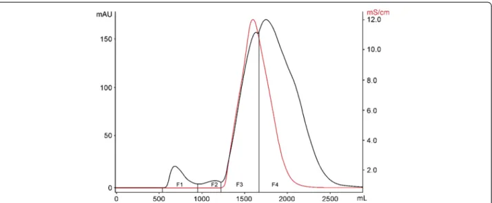
by gel filtration in a Sephadex G-50 M column and eluted with 0.3 M acetic acid buffer at a flow rate of 2 mL min-1. Four fractions–F1, F2, F3 and F4–were obtained. There-after, fractions were vacuum-concentrated and lyophilized (Figure 1). Table 1 shows the protein content of CV, F1, F2, F3 and F4 quantified by the Bradford method [17].
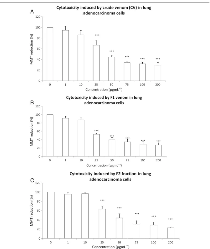
The cytotoxic effects of CV as well as the F1, F2, F3 and F4 fractions were tested on a human lung adenocarcinoma cell line using an MTT assay. The exposure to 1μgmL-1or
10μgmL-1 to CV did not induce changes in MTT
reduc-tion. However, CV at 25 μgmL-1, 50μgmL-1, 75 μgmL-1,
100μgmL-1and 200μgmL-1decreased cell viability by the
following percentages: 33.37, 55.46, 65.8, 68.36 and 70.44, respectively (Figure 2A). The exposure to F1 at 1 μgmL-1
or 10 μgmL-1 did not show a cytotoxic effect; however,
25 μgmL-1, 50 μgmL-1, 75 μgmL-1, 100 μgmL-1 and
200μgmL-1induced respective decreases in cell viability of
47.11%, 60.21%, 65.27%, 70.87% and 72.35% (Figure 2B). The exposure to F2 at 1μgmL-1or 10μgmL-1did not
pro-duce any change in MTT reduction, but 50 μgmL-1,
75 μgmL-1, 100 μgmL-1 and 200 μgmL-1 induced
cell-viability diminutions of 36.49%, 55.83%, 69%, 71.27% and 77.13%, respectively (Figure 2C). The F3 and F4 did not Figure 1Gel filtration chromatography Sephadex G-50 M of CV (4 g) fromB. globulifera.The column was equilibrated and eluted with 0.3 M acetic acid buffer at a flow rate of 2 mL min-1to obtain the fractions F1, F2, F3 and F4.
Table 1 Protein quantification of CV and fractions isolated fromB. globuliferaat 1 mgmL-1using the Bradford method
Sea anemone toxins Protein (μg)
Crude venom 32.4
Fraction F1 157.7
Fraction F2 152.6
Fraction F3 20.4
Figure 2Cytotoxicity induced by CV and the fractions F1 and F2 fromB. globuliferaat the concentrations 1, 10, 25, 50, 75, 100 and 200μgmL-1on lung adenocarcinoma cell culture.Effect of: (A) CV, (B) F1 fraction, (C) F2 fraction. Graphs show the percentage of MTT
reduction. Data are expressed as the mean ± SD of four independent experiments. Significance was defined as ***p< 0.001 (ANOVA, Bonferroni’s post test) in comparison with the untreated control values (100%).
alter MTT reduction (data not shown) and thus were not considered for further experiments in the present work.
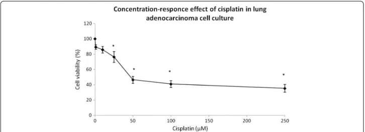
Inhibitory concentration (IC50) values for cisplatin
The cisplatin concentration that inhibited cell growth by 50% (IC50) was determined in a human lung adenocarci-noma cell line. A cisplatin concentration between 1 and 250μM was used in cell culture for 24 hours in three
inde-pendent experiments. After treatment, cytotoxicity was tested by MTT reduction and the IC50 found to be 50μM
(Figure 3). However, concentrations below IC50 were used to test whether CV and its fractions could enhance the cis-platin cytotoxicity. Human lung adenocarcinoma cells ex-posed to cisplatin at 1 and 10 μM remained without
changes in MTT reduction; however, cisplatin concentra-tions of 25, 50, 100 and 250μM induced respective
cell-viability decreases of 23.64%, 53.59%, 58.98% and 64.64% (Figure 3). We selected cisplatin concentrations of 10μM
and 25μM, which are below the IC50.
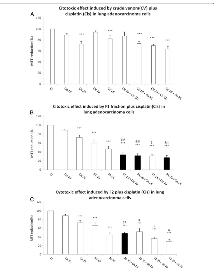
CV, F1 and F2 enhanced cisplatin-induced cytotoxicity
Lung adenocarcinoma cells were pre-exposed to CV, F1 or F2 at 10μgmL-1or 25μgmL-1for 24 hours. After this
time, the medium was removed and cell culture was washed and exposed to cisplatin at 10 μM or 25 μM.
The abovementioned strategy was executed in order to increase cisplatin cytotoxicity by pretreatment with CV, F1 or F2 at low concentration.
The pretreatment with CV at 10μgmL-1plus cisplatin at
25 μM yielded a 26.47% decrease in cell viability; the
pre-treatment with 25 μgmL-1 CV plus 10 μM cisplatin
aug-mented the cell-viability decrease to 30.22%. But the maximum effect was produced by 25 μgmL-1 CV plus
25μM cisplatin (Figure 4A). However, the pre-exposure to
CV did not increase the cisplatin-induced cytotoxicity (Figure 4A).
The pre-exposure to F1 at 10μgmL-1caused a decrease
in MTT reduction induced by 10μM and 25 μM of
cis-platin to 66.39% and 68.4%, respectively (Figure 4B). The pre-exposure to 25μgmL-1F1 had similar effect; it reduced
cell viability down to 72.55% after exposure to 25μM
cis-platin (Figure 4B). The 10μgmL-1 F2 increased the
cyto-toxicity induced by 10μM cisplatin to 51.56% (Figure 4C).
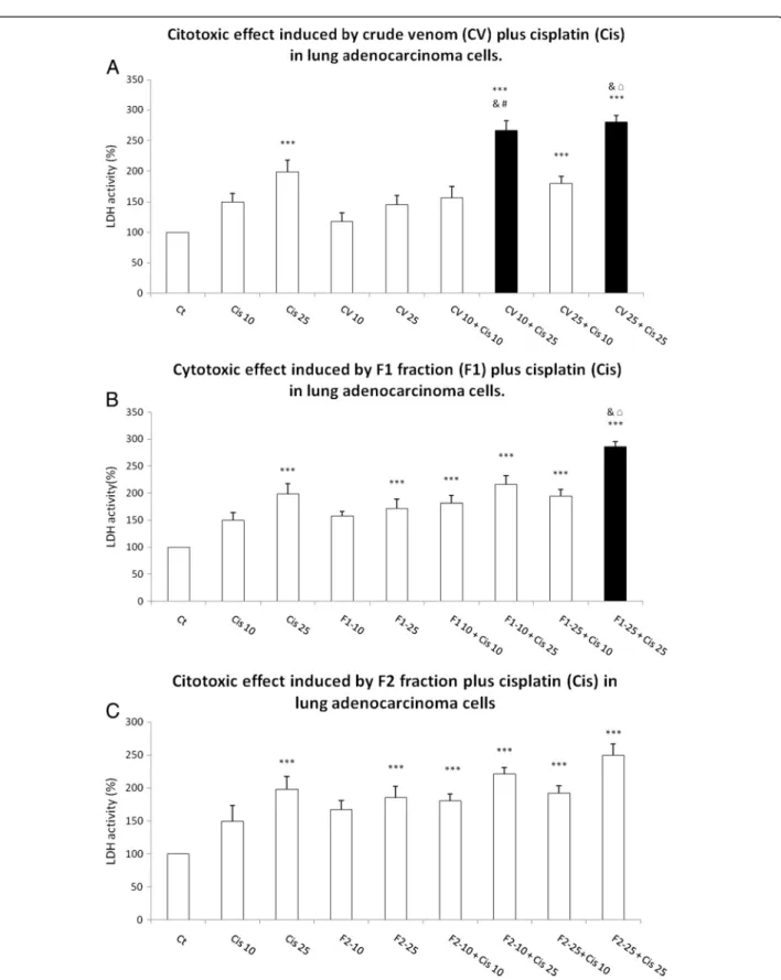
Then, while evaluating the LDH release into the cell cul-ture medium as a loss of membrane integrity, we observed that 10μgmL-1CV plus 25 μM cisplatin and 25μgmL-1
CV plus 25 μM cisplatin induced significant increases
of 2.665- and 2.799-fold in LDH release, respectively (Figure 5A). The pre-exposure to F1 at 25 μgmL-1 plus
cisplatin at 25 μM produced a significant 2.858 fold
in-crease in LDH release (Figure 5B). Pre-exposure to 10 and 25μgmL-1F2 showed no statistical increase in cytotoxicity
induced by 25 and 10μM cisplatin (Figure 5C).
Discussion
The present work showed that CV ofB. globuliferaand F1 and F2 obtained from pre-purification by Sephadex G-50 M gel filtration chromatography induced cytotoxicity in a human lung adenocarcinoma cell line. Exposure to CV at 50μgmL-1induced a reduction of approximately 50% in
cell viability, while a similar cytotoxic effect was observed when cell culture was exposed to F1 at 25μgmL-1or F2 at
50μgmL-1.
This cytotoxic effect has been also presented by other sea anemones, such as crude venoms of Heteractis magnifica, Stichodactyla haddoni and Parachondylactis sinensison mouse fibroblast cell line L929 and leukemia cell line P388. In those studies, the decrease in cell via-bility was concentration-dependent [19]. EqTX-II cytoly-sin toxin from the sea anemone Actinia equina also induces a decrease in the viability of V-79-379 A cells (diploid lung fibroblast from Chinese Hamster) in a Figure 3Concentration-response effect of cisplatin on lung adenocarcinoma cell culture: cell viability was measured by MTT assay.
Figure 4Cytotoxic effect induced by CV, F1 and F2 fraction fromB. globuliferaon adenocarcinoma lung cell culture.(A) CV, (B) F1 fraction plus cisplatin, (C) F2 fraction plus cisplatin. Graphs show the percentage of MTT reduction and data are expressed as the mean ± SD of four independent experiments. ***p< 0.001 vs. control,$p< 0.001 vs. Cis 10,&p< 0.001 vs. Cis 25,#p< 0.001 vs. CV 10 or F1-10 or F2-10, □p< 0.001 vs. CV 25 or F1-25 or F2-25 (One-way analysis of variance plus Bonferroni
’s test). Gray bars show the ability of (B) F1 and (C) F2 to augment the cisplatin decrease in MTT reduction.
Figure 5Cytotoxic effect induced by CV, F1 and F2 fraction fromB. globuliferaon adenocarcinoma lung cell culture: (A) CV, (B) F1 fraction plus cisplatin, (C) F2 fraction plus cisplatin.Graphs show the percentage of LDH release, with data expressed as the mean ± SD of four independent experiments. ***p< 0.001 vs. control,$p< 0.001 vs. Cis 10,&p< 0.001 vs. Cis 25,#p< 0.001 vs. CV 10 or F1-10 or F2-10, □p< 0.001 vs. CV 25 or F1-25 or F2-25 (one-way ANOVA, Bonferroni’s post test)(a)CV plus cis,(b)F1 plus cis,(c)F2 plus Cis. Gray bars showed
concentration-dependent manner [20]. RTX-A cytolysin from the sea anemoneHeteractis crispareduced cell via-bility of JB6 P+ Cl41 cells, Hela, THP-1, MDA-MB-231 and snu-c4 human tumor cell lines [14].
These results have shown that sea anemone toxins and some proteins derived from them exert cytotoxic activity on a non-small lung cancer cell lines, although the cyto-toxic mechanisms induced by the sea anemone toxins have been sparsely investigated. In this regard, Soletti et al. [8] showed that the cytolysins EqTx-II and Bc2 from sea anem-ones potentiated the cytotoxicity induced by low-dose con-centrations of the chemotherapeutic drugs against human glioblastoma cells through necrosis-like cell death [8].
In the present work, it has been observed that CV, F1 and F2 fractions from B. globulifera increase the cyto-toxic effect induced by cisplatin under specific condi-tions. The exposure to F1 at 10 μgmL-1 induced a
decrease of 40.02% in MTT reduction versus 11.12% by cisplatin. However, the pre-exposure to F1 (10 μgmL-1)
provoked a decrease in MTT reduction of 66.30% (Figure 4B). The combination of F1 (10 μgmL-1) and
25 μM cisplatin decreased the MTT reduction to
67.57%, which may indicate the absence of a difference between 10 μM and 25 μM cisplatin (Figure 4B). The
exposure to 25μgmL-1F1 induced a decrease of 53.42%
in MTT reduction but in combination with 25 μM
cisplatin, which by itself induced a decrease of 25.55% in MTT, there was a decrease of 72.55% (Figure 4B).
A similar effect was observed when lung adenocarci-noma cell line was exposed to 25μgmL-1F2 followed by
10μM cisplatin, which induced a decrease of 51.56% in
MTT reduction, while 25μgmL-1F2 induced only a
de-crease of 33.48% and 10 μM cisplatin a decrease of
11.12% in MTT reduction (Figure 4C).
LDH activity was measured in a cell culture medium, since this enzyme is released under conditions of cell damage, wherein we observed that 10 μgmL-1 CV
in-duced an increase of 17.5% of activity versus 98% by 25μM cisplatin; the combination induced 166.59% of
in-crease (Figure 5A). The augmentation of LDH activity was 45% after exposure solely to CV at 25 μgmL-1, but
rose to 1423.3% when this CV was combined with 25μM cisplatin (Figure 5A). This finding may signify the
absence of a real difference between 10 and 25 μgmL-1
CV. Similarly, the 71.6% increase in LDH activity pro-duced by treating cell culture with only F1 at 25μgmL-1
rose to 185.8% when the fraction was combined with 25μM cisplatin (Figure 5B).
Based on the results of the present work, we suggest not only that CV, F1 and F2 may present different mech-anisms of cell damage in lung adenocarcinoma cells, but also that CV may augment cell damage by compromi-sing the membrane, given the evident changes in LDH release (Figure 5A), and F2 fraction, which could cause
higher mitochondrial damage, since MTT reduction is related to mitochondrial activity (Figure 5C). However, F1 fraction may produce greater mitochondrial alter-ations and membrane damage (Figures 4B and 5B) than CV and F2 fraction.
Interestingly, this study proposes the usage of cisplatin at a dose below IC50, which in turn might represent fewer side effects. It is also interesting that cisplatin causes hardly any mitochondrial damage below 25 μM;
however, the F1 and F2 fractions caused more damage, as measured by MTT reduction, than cisplatin alone while their combination seems to produce an additive ef-fect (Figure 4B and C). It is also compelling that the combination of CV and F1 fraction with cisplatin ap-pears to have synergistic effect, specifically in LDH re-lease (Figure 5A and B).
Based on the data of the present work, we suggest that more studies are needed to investigate the cytotoxicity mechanism of CV, F1 and F2, which could help to de-sign a combinatory treatment to increase the effects of antineoplasic agents, including cisplatin. We also suggest that these compounds may execute their toxicity though a different cellular mechanism that includes mitochon-drial damage and alterations of the cell membrane.
Conclusions
We observed that CV of B. globuliferaand its F1 and F2 fractions obtained by Sephadex G-50 M gel filtration have a cytotoxic effect on the A549 lung cancer cell line. The F1 fraction had the greatest effect compared with CV and F2. The combination of antineoplastic drugs and sea anemone toxins might allow a reduction of chemotherapeutic doses and thus mitigate side effects. It will be necessary to evalu-ate not only the damage and action mechanisms in lung adenocarcinoma cells, but also whether these compounds are cytotoxic to different types of lung cancer cells and other cancer cell lines, including breast, cervix and colon cancers, each of which has a high incidence and prevalence.
Competing interests
The authors declare that there are no conflicts of interest.
Authors’contributions
HIME performed experiments; YIC designed experiments and drafted the manuscript; IESM designed experiments and drafted the manuscript; JSR conceived, designed, coordinate the study and drafted the manuscript. All authors read and approved the final manuscript.
Acknowledgments
This study was supported by the grant from Dirección General de Asuntos del Personal Académico (DGAPA) of the Universidad Nacional Autónoma de México (UNAM), grant number IN 202309 and IB 201112 and CONACyT grant number 127656. We thank the CONACYT for scholarship to HIME 190209, Rebeca Candia Zulbarán, Sergio Guendolain, Fernando Negrete-Soto, Cecilia Barradas, and Laura Celis Gutiérrez for their help in field and laboratory work. Thank Fanny Bernal for her support with cell cultures.
Author details
1Facultad de Ciencias Marinas, Universidad Autónoma de Baja California,
Ensenada, Baja California State, Mexico.2Unidad de Biomedicina, Facultad de Estudios Superiores Iztacala, Universidad Nacional Autónoma de México, Tlalnepantla, Estado de México, Mexico.3Unidad Académica de Sistemas Arrecifales, Puerto Morelos, Instituto de Ciencias del Mar y Limnología, Universidad Nacional Autónoma de México, Puerto Morelos, Quintana Roo State, Mexico.
Published: 7 May 2013
References
1. Travis WD, Rambilla E, Müller-Hermeling K, Harris CC (Eds):World Health Organization classification of tumours. Pathology and genetics of tumours of the lung, pleura, thymus and heart.Lyon: IARC Press; 2004:344.
2. Shen H, Hu D, Du J, Wang X, Liu Y, Wang Y, Wei J, Ma D, Wang P, Li L:
Paclitaxel-octreotide conjugates in tumor growth inhibition of A549 human non-small cell lung cancer xenografted into nude mice.Eur J Pharmacol2008,601(1–3):2329.
3. Panchal RG:Novel strategies to selectively kill cancer cells.Biochem Pharmacol1998,55(3):247–252.
4. Natarajan N, Thamaraiselvan R, Lingaiah H, Srinivasan P, Periyasamy BM:
Effect of flavonone hesperidin on apoptosis of human mammary carcinoma cell line MCF-7.Biomed Prev Nutr2011,1(3):207–215. 5. Köberle B, Tomicic MT, Usanova S, Kaina B:Cisplatin resistance: preclinical
findings and clinical implications.Biochim Biophys Acta2010,1806(2):172–182. 6. Pyo JS, Ju HK, Park JH, Kwon SW:Determination of volatile biomarkers for
apoptosis and necrosis by solid-phase microextraction-gas
chromatography/mass spectrometry: a pharmacometabolomic approach to cisplatin’s cytotoxicity to human lung cancer cell lines.J Chromatogr B Analyt Technol Biomed Life Sci2008,876(2):170–174.
7. Miller RP, Tadagavadi RK, Ramesh G, Reeves WB:Mechanisms of cisplatin nephrotoxicity.Toxins2010,2(11):2490–2518.
8. Soletti RC, de Faria GP, Vernal J, Terenzi H, Anderluh G, Borges HL, Moura-Neto V, Gabilan NH:Potentiation of anticancer-drug cytotoxicity by sea anemone pore-forming proteins in human glioblastoma cells.Anticancer Drugs2008,19(5):517–525.
9. Anderluh G, Macek P:Cytolytic peptide and protein toxins from sea anemones (Anthozoa: Actinaria).Toxicon2002,40(2):111–124. 10. Maček P, Zecchini M, Stanek K, Menestrina G:Effect of
membrane-partitioned n-alcohols and fatty acids on pore-forming activity of a sea anemone toxin.Eur Biophys J1997,25(3):155–162.
11. Tejuca M, Dalla Serra M, Potrich C, Álvarez C, Menestrina G:Sizing the radius of the pore formed in erythrocytes and lipid vesicles by the toxin Sticholysin I from the sea anemoneStichodactyla helianthus.J Membr Biol
2001,183(2):125–135.
12. Tejuca M, Anderluh G, Dalla Serra M:Sea anemone cytolysins as toxic components fo immnunotoxins.Toxicon2009,54(8):1206–1214. 13. Pentón D, Pérez-Barzaga V, Díaz I, Reytor ML, Campos J, Fando R, Calvo L,
Cilli EM, Morera V, Castellanos-Serra LR, Pazos F, Lanio ME, Álvarez C, Ponds T, Tejuca M:Validation of a mutant of the pore-forming toxin sticholysin-I for the construction of proteinase-activated immunotoxins.Protein Eng Des Sel2011,24(6):485–493.
14. Fedorov S, Dyshlovoy S, Monastyrnaya M, Shubina L, Leychenko E, Kozlovskaya E, Jin JO, Kwaj JY, Bode AM, Dong Z, Stonick V:The anticancer effects of actinoporin RTX-A from the sea anemoneHeteractis crispa
(=Radianthus macrodactylus).Toxicon2010,55:811–817.
15. Milla L, Sánchez-Rodríguez J, Segura-Puertas L:Dermatitis por contacto con
Bunodeopsis globulifera(Cnidaria anthozoa).Dermatología Rev Mex2003,
47:140–141.
16. Mosmann T:Rapid colorimetric assay for cellular growth and survival: application to proliferation and cytotoxicity assays.J Immunol Methods
1983,65(1–2):55–63.
17. Bradford MM:A rapid and sensitive method for the quantitation of microgram quantities of protein utilizing the principle of protein dye-binding.Anal Biochem1976,72:248–254.
18. Laemmli UK:Cleavage of structural proteins during the assembly of the head of bacteriophage T4.Nature1970,227(5259):680–685.
19. Ravindran VS, Kannan L, Venkateshvaran K:Biological activity of sea anemone proteins: II. Cytolysis and cell line toxicity.Indian J Exp Biol
2010,48(12):1233–1236.
20. Batista U, Macek P, Sedmak B:The cytotoxic and cytolytic activity of Equinatoxin II from the sea anemoneActinia equina.Cell Biol Int Rep1990,
14(11):1013–1024.
doi:10.1186/1678-9199-19-12
Cite this article as:Monroy-Estradaet al.:Toxins from the Caribbean sea anemone Bunodeopsis globulifera increase cisplatin-induced cytotoxicity of lung adenocarcinoma cells.Journal of Venomous Animals and Toxins including Tropical Diseases201319:12.
Submit your next manuscript to BioMed Central and take full advantage of:
• Convenient online submission
• Thorough peer review
• No space constraints or color figure charges
• Immediate publication on acceptance
• Inclusion in PubMed, CAS, Scopus and Google Scholar
• Research which is freely available for redistribution