Taxonomic and Environmental Variability
in the Elemental Composition and
Stoichiometry of Individual Dinoflagellate
and Diatom Cells from the NW
Mediterranean Sea
Mariona Segura-Noguera¤*☯, Dolors Blasco☯, José-Manuel Fortuño☯
Department of Marine Biology and Oceanography, Institut de Ciències del Mar (CSIC), Barcelona, Catalonia
☯These authors contributed equally to this work.
¤ Current address: ChELSI Institute, Advanced Biomanufacturing Centre, Department of Chemical and Biological Engineering, The University of Sheffield, Sheffield, United Kingdom
Abstract
Here we present, for the first time, the elemental concentration, including C, N and O, of sin-gle phytoplankton cells collected from the sea. Plankton elemental concentration and stoi-chiometry are key variables in phytoplankton ecophysiology and ocean biogeochemistry, and are used to link cells and ecosystems. However, most field studies rely on bulk tech-niques that overestimate carbon and nitrogen because the samples include organic matter other than plankton organisms. Here we used X-ray microanalysis (XRMA), a technique that, unlike bulk analyses, gives simultaneous quotas of C, N, O, Mg, Si, P, and S, in single-cell organisms that can be collected directly from the sea. We analysed the elemental com-position of dinoflagellates and diatoms (largelyChaetocerosspp.) collected from different
sites of the Catalan coast (NW Mediterranean Sea). As expected, a lower C content is found in our cells compared to historical values of cultured cells. Our results indicate that, except for Si and O in diatoms, the mass of all elements is not a constant fraction of cell vol-ume but rather decreases with increasing cell volvol-ume. Also, diatoms are significantly less dense in all the measured elements, except Si, compared to dinoflagellates. The N:P ratio of both groups is higher than the Redfield ratio, as it is the N:P nutrient ratio in deep NW Mediterranean Sea waters (N:P = 20–23). The results suggest that the P requirement is
highest for bacterioplankton, followed by dinoflagellates, and lowest for diatoms, giving them a clear ecological advantage in P-limited environments like the Mediterranean Sea. Finally, the P concentration of cells of the same genera but growing under different nutrient conditions was the same, suggesting that the P quota of these cells is at a critical level. Our results indicate that XRMA is an accurate technique to determine single cell elemental quo-tas and derived conversion factors used to understand and model ocean biogeochemical cycles.
a11111
OPEN ACCESS
Citation:Segura-Noguera M, Blasco D, Fortuño J-M (2016) Taxonomic and Environmental Variability in the Elemental Composition and Stoichiometry of Individual Dinoflagellate and Diatom Cells from the NW Mediterranean Sea. PLoS ONE 11(4): e0154050. doi:10.1371/journal.pone.0154050
Editor:Jiang-Shiou Hwang, National Taiwan Ocean University, TAIWAN
Received:November 24, 2015
Accepted:April 7, 2016
Published:April 25, 2016
Copyright:© 2016 Segura-Noguera et al. This is an open access article distributed under the terms of the Creative Commons Attribution License, which permits unrestricted use, distribution, and reproduction in any medium, provided the original author and source are credited.
Data Availability Statement:All relevant data are within the paper and its Supporting Information files.
Introduction
The C:N:P:Si ratio as well as nutrient quotas or concentrations in marine phytoplankton are routinely used in ocean biogeochemistry models to explain global patterns of plankton distri-bution and to predict primary production both qualitatively (in terms of elemental and bio-chemical composition) and quantitatively. Hence, these parameters are of critical importance to study, understand, model and predict ocean biogeochemical cycles [1,2,3]. Field studies have shown that these parameters may vary considerably in the ocean [4,5]. Furthermore, experimental work has revealed taxonomic differences in macronutrient ratios in phytoplank-ton related to fundamental biochemical differences, or unique phenotypic strategies in response to their environment [6,7,8,9,10].
Several hypotheses have been put forward to explain the variability observed in the ocean, and to reconcile phytoplankton dynamics with the ratios of major nutrients in the water (e.g.
[3,11]). Unfortunately, data on the elemental composition of plankton in nature is still too
sparse to validate these hypotheses [9,12], specially for particulate phosphorus [13]. Two rela-tively new methods, energy dispersive X-ray microanalysis (XRMA, also abbreviated as EDX or EDS for energy dispersive X-ray spectroscopy) [14,15] and synchrotron–based X-ray fluo-rescence microprobe (SXRF) [16], show promise to overcome this scarcity. However, these methods are not yet routinely applied and the existing data is still limited, available only for a few taxonomic groups and environmental conditions. Furthermore, very few studies have pro-vided quantitative data (mass per unit volume), and used the same instruments and techniques to simultaneously measure all elements [17]. XRMA can overcome this problem, because, unlike other single-cell methods, it allows the simultaneous identification and quantification of all the elements (C, N, O, Na, Mg, Al, Si, P, S, Cl, K and Ca) present in the cell.
In this study, we have used XRMA to simultaneously determine the mass of C, N, O, Mg, Si, P and S in individual field marine dinoflagellate and diatom cells collected from different envi-ronments, in terms of nutrients availability and water column stratification, along the coast of the Catalan Sea (NW Mediterranean Sea). The species analysed during this study ( Prorocen-trumsp.,Dinophysisspp.,Tripossp.,Protoperidiniumspp.,Rhizosoleniasp.,Pseudo-nitzschia
sp. andChaetocerosspp.) are among the most abundant species in the NW Mediterranean Sea
[18,19], and are all major components of the phytoplankton exported to the deep ocean [20].
This allowed us to compare the average stoichiometry of our cells with nutrients stoichiometry in deep NW Mediterranean Sea. Species assemblages varied accordingly from one site to another [21]. However, a few genera were present at different sites, which gave us the opportu-nity to observe the influence of the environment on the elemental composition of cells of the same species or genera.
To our knowledge, this is the first study that shows the elemental composition of single microphytoplankton cells collected from the field, including light elements (C, N and O). We focused on the inter-taxonomic variability of both the C:N:P ratios and the elemental concen-tration of these elements, as well as of O, Mg, Si and S. Our data indicate that environmental conditions affect the intracellular concentration of different elements independently, and therefore have an impact on the stoichiometry of both cells and populations.
Methods
Ethics Statement
No specific permits were required for the described field studies. No specific permissions were required for these locations/activities. The sampling sites are not privately-owned or protected, and the field studies did not involve endangered or protected species.
Sampling sites and biogeochemical analyses
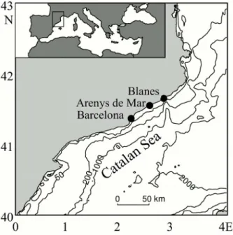
Four surface water samples were taken at three different locations along the Catalan coast (Fig 1) with different environmental characteristics (Table 1).
The first sample was taken inside the harbour of Arenys de Mar (41°34’N, 2°32’E) during a routine sampling of the SEED program (GOCE-CT-2005-003875, EC). This 2-meter deep har-bour is a good example of a heavily eutrophicated area in the Mediterranean, where red tides occur often. The sample (from now on“Harbour”) was taken at the end of the exponential growth phase of a bloom of the dinoflagellateAlexandrium minutum[22]. Nutrient concentra-tions (Table 1) were high compared to usual values observed in the Catalan Sea [23]. Most phy-toplankton cells were dinoflagellates of the speciesA.minutum,Scrippsiellasp. andDinophysis
cf.punctata.
The second sample was taken at the Blanes Bay Microbial Observatory (BBMO, NW Medi-terranean 41°40’N, 2°48’E, from now on“Bay”). The Blanes Bay is a well-studied (e.g. [24]) open oligotrophic bay located at the Catalan coast, and it is a good example of the oligotrophic Mediterranean coastal ecosystem, directly impacted by human and terrestrial influences from the nearby coast. The site is about 1 km offshore (41°40’N, 2°48’E) with a depth between 20 and 24 meters. The sample was taken in May, at the end of a typical spring bloom when chloro-phyllaconcentration was still high but nutrients were already low (Table 1). The microphyto-plankton population was quite diverse, although dinoflagellates dominated, as is characteristic for this region during spring [25]. The cells analysed from this sample were dinoflagellates of the speciesTripos fusus,Tripos furca,D. cf.punctata,Dinophysiscf.acuta,Prorocentrumcf.
micansandProtoperidiniumspp., and some diatoms of the generaChaetocerosspp., Pseudo-Nitzschiaspp. andRhizosoleniasp.
The remaining two samples were both taken under contrasting environmental conditions on the continental shelf at the Coastal Monitoring Station of Barcelona (Estació Litoral de Bar-celona, 41°53’N, 2°14’E). The sampling site is about 2.5 km offshore the city of Barcelona, on a 40 m deep water column. The first sample was taken in January 2006, under mixed water
Fig 1. Map of the localization of the sampling sites at the Catalan coast.The inlet map shows the position of the Catalan Sea in the Mediterranean Sea.
column conditions, and the second on May 2006, after the thermocline was already established. In January (from now on,“CS Mixed”for Continental Shelf Mixed site), the surface tempera-ture as well as the nutrient and chlorophyllaconcentration indicated typical Mediterranean well mixed winter conditions (Table 1). The microphytoplankton population was composed mostly by diatoms and nanoflagellates, as is typical for this time of the year [26]. The cells were diatoms of the generaChaetocerosspp.,Pleurosigmasp. andThalassiosiraspp. During the sam-pling in May (from now on,“CS Stratified”for Continental Shelf Stratified site), a clear vertical gradient of salinity, temperature and density was observed, with low nutrients and low chloro-phylla(Table 1). The microphytoplankton population present was a mixture of dinoflagellates and diatoms, all of them at very low concentrations. The cells from this sample were the genera
D. cf.punctata,P. cf.micansandProtoperidiniumspp., as well as diatoms of the genera Chaeto-cerosspp. andPseudo-nitzschiaspp.
Temperature, salinity, and other basic parameters such as chlorophylla(Chla) concentra-tion and dissolved inorganic nutrients (NO3-, NO2-, NH4+, PO43-and Si[OH]4) were measured
at all sites (Table 1). A full description of the methods used to analyse those parameters at the Harbour, Bay, and Continental Shelf sites are given in Alonso-Sáez et al. [24], Van Lenning et al. [27] and Romero et al. [28], and Arin et al. [26], respectively.
Elemental analysis of single cells
Water samples for XRMA analysis were collected in 5 to 8 L plastic bottles after pre-screening with a 200μm nylon mesh to remove large mesozooplankton. A complete description of the methodology followed to collect, prepare, and analyse the elemental composition of microphy-toplankton cells using XRMA can be found in Segura-Noguera et al. [15]. Briefly, the cells were concentrated onto a 20μm nylon mesh and washed to remove the salt. After washing, the cells were centrifuged at 15°C over a 10 mm diameter Cu grid coated with formvar. The grid was air-dried and stored in a vacuum chamber until analysis. Cells were viewed and analysed for elements in a Hitachi S-3500N Scanning Electron Microscope (SEM), equipped with an energy-dispersive spectrometer Si(Li) detector (Bruker AXS). The detector processes X-rays
Table 1. Physical and biogeochemical characteristics of the different sampling sites.
Location Barcelona Arenys de Mar Blanes Bay Barcelona
Id. location CS M Harbour Bay CS S
Date 26 January 2006 9 March 2006 16 May 2006 25 May 2006
Position 41°34’N, 2°32’E 41°34’N, 2°32’E 41°34’N, 2°32’E 41°34’N, 2°32’E
Bottom depth (m) 40 2 20–24 40
Sampling depth (m) surface surface 0.5 surface
Distance from coast (m) 2500 inside the harbour 800 2500
Temperature (°C) 12.6 12.3 18.4 19.0
Salinity 38.3 38.9 37.8 37.8
Density (σ-T, Kg m-3) 29.0 29.6 27.3 27.2
Chlorophylla(μg L-1) 0.75 0.65 2.45 0.27
Phosphate (μmol L-1) 0.09 0.09 0.10 0.07
Nitrate (μmol L-1) 3.02 4.92 0.54 0.12
Nitrite (μmol L-1) 1.19 0.08 0.14 0.05
Ammonium (μmol L-1) 0.89 0.10 0.13 0.07
Silicate (μmol L-1) 1.81 3.07 0.13 0.22
CS M: Continental Shelf Mixed, H: Harbour, B: Bay, CS S: Continental Shelf Stratified.
with atomic number>2, thus including light elements, and has a resolution129 eV (cali-brated with Mn Kαlines). To avoid interference of holder material with sample elements, as
well as with the X-ray spectra, the sample grid was mounted over a customized high-resolution holder (Hitachi) at a distance of 17 mm over the holder bottom [15]. With this modification, the thin-film analysis conditions of the Scanning Transmission Electron Microscope (STEM) were obtained in the SEM. The microscope was operated at 15 kV of electron beam energy, 15 mm of working distance, accumulation time of 100 sec (live time) per analysis, and 15° of tilt angle. The X-ray spectrum was recorded from an area that circumscribes the specimen [14]. The software used to acquire the spectra and to obtain the counts per second of each ele-ment present in the spectra was QUANTAX 1.6 (Bruker AXS).
Cells were identified by the imaging system, and two perpendicular axes of the cell were measured from the SEM pictures of the analysed cells using the software QUARTZ PCI 5.1. After, cell volumes were calculated following Sun and Liu [29], assuming depth to be half the width, except inProtoperidiniumsp. andTripossp., for which depth was assumed to be equal to width.
Latex beads (Agar Scientific) were used to calibrate carbon, and the calibration constants for other elements were obtained following Norland et al. [14]. The detection limit of the method is 0.16% [15]. In the present study, only elemental quotas above the detection limit, which are more than 98% of the quotas measured, are shown and used to calculate ratios. The standard error of the elements quantification are 7–8% for C, O, Mg, P, S, Ca and 10–11% for N and K [15]. Total cellular dry weight per cell was calculated adding the molecular weight of all the quantified elements (C, N, O, Na, Mg, Al, Si, P, S, Cl, K and Ca), and assuming 1.6 mols of H per each mol of C (from equation 2 in Fraga [30]). Data of our single-cell analysis is shown in S1 Table.
Statistical analyses
Elementvs. volume regressions were studied first normalizing the variables by log transforma-tion. Conversion factors to predict cellular carbon from cell volumes were estimated as the slope of the regression between element mass and volume, using Ordinary Least Squares (OLS) regression model type I [31,32]. Elemental ratios can be estimated as the average of individual ratios in a given population, or as the slope of the linear regression of each element with P (e.g.
[33,34,35]). The linear regression method minimizes the error between ratios when the
con-centrations are low [36], and so it is used in this study to compare ratios between phylogenetic groups. Both estimates are similar when the intercept of the regression is close to 0.
Elementvs. element regressions were estimated using Standard Major Axis (SMA), a type II regression model that takes into account differences in the scale of both axes (e.g. C:P relation-ship). Extreme values were identified using the Quartile method (values smaller/higher than the first/third quartile minus/plus three times the interquartile range) and not used to calculate regression lines. The stoichiometries were first calculated independently for each group. After-wards, tests for differences between y-intercepts and slopes of diatoms and dinoflagellates were performed. The software SMATR v2.0 [37] was used to calculate both OLS and SMA slopes and intercepts, along withp-values and confidence intervals, as well as differences and shifts in slopes between dinoflagellates and diatoms.
Results and Discussion
Single-cell
vs
. published elemental concentrations
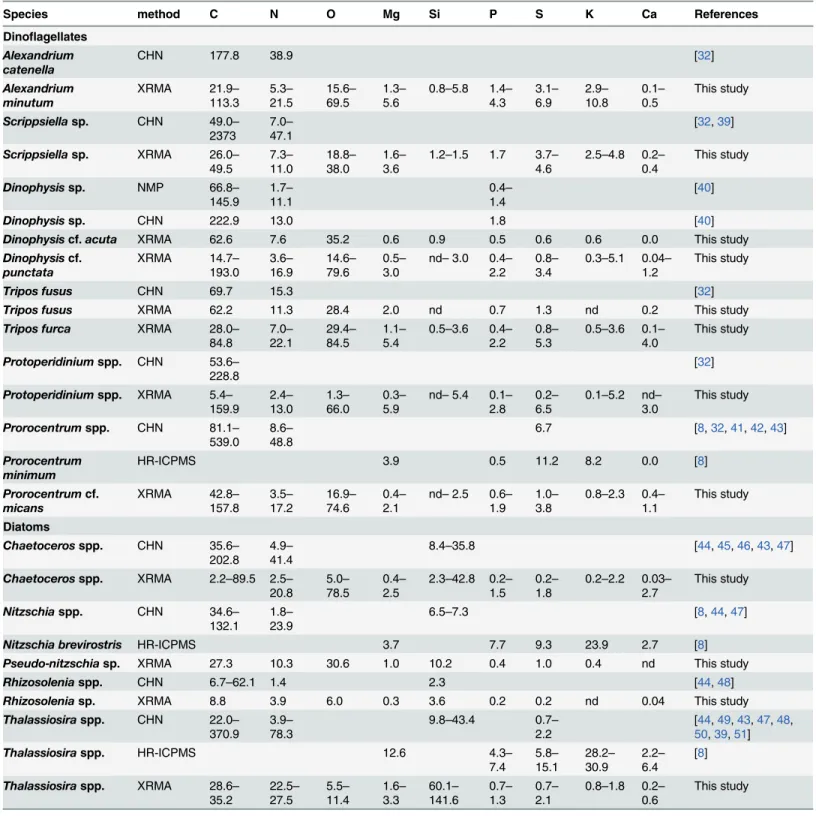
The elemental concentrations of our single-cell analyses, along with data from cultured cells of the same or close genera obtained with bulk analysis methods, are shown inTable 2. Our C, N and P concentrations are within the ranges of published data for cultures of the same or close genera, although, mostly, fall at the low end of these ranges. This can also be observed in Figs2 and3for dinoflagellates and diatoms, respectively, which include all our cells and additional genera from the literature. The average mass of C, N, O, Si, P, and S per unit volume for the cells of the same species or genera, and of the same site are shown inS2 Table, which also includes the mean cell size and dry weight.
Relatively lower concentrations compared to published values were expected for two rea-sons: first, we used a single-cell method that excluded from the analysis extraneous material, like dead cells, detritus, or other organic extracellular material in the analysis, which are partic-ularly rich in C and N [52]. The extracellular material significantly changes the intercepts but not the slopes of C and N massvs. volume compared to published data [15]. And second, in contrast to cultured cells, which are usually harvested on exponential phase, our cells collected from the field had experienced different environmental histories and could be at any growth phase, even within the same sample. This variability translates into different biochemical com-positions and hence elemental concentrations (e.g. [4]).
Low C concentrations are unlikely to be the result of methodological constraints: histori-cally, XRMA has been deemed unsuitable for microphytoplankton cells because the calculated interaction volume (that is, the volume of sample where X-rays are generated) is smaller than the theoretical cell thickness (at least 20μm). However, diatoms and dinoflagellates, with dif-ferent thicknesses and densities, can be overpenetrated by a 15 kV electron beam as a result of drying the cells during SEM sample preparation [15]. All the cells analysed had a mass to area ratio below the most restrictive theoretical limits of beam penetration (4.8 and 5.7 pgμm-2for dinoflagellates and diatoms, respectively [15]). Finally, the completeness of the beam penetra-tion into the largest diatoms was also verified by the good agreement of the Si concentrapenetra-tions with data of Brzezinski [44] (Table 2,Fig 3E).
Some of our single-cell C concentrations, mostly in diatoms, were even lower than the pub-lished values: highly silicified diatom cells from the CS Mixed sampling site in January (mostly
ThalassiosiraandPleurosigma, but also someChaetoceros), as well as thin diatoms from the Bay in May (Pseudo-nitzschiaandRhizosolenia). Some of these diatoms also had a P content below detection limit, which suggests us that they were not viable at the moment of sampling (twoChaetoceroscells, allPleurosigmaand the diatoms not identified).Thalassiosiracells, on the other hand, had an unusually high N and O content (Fig 3B and 3C), which suggests that those cells could had been storing nitrate.
The concentrations of Mg, K and Ca in the cells analysed are also lower, although of the same order of magnitude, than in cells of the same genera analysed using high-resolution induced coupled plasma mass spectrometry (HR-ICPMS, [8]) (Table 2). Our data is within the range of published S concentrations [53,49,41,54,17,8], although again at the low end. How-ever, our S and O results for dinoflagellates (2.33 ± 1.43 fgμm-3and 40.12 ± 14.49 fgμm-3, respectively) and diatoms (O = 37.46 ± 38.43 fgμm-3) are comparable with other XRMA data ofProchlorococcus(2.61 ± 0.92 fgμm-3and 40.65 ± 3.96 fgμm-3, respectively) and
Table 2. Comparison between the elemental concentration (fgμm-3) of diatoms and dinoflagellates of the same or close genera analysed with bulk
analysis techniques (CHN and HR-ICPMS) and single-cell methods (NMP and XRMA). Single-cell data presented in this table corresponds to the range of individual measurements of cells of the same genera.
Species method C N O Mg Si P S K Ca References
Dinoflagellates Alexandrium catenella
CHN 177.8 38.9 [32]
Alexandrium minutum
XRMA 21.9–
113.3 21.55.3– 15.669.5– 1.35.6– 0.8–5.8 4.31.4– 3.16.9– 2.910.8– 0.10.5– This study
Scrippsiellasp. CHN 49.0–
2373 7.047.1– [32,39]
Scrippsiellasp. XRMA 26.0–
49.5 7.311.0– 18.838.0– 1.63.6– 1.2–1.5 1.7 3.74.6– 2.5–4.8 0.20.4– This study
Dinophysissp. NMP 66.8–
145.9 1.711.1– 0.41.4– [40]
Dinophysissp. CHN 222.9 13.0 1.8 [40]
Dinophysiscf.acuta XRMA 62.6 7.6 35.2 0.6 0.9 0.5 0.6 0.6 0.0 This study
Dinophysiscf. punctata
XRMA 14.7–
193.0 3.6– 16.9 14.6– 79.6 0.5– 3.0
nd–3.0 0.4–
2.2
0.8–
3.4
0.3–5.1 0.04–
1.2
This study
Tripos fusus CHN 69.7 15.3 [32]
Tripos fusus XRMA 62.2 11.3 28.4 2.0 nd 0.7 1.3 nd 0.2 This study
Tripos furca XRMA 28.0–
84.8 22.17.0– 29.484.5– 1.15.4– 0.5–3.6 2.20.4– 0.85.3– 0.5–3.6 0.14.0– This study
Protoperidiniumspp. CHN 53.6–
228.8 [32]
Protoperidiniumspp. XRMA 5.4–
159.9 13.02.4– 1.366.0– 0.35.9– nd–5.4 2.80.1– 0.26.5– 0.1–5.2 nd3.0– This study
Prorocentrumspp. CHN 81.1–
539.0 8.648.8– 6.7 [8,32,41,42,43]
Prorocentrum minimum
HR-ICPMS 3.9 0.5 11.2 8.2 0.0 [8]
Prorocentrumcf. micans
XRMA 42.8–
157.8 17.23.5– 16.974.6– 0.42.1– nd–2.5 1.90.6– 1.03.8– 0.8–2.3 0.41.1– This study
Diatoms
Chaetocerosspp. CHN 35.6–
202.8 4.941.4– 8.4–35.8 [44,45,46,43,47]
Chaetocerosspp. XRMA 2.2–89.5 2.5–
20.8 78.55.0– 0.42.5– 2.3–42.8 1.50.2– 0.21.8– 0.2–2.2 0.032.7 – This study
Nitzschiaspp. CHN 34.6–
132.1 1.823.9– 6.5–7.3 [8,44,47]
Nitzschia brevirostris HR-ICPMS 3.7 7.7 9.3 23.9 2.7 [8]
Pseudo-nitzschiasp. XRMA 27.3 10.3 30.6 1.0 10.2 0.4 1.0 0.4 nd This study
Rhizosoleniaspp. CHN 6.7–62.1 1.4 2.3 [44,48]
Rhizosoleniasp. XRMA 8.8 3.9 6.0 0.3 3.6 0.2 0.2 nd 0.04 This study
Thalassiosiraspp. CHN 22.0–
370.9 78.33.9– 9.8–43.4 0.72.2– 50[44,,3949,,5143],47,48,
Thalassiosiraspp. HR-ICPMS 12.6 4.3–
7.4 5.815.1– 28.230.9– 2.26.4– [8]
Thalassiosiraspp. XRMA 28.6–
35.2 27.522.5– 5.511.4– 1.63.3– 60.1141.6– 1.30.7– 0.72.1– 0.8–1.8 0.20.6– This study
CHN: Carbon+Hydrogen+Nitrogen elemental analysis; XRMA: X-Ray Microanalysis; NMP: Nuclear Microprobe; HR-ICPMS: High-Resolution Inductively Coupled Plasma Mass Spectrometry.n= number of cells. nd = not detected.
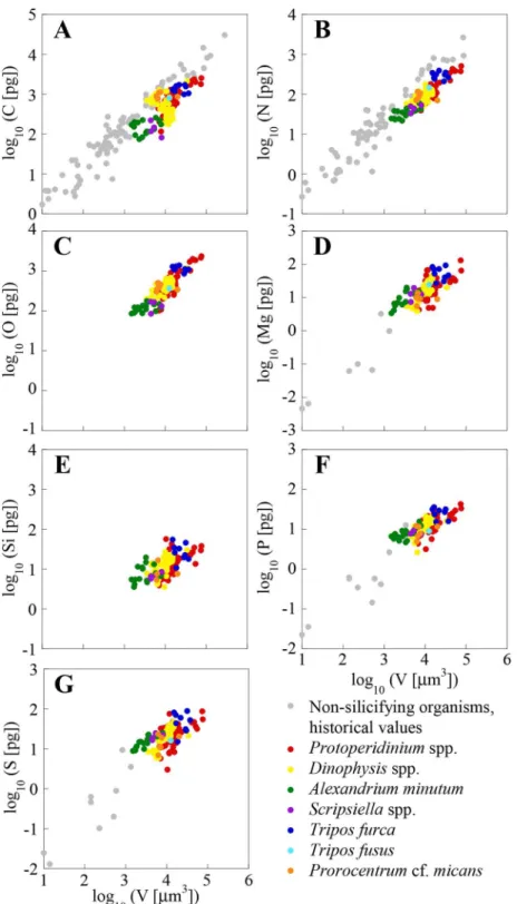
Fig 2. Log-log plots of the mass of C, N, O, Mg, Si, P and S (pg cell-1)vs. volume (
μm3cell-1).
Dinoflagellate single-cell values from the NW Mediterranean Sea are presented along with historical values of cultured non-silicifying planktonic organisms obtained using bulk analysis techniques. Note that vertical axes vary between elements.
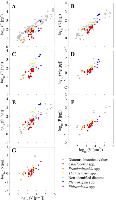
Figs2and3show a large variability in both bulk analysis published values and our single-cell analysis. This is likely the consequence of differences in single-cell size, as well as of genotypic,
Fig 3. Log-log plots of the mass of C, N, O, Mg, Si, P and S (pg cell-1)vs. volume (
μm3cell-1).Diatom
single-cell values from the NW Mediterranean Sea are presented along with historical values of cultured diatom cells obtained using bulk analysis techniques. Note that vertical axes vary between elements.
environmental and methodological differences. The variability in the elemental concentra-tion between cells of the same genera, and from the same sample (e.g.,A.minutum,T.furca) is larger than the error of the analysis, as found by other authors using XRMA of individual organisms from the field [54,55]. As pointed out by these authors, this variability is probably due to differences in life cycle and growth status between individual cells, which in field populations are most certainly not synchronized. Other researchers using the same or other single-cell analysis techniques to measure C, N, P, and S, have observed the existence of intra-population variability in clonal cultures [17,56].
Differences in the elemental concentration between diatoms and
dinoflagellates
We explored the differences in elemental concentration among elements and between plankton groups by comparing the slopes of the OLS regressions of the single-cell measurements (Figs2
and3). All elementvs. volume slopes, shown inTable 3, are significantly different from 0
(p0.001). No significant differences in elementvs. volume slopes were found between dia-toms and dinoflagellates, except for Si (p0.01). Menden-Deuer and Lessard [32], using pub-lished data available at the time, exhaustively examined C per volume relationships in
plankton, concluding that dinoflagellates are significantly C denser than diatoms. Our results show them to be denser also in N, O, Mg, P and S, since there are significant elevation shifts between groups for all these elements (p0.001). Were these results extrapolated beyond the limited set of species and cell volumes analysed in the present study (between 2000 and
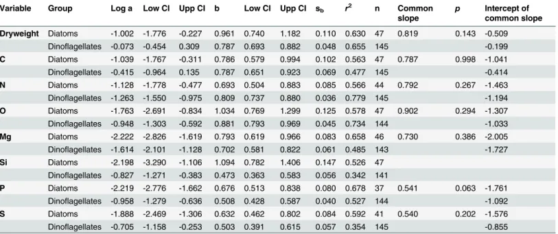
Table 3. Results of the ordinary least squares regression of log10transformed element concentration (pg cell-1)vs. log10transformed volume
(μm3cell-1) of diatoms and dinoflagellates from the Catalan Sea.
Variable Group Log a Low CI Upp CI b Low CI Upp CI sb r2 n Common
slope
p Intercept of common slope
Dryweight Diatoms -1.002 -1.776 -0.227 0.961 0.740 1.182 0.110 0.630 47 0.819 0.143 -0.509 Dinoflagellates -0.073 -0.454 0.309 0.787 0.693 0.882 0.048 0.655 145 -0.199
C Diatoms -1.039 -1.767 -0.311 0.786 0.579 0.994 0.102 0.563 47 0.787 0.998 -1.041 Dinoflagellates -0.415 -0.964 0.135 0.787 0.651 0.923 0.069 0.477 145 -0.414
N Diatoms -1.128 -1.778 -0.477 0.693 0.504 0.883 0.085 0.566 44 0.792 0.267 -1.463 Dinoflagellates -1.263 -1.550 -0.975 0.809 0.737 0.880 0.036 0.779 145 -1.194
O Diatoms -1.763 -2.691 -0.834 1.034 0.769 1.299 0.125 0.578 47 0.902 0.294 -1.307 Dinoflagellates -0.948 -1.303 -0.592 0.881 0.793 0.969 0.045 0.734 144 -1.033
Mg Diatoms -2.222 -2.826 -1.619 0.793 0.619 0.966 0.083 0.658 46 0.730 0.386 -2.005 Dinoflagellates -1.614 -2.101 -1.128 0.702 0.581 0.822 0.061 0.485 143 -1.727
Si Diatoms -2.198 -3.290 -1.106 1.094 0.782 1.406 0.147 0.526 47 Dinoflagellates -0.827 -1.271 -0.383 0.473 0.363 0.583 0.056 0.342 141
P Diatoms -2.219 -2.776 -1.662 0.676 0.513 0.838 0.080 0.678 37 0.541 0.063 -1.761 Dinoflagellates -0.958 -1.279 -0.636 0.508 0.428 0.587 0.040 0.527 144 -1.092
S Diatoms -1.888 -2.469 -1.306 0.632 0.462 0.802 0.084 0.592 41 0.540 0.202 -1.576 Dinoflagellates -0.705 -1.158 -0.253 0.503 0.391 0.615 0.057 0.354 145 -0.855
Shown in this table are the y-intercept (log a) and the slope (b) of the regression equations, the interval of confidence (Low CI and Upp CI), the standard error of the slope (sb), the coefficient of determination (r2), the number of data points included (n), the common slope (when there are no significant differences between the slope of both groups), itsp-value, and the intercept of each group with the common slope. The elemental quota can be
determined from volume based on the equation log10element (pg cell-1) = log a + b x log10volume (μm3). Same element slopes of diatoms and
dinoflagellates are not statistically different, except for Si (p= 0.002). The intercepts of the dinoflagellates regression lines are larger than those of diatoms for the same element, except for N (p0.001). However, this difference disappears when the common slope is calculated.
40000μm3), important errors in the prediction of C, N, O, P and S from volume measurements may occur when the same conversion equations are used for both groups.
All elements, except O and Si in diatoms, had volume-scaling factors (i.e. slopes) signifi-cantly lower than 1.0 (p0.001,Table 3). This indicates that the masses of C, N, Mg, P or S, as well as O and Si in dinoflagellates, are not a constant fraction of cell volume, but rather decrease with increasing cell volume. Thus, small cells have higher elemental content per vol-ume than large cells. Slopes for C, N, O, Mg, as well as Si in diatoms, were not statistically dif-ferent (p>0.05). C and N slopes of our dinoflagellates were close to values compiled by
Menden-Deuer and Lessard [32] (0.82 and 0.85, for C and N). Interestingly, P and S slopes, as well as Si in dinoflagellates, were significantly lower than the slopes for other elements both in diatoms and dinoflagellates (p0.05), indicating that the size effect on P and S is important in these organisms.
Thingstad et al. [57] hypothesized that some osmotrophic microorganisms may get a com-petitive advantage by using a non-limiting nutrient to increase size, and thus reduce predation stress without increasing nutrient requirements. This is the strategy of diatoms, which have frustules conformed by elements with slopes larger than 1 (Table 3). However, the fact that all elements, except O and Si in diatoms, had slopes lower than 1, suggests that decreases in the concentration of an element are not always offset by increases in another element. Thus, these decreases could be achieved through an increase in water cell content, either by increasing cyto-plasm volume or by enlarging vacuoles [58]. In such case, decreases in element content with increasing cell size will not necessarily lead to stoichiometric changes. Our results suggests that this“dilution effect”may be a possible mechanism for dinoflagellates to mitigate, to a certain point, the negative effect that the increase in size has on nutrient uptake.
The average P concentration for bacterioplankton,ProchlorococcusandSynechococcusis 2.93–3.17, 2.92 and 3.85 fgμm-3respectively (calculated from data in [54,59,17]). The average P concentration in dinoflagellates and diatoms from our own results is 1.30 ± 0.68 and
0.58 ± 0.41 fgμm-3respectively. This indicates that the P requirement is highest for bacterio-plankton, followed by dinoflagellates, and lowest for diatoms. Low P quotas represent a clear ecological advantage in P-limited environments like the Mediterranean Sea.
The analysed diatoms have around 40–60% less C, P and S concentration than dinoflagel-lates, but N and O concentrations are only around 20% lower. A relatively low C concentration in diatoms with respect to other plankton groups has been previously observed [60,32] and it has been linked to the presence of large intracellular vacuoles [58]. However, the presence of vacuoles does not explain why N is not proportionally reduced, unless N is stored inside them as nitrate (e.g. [43]). Such hypothesis is consistent with the observation that N concentration in diatoms collected in the nutrient-rich environment (CS Mixed) is higher than in the relatively low-nutrient environment (Bay). Additionally, the observed differences in C concentration between both groups could be due to the contribution of the carbohydrate theca of dinoflagel-lates. On the other hand, observed differences in P could be explained by a larger amount of DNA in the dinoflagellate nucleus compared to other eukaryotic cells (2.2–200 pg per nucleus [61]).
Finally, it should be noted that the extrapolation to other sizes or groups of the results pre-sented here can be misleading. For example, extrapolating the C to V regression for dinoflagel-lates inTable 3to picophytoplankton volumes yields lower values than those obtained for
cellular biomass in models and predictions, the confidence intervals of the equations being used should be explicitly stated, and the possible effect of errors should be considered in both the results and the conclusions.
Elemental stoichiometry
The elemental composition of plankton can be expressed as the proportion of each element to total dry weight (percentage) or to one element. Both ratios are independent of the cellular size or mass and therefore allow for direct comparisons among species, plankton groups and eco-systems [62,63]. However, they do not provide information on which element is controlling any observed change. Although P normalized numbers are most often used by oceanographers to quantitatively link the marine nutrient cycles in numerous biogeochemical applications, empirical and theoretical studies, as well as our individual cell values (S3 Table) show that ele-mental ratios vary greatly with taxa and growth conditions [4,8].
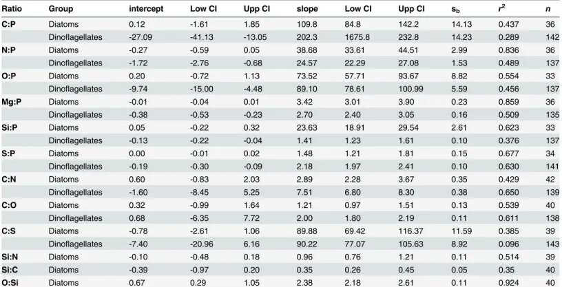
Individual values of dinoflagellate and diatom cells, with the cells grouped by sampling site, are shown in Figs4and5, respectively. Slopes and intercepts of the regression lines (SMA) for dinoflagellates and diatoms are shown inTable 4. Significant differences (p0.01) between diatoms and dinoflagellates were found for all slopes, except O:P and C:S. Note the tight rela-tion that exists in the O:Si regression line (r2= 0.92), which is reflecting the composition of the frustules. Based on the slope of the best fit regression lines, the overall elemental ratio C:N:O:P: S of the analysed dinoflagellates was 202.3±14.2: 24.6±1.5: 89.1±5.6: 1: 2.1±0.1 (slope ± standard error). The C:N:O:P:S:Si for diatoms was 109.8±14.1: 38.9±3.0: 73.53±8.8: 1: 1.5±0.2: 23.6±2.6.
The C:P slope for dinoflagellates, as well as the N:P slopes for both groups (Table 4), are higher than the canonical Redfield ratio (116 and 16), but within the ranges of published values
[4,17]. The C:P slope for diatoms is not statistically different from the Redfield ratio (p= 0.787).
In our study, the only dinoflagellate C:P and N:P values below the Redfield ratio have been found inA.minutum, andScrippsiellasp. cells collected from the Harbour site. Both dinoflagellates were in exponential growing phase [22] in a high nutrient environment (Table 1). Thus, the low C:P and N:P ratios were likely caused by the relatively high P concentration, as expected in actively growing cells with high contents in nucleic acids [4].
The average C:S values for diatoms and dinoflagellates are similar to assumed phytoplank-ton values [64,65]. However, the low coefficient of determination in dinoflagellates (Table 4, see alsoFig 4F), casts doubt on the use of C as conversion factor for S, or vice-versa.
Because it is not yet possible to measure O with direct analytical methods, O:P ratios and C: O ratios in phytoplankton have been rarely reported in the literature, and most often the num-bers provided rely on indirect estimates [66]. Very low C:O ratios have been occasionally found both in dinoflagellates and diatoms (S3 Table), possibly reflecting a low C, rather that high O concentrations. Such low values contradict our knowledge of the average elemental composition and proportions of planktonic biomolecules [67,68,4]. All our cells with a C: O<1 also had a C:N<3.7, a value that corresponds to the average ratio of proteins, pigments
Fig 4. Molar relationships between C, N, O and Svs. P, Cvs. N and S, and Sivs. P of individual dinoflagellates from the NW Catalan Sea.Cells from the same location are plotted with the same colour.
Note that axes change.
Fig 5. Molar relationships between C, N, O and Svs. P, Cvs. N and S, and Sivs. P of individual diatoms from the NW Catalan Sea.Cells from the same location are plotted with the same colour. Note that axes
change.
S:P = 1.7 [67], to the average value of 1.71 ± 0.70 for cyanobacteria [17], and within the range of published values for dinoflagellates and diatoms [8,65].
Interestingly, Si was detected in both groups. In diatoms this element is a well known con-stituent of the exoskeleton [72]. The presence of Si has already been reported in the dinoflagel-latesCeratium hirundinella[55] andExuviaellasp. [45]. Recently, Si has also been detected in
Synechococcusin concentrations of the same order than our dinoflagellates and with similar Si: P values (1.54 ± 1.18, [73]). However, the physiological role of Si in these organisms is still unclear. The Si:P ratio of our diatoms (Table 4) is higher than the Redfield-Brezinski ratio of 15:1 (derived from [44]), and of that found in chemostat studies (5.4, 6.7 and 3.8 [46]; 5.9 ± 1.3 [74]). However, it is consistent with ratios reported for diatoms in the Equatorial Pacific [73], which also had a low P quota. The tight relationship between Si and P is remarkable consider-ing the well known inter-specific variability in frustule thicknesses, and the high variability of the Si content per cell [44].
The C:N slope in dinoflagellates (7.5 ± 0.4,Table 4) is within the range of ratios reported in the literature [4], close to Redfield's original ratio of 6.6 [75], and similar to particulate matter values in the NW Mediterranean Sea (C:N = 6–7 [36,76]). Higher values (8.2 to 10) have been reported for cyanobacteria in cultures using the same single-cell methodology [17], and the dif-ference has been attributed to cyanobacteria being relatively denser in C than N, which would explain also the higher C:P reported for cyanobacteria. The N:P ratio of our dinoflagellates and diatoms is more variable than the C:N ratio, as previously reported [4]. In diatoms, the C:N
Table 4. Results of the least square regression of different molar ratios (elementvs. element) of single phytoplankton cells.
Ratio Group intercept Low CI Upp CI slope Low CI Upp CI sb r2 n
C:P Diatoms 0.12 -1.61 1.85 109.8 84.8 142.2 14.13 0.437 36
Dinoflagellates -27.09 -41.13 -13.05 202.3 1675.8 232.8 14.23 0.289 142
N:P Diatoms -0.27 -0.59 0.05 38.68 33.61 44.51 2.99 0.836 36
Dinoflagellates -1.72 -2.76 -0.68 24.57 22.29 27.08 1.53 0.489 137
O:P Diatoms 0.20 -0.72 1.13 73.52 57.71 93.67 8.82 0.554 33
Dinoflagellates -9.74 -15.00 -4.48 89.10 78.61 100.99 5.59 0.456 137
Mg:P Diatoms -0.01 -0.04 0.01 3.42 3.01 3.90 0.23 0.859 36
Dinoflagellates -0.38 -0.53 -0.23 2.70 2.40 3.05 0.16 0.509 135
Si:P Diatoms 0.05 -0.22 0.32 23.63 18.91 29.54 2.61 0.623 33
Dinoflagellates -0.13 -0.22 -0.04 1.41 1.23 1.61 0.10 0.376 137
S:P Diatoms 0.00 -0.01 0.02 1.48 1.21 1.81 0.15 0.677 34
Dinoflagellates -0.19 -0.30 -0.09 2.18 1.97 2.41 0.10 0.630 141
C:N Diatoms 0.60 -0.83 2.03 2.89 2.28 3.67 0.35 0.429 42
Dinoflagellates -1.60 -8.45 5.25 7.51 6.80 8.30 0.38 0.650 139
C:O Diatoms 0.32 -0.99 1.64 1.21 0.97 1.51 0.13 0.539 40
Dinoflagellates 0.68 -6.35 7.72 2.00 1.80 2.19 0.11 0.611 138
C:S Diatoms -0.78 -2.61 1.06 89.88 69.42 116.37 11.59 0.385 39
Dinoflagellates -7.40 -20.96 6.16 90.22 77.07 105.63 8.92 0.096 143
Si:N Diatoms -0.10 -0.48 0.18 0.96 0.76 1.21 0.11 0.514 39
Si:C Diatoms -0.39 -0.97 0.20 0.35 0.26 0.45 0.05 0.35 40
O:Si Diatoms 0.67 0.29 1.05 2.38 2.18 2.61 0.11 0.924 40
Shown are the y-intercept and the slope of the regression equations, the interval of confidence (Low CI and Upp CI), the standard error of the slope (sb), the coefficient of determination (r2), and the number of data points included (n). All slopes are significantly different from zero (p0.001). C:S and O:P slopes of dinoflagellates and diatoms are not statistically different (p= 0.983 andp= 0.149, respectively).
slope (2.9 ± 0.4) is in the lower side of the broad range of C:N ratios reported by Sarthou et al. [74] (range = 2.7–29.7). Our results show that the diatoms are N enriched compared to C, but some highly enriched cells, which are however not detected as extreme values, force the slope towards such low value (Fig 5E). If cells with N quota values above 4 pmol are removed from the regression, remaining onlyChaetocerosandPseudo-nitzschiacells, the C:N ratio increases to 5.4 ± 0.6 (n = 31), which is within the range of 85% of the diatom species gathered by Sarthou et al. [74] (range = 5.0–9.7). This evidences the need of more single-cell analyses to define the range of C:N values in the nature. Interestingly, the N:P ratio drops from 38.7 to 26.5 when these same diatom cells with high N quota are removed from the analysis. This ratio is not significantly different than the one from dinoflagellates (p= 0.571), and the common slope is N:P = 25.1 (n = 168).
Phytoplankton single-cell stoichiometry and Western Mediterranean Sea
biogeochemistry
The Mediterranean Sea has long been known as a nutrient-depleted basin in which, in contrast with other oligotrophic oceanic regions, P is the main limiting nutrient (e.g. [77,78]). The main inputs of macronutrients in the NW Mediterranean Sea are river and groundwater dis-charges, and atmospheric deposition. They provide a total nutrient budget in terms of N and P significantly higher than the 16:1 Redfield ratio [79,80]. It is well known that nutrient supply sets an upper limit to the biological production, but planktonic organisms exert a tight control on the elemental distribution particularly in deeper layers [81] because the main source of nitrate, orthophosphate and orthosilicate in the deep sea waters is the remineralization of sink-ing biological material. The dissolved inorganic nitrate:phosphate ratio of deep Western Medi-terranean waters is between 20–23, e.g. [82,83]. This high ratio, compared to the Redfield ratio is still difficult to explain and has been the object of different hypotheses [33,84]. Correspond-ingly, our cells also show a high N:P ratio. This is consistent with studies performed in the NW Mediterranean Sea, which also found higher than Redfield ratios in picoplanktonic cultured cells (N:P = 21.2–62.3, [85]) and detritic matter (N:P = 32, [86]), both studies using XRMA methods. Also, bulk analysis studies of particulate organic mater from surface waters of the Mediterranean Sea found again N:P ratios higher than Redfield's (N:P = 18.8–23.2, [35,36]).
A high N:P ratio could be a result of either high N or low P content. The fact that the Si:P in the diatoms analysed (27.8 ± 3.4) is also high, while the Si:N ratio (1.06 ± 0.08) is similar to what has been reported for diatoms (e.g. Si:N = 1.12 ± 0.33, range: 0.5–1.5 [44]; Si:N = 0.8 ± 0.3 [74]), as well as for the Western Mediterranean Sea (e.g. Si:N = 1.13 [87]; Si:N = 1.0 [83]), sug-gests that the high ratio is due to a lower P cell content. Unfortunately, there are very few pub-lished data to compare P with (Figs2Fand3F). The consistently high observed cell N:P ratios can be an adaptation to low P availability in Mediterranean surface waters. One possible mech-anism of adaptation to phosphorus limitation is the use of non-phosphorus lipids [88]. Phos-pholipid substitutions appear to be an important biochemical mechanism for cyanobacteria and eukaryotic phytoplankton to maintain photosynthesis in environments where phosphorus is scarce [10]. In cyanobacteria the substitute is the non-phosphorus membrane lipid sulpho-quinovosyldiacylglycerol (SQDG). In eukaryotes the substitute are the non-phosphorus‘ beta-ine’lipids. A preferential synthesis of non-phospholipds has been observed in the P-deprived phytoplankton of the Adriatic Sea at the Western Mediterranean Sea [89].
Chaetocerosspp. are among the most abundant species in the NW Mediterranean Sea [18,19], and are all major components of the phytoplankton exported to the deep ocean [20]. So we can assume that our data, although scarce, is representative of the phytoplankton of this area. Con-sequently, and following Redfield's theory [67], the remineralization of the cells with high N:P, like the ones measured in this study, should be the source of the high N:P ratio observed in the deeper waters of the Western Mediterranean sea. Further single-cell XRMA of phytoplankton, including other abundant phytoplankton groups such as coccolithophorids, are needed to vali-date this hypothesis.
Phylogenetic and environmental differences in elemental composition
and stoichiometry between genera
The evidence of phylogenetic differences between phytoplankton groups in elemental concen-trations or quotas has gained a lot of attention in recent years [8,90]. Our dataset allowed us to study inter-generic differences of cells sampled under the same environmental conditions, as well as intra-generic differences of cells from contrasting environments.
Inter-generic differences were found between the dinoflagellates (1)A.minutumandD. cf.
Punctatafrom the Harbour sample; (2)T.furcaandProtoperidiniumspp. from the Bay site, the last genera grouped into two very different size classes (<25000μm3and>45000μm3);
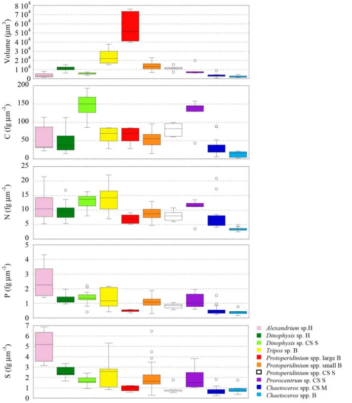
and (3)P.micans,D. cf.punctataandProtoperidiniumspp. in the CS Stratified sample. Dinoflagellates sampled in the Harbour and in the Bay had significant differences in sizes (p0.005) (Fig 6), so we expected to find higher elemental concentrations in smaller cells (i.e.
A.minutumin the Harbour and smallProtoperidiniumcells andT.furcain the Bay site). We found that P and S followed that trend (p0.01), but not C neither N. As a result, the N:P ratio was significantly different (p0.01) between our dinoflagellates of different sizes sam-pled at the same site (Fig 7). Often, low N:P is explained by an accumulation of inorganic P storage products [91], however, inA.minutumcells low N:P could indicate a high investment in the growing machinery of cell [92], as would be expected from a fast growing red tide bloom-ing phytoplankton. On the other hand, the lower N concentration (although not significant
p= 0.017) in the small heterotrophicProtoperidiniumcompared to the mixotrophicT.furca
could be reflecting extra N associated with photosynthetic pigments.
Dinoflagellates of similar sizes sampled from the CS Stratified site also showed inter-generic differences: C and N were significantly higher inD. cf.punctatacompared toProtoperidinium
(p0.01,Fig 6). The same trend, albeit not significant, was observed in the rest of elements, and thus the ratios C:P, N:P, O:P and S:P were not significantly different (p>0.01). Only the
C:N ratio was significantly different betweenProtoperidiniumand the other two genera (p 0.01,Fig 7).
These inter-generic differences highlight the variability in elemental concentration at the smallest level of observation. Thus, while the whole dataset, which includes a variety of species, sizes and environmental conditions representative of the NW Mediterranean Sea, shows the expected general trends (e.g. a scaling factor below 1 between C, N and P contentvs. cell vol-ume), environmental and phylogenetic differences can blur or even reverse these trends in small samples. This phenomenon is common in observations in nature when we go from large scale to micro or individual scale and are often the basis of contradictory conclusions in publications.
significantly between three different mesoscale eddies in the Sargasso Sea [93]. To our knowl-edge, our study is the first to report intra-specific comparison of the elemental composition of single microphytoplankton cells sampled from the field, under contrasting environmental
Fig 6. Box plots comparing cell volume (inμm3) and C, N, P and S elemental concentrations (in fgμm-3) of dinoflagellates and diatoms from the
NW Mediterranean Sea.H: Harbour; B: Bay; CS S: Continental Shelf Stratified water column; CS M: Continental Shelf Mixed water column.
conditions (Fig 1,Table 1), including C, N and O. Our sampling sites represent a gradient in nutrients availability and physical conditions: high nutrient concentrations and high stratifica-tion in the Harbour site, high nutrient concentrastratifica-tions and mixed water column in the CS
Fig 7. Box plots comparing different molar ratios (C:P, N:P, O:P, S:P, and C:N) of dinoflagellates and diatoms from the NW Mediterranean Sea.H: Harbour; B: Bay; CS S: Continental Shelf Stratified water column; CS M: Continental Shelf Mixed water column.
Mixed site, and high stratification and low nutrient concentrations in the CS Stratified site. The Bay site represents an intermediate nutrient state between the last two (Table 1), however, Chla concentration was unusually high for the time of the year (more than 4 times higher than the average value for May for the last 5 years, with values closer to those found in winter (Novem-ber to March) [24], when mixing with deeper water fertilizes the surface of the Catalan Sea), and was probably associated to a relatively recent nutrient input through a flood from the nearby river Tordera [94]. While the species assemblage varied accordingly among samples [21], few genera were common in two of the samples, giving us the chance to observe the effect of the environment on the elemental concentration and stoichiometry.
Two dinoflagellates genera (D. cf.punctataandProtoperidiniumspp.) and one diatom (Chaetocerosspp.) were sampled in different sites. We found that, independently of changes in the size of dinoflagellates (D. cf.punctatawere significantly lower (p0.0001), but Protoperi-diniumspp. cells had similar (p= 0.20) size), cells collected in nutrient poor environments had significantly lower S and higher C concentration (p0.01), while P concentration did not change (p>0.05 to 0.23) (Table 2,Fig 6). N concentrations were higher (p0.001) inD. cf.
punctatafrom the nutrient rich Harbour, but were not different inProtoperidiniumspp. (p= 0.41). This resulted in significantly higher C:P (p0.01), C:N (p0.001), and lower S:P (p0.01) ratios in cells living in low nutrient waters, as well as larger N:P (p0.01) inD. cf.
punctata. In the case of the diatomChaetocerosspp., with similar cell sizes in both sites (p= 0.12), we found larger C (p0.01), N (p0.0001) and Si concentrations (p0.0001) in cells sampled from nutrient rich conditions (Fig 6). Like in dinoflagellates, there was no differ-ence in the P concentration between the two groups (p= 0.35). Because changes in concentra-tion of all elements were of the same sign, the stoichiometric ratios were not significantly affected, except for N:P (p<0.01), and for Si:P (p0.001). Larger C concentration in cells growing under rich nutrient conditions is opposite to what we found in dinoflagellates and what is known in cultured cells [4]. However, since the diatoms were not growing in such con-trasting environments in terms of nutrients, as the dinoflagellates compared (nutrient rich environments -CS M or H- versus low nutrient environments -CS S-), our diatoms results could be reflecting the environmental history of the sampling sites, with elemental composition typical of nutrient-rich environments.
The plasticity of the C:N:P ratio of phytoplankton in the field and in laboratory cultures has been subject of interest to biochemists and physiologists for decades (e.g., [4]). In N-limited cultures, increases in C:N and decreases in N:P are usually observed. In P-limited cells, on the other hand, both C:P and N:P ratios increase. The changes observed in C:P and N:P ratios of the dinoflagellates in this study fit the paradigm of P-limited cells. It is interesting to remark that nutrients scarcity does not affect the P concentration in any of the genera compared ( Dino-physis,ProtoperidiniumandChaetoceros), suggesting that the concentration of P in these cells is already at the minimum critical level adapted to the low phosphate concentrations typically found in the Mediterranean Sea.
improve the understanding and parametrization of variations in phytoplankton physiology, observed or modelled, in the marine system.
Conclusions
To our knowledge, this study presents the first data of C, N, O, Mg, Si, P and S elemental con-centration and stoichiometry of single dinoflagellate and diatom cells collected from the sea. These values have been obtained using X-ray microanalysis, the only single-cell method that can simultaneously identify and quantify all these elements in individual cells. We have vali-dated this new methodology by putting the data obtained from individual cells from the NW Mediterranean Sea within the frame of historical data on phytoplankton elemental composi-tion and stoichiometry. Our results indicate that, except for Si and O in diatoms, dinoflagellates are denser in all the elements compared to diatoms, and that the masses of C, N, Mg, P and S are not a constant fraction of cell volume but rather decrease with increasing cell volume. If these results turn out to be extrapolated beyond the limited set of species analysed important errors in the prediction of C, N, O, P and S from volume measurements may occur if the same conversion equations are used for both groups.
The elemental composition and stoichiometry of the cells analysed reflects the nutrient composition of deep Western Mediterranean Sea, a largely recognized P-limited system. Firstly, the N:P (slope ± standard error) for both dinoflagellates (24.6 ± 1.5) and diatoms (38.9 ± 3.0) is higher than the canonical 16:1 Redfield ratio, and closer to the nutrients ratio in deep NW Mediterranean waters (N:P = 20–23). Secondly, a comparison of the P concentration of our cells with other published results of bacterioplankton show that the P requirement is highest for bacterioplankton, followed by dinoflagellates, and lowest for diatoms. A low P-requirement represents a clear ecological advantage in this P-limited environment. And finally, the intra-generic comparison of cells sampled under different conditions show that there are changes in the C, N and S concentration between sampling sites, but not in the P concentration in any case, which suggests that the P quota of these cells is at the critical level, and determined by their phylogenetic biochemical characteristics. Consequently, and following Redfield's the-ory [67], the remineralization of the cells with high N:P, like those measured in this study, should be the source of the high N:P ratio observed in the deeper waters of the Western Medi-terranean sea. Further XRMA single-cell analyses, not only of diatoms and dinoflagellates but also of other groups whose contribution to the export of organic material to deep waters is rele-vant, are needed to test this hypothesis.
Supporting Information
S1 Table. Elemental quota (pg cell-1) and volume (V,μm3cell-1) of single dinoflagellates and diatom cells from the Catalan Sea (NW Mediterranean Sea).
(XLS)
S2 Table. Average ± standard deviation elemental concentrations (fgμm-3), dry weight (fgμm-3) and volume (V,μm3cell-1) of alive dinoflagellate and diatom cells from the Cata-lan Sea.
(DOC)
Continental Shelf Mixed.n: number of cells analysed. (DOC)
Acknowledgments
We thank Sílvia Anglès, Albert Reñé and Esther Garcés for providing the water sample from Arenys de Mar harbour and temperature, salinity and chlorophylla, nutrient and cell counts data; Josep-Maria Gasol, Ramon Massana, Vanessa Balagué and Clara Cardelús for providing the Blanes water sample and temperature, salinity, chlorophyllaand nutrient data; Laura Arin for helping with the Barcelona water samples and temperature, salinity, and chlorophylladata analysis; Nagore Sampedro and Esther Garcés for assistance on species identification of the cells; and Jane Morris and Michael Orchard for kindly reviewing the English language of this manuscript.
Author Contributions
Conceived and designed the experiments: MSN DB. Performed the experiments: MSN J-MF DB. Analyzed the data: MSN MF DB. Contributed reagents/materials/analysis tools: MSN J-MF DB. Wrote the paper: MSN J-J-MF DB.
References
1. Paulmier A, Kriest I, Oschlies A. Stoichiometries of remineralisation and denitrification in global biogeo-chemical ocean models. Biogeosciences. 2009; 6(5):923–35.
2. Flynn KJ. Ecological modelling in a sea of variable stoichiometry: Dysfunctionality and the legacy of Redfield and Monod. Prog Oceanogr. 2010; 84(1–2):52–65.
3. Weber TS, Deutsch C. Ocean nutrient ratios governed by plankton biogeography. Nature. 2010; 467 (7315):550–4. doi:10.1038/nature09403PMID:20882009
4. Geider RJ, Roche JL. Redfield revisited: variability of C:N:P in marine microalgae and its biochemical basis. Eur J Phycol. 2002; 37(01):1–17.
5. Deutsch C, Weber T. Nutrient Ratios as a Tracer and Driver of Ocean Biogeochemistry. Annu Rev Mar Sci. 2012; 4(1):113–41.
6. Rhee G-Y. Effects of N:P atomic ratio and nitrate limitation on algal growth, cell composition and nitrate uptake. Limnol Oceanogr. 1978; 23(1):10–25.
7. Burkhardt S, Zondervan I, Riebesell U. Effect of CO2 concentration on the C:N:P ratio in marine
phyto-plankton: A species comparison. Limnol Oceanogr. 1999; 44(3):683–90.
8. Ho T-Y, Quigg A, Finkel ZV, Milligan AJ, Wyman K, Falkowski PG, et al. The elemental composition of some marine phytoplankton. J Phycol. 2003; 39(6):1145–59.
9. Quigg A, Finkel ZV, Irwin AJ, Rosenthal Y, Ho T-Y, Reinfelder JR, et al. The evolutionary inheritance of elemental stoichiometry in marine phytoplankton. Nature. 2003; 425(6955):291–4. PMID:13679916
10. Van Mooy BAS, Fredricks HF, Pedler BE, Dyhrman ST, Karl DM, Koblížek M, et al. Phytoplankton in the ocean use non-phosphorus lipids in response to phosphorus scarcity. Nature. 2009; 458(7234):69–
72. doi:10.1038/nature07659PMID:19182781
11. Martiny AC, Pham CTA, Primeau FW, Vrugt JA, Moore JK, Levin SA, et al. Strong latitudinal patterns in the elemental ratios of marine plankton and organic matter. Nat Geosci. 2013; 6(4):279–83.
12. Beardall J, Allen D, Bragg J, Finkel ZV, Flynn KJ, Quigg A, et al. Allometry and stoichiometry of
unicellu-lar, colonial and multicellular phytoplankton. New Phytol. 2009; 181(2):295–309. doi: 10.1111/j.1469-8137.2008.02660.xPMID:19121029
13. Sterner RW, Andersen T, Elser JJ, Hessen DO, Hood JM, McCauley E, et al. Scale-dependent carbon: nitrogen:phosphorus seston stoichiometry in marine and freshwaters. Limnol Oceanogr. 2008; 53 (3):1169–80.
14. Norland S, Fagerbakke KM, Heldal M. Light element analysis of individual bacteria by X-Ray
Microanal-ysis. Appl Environ Microbiol. 1995; 61(4):1357. PMID:16534992
and anion concentrations in single natural marine microplankton cells. Limnol Oceanogr Methods. 2012; 10(9):666–80.
16. Twining BS, Baines SB, Fisher NS, Maser J, Vogt S, Jacobsen C, et al. Quantifying trace elements in individual aquatic protist cells with a synchrotron X-ray fluorescence microprobe. Anal Chem. 2003; 75 (15):3806–16. PMID:14572047
17. Heldal M, Scanlan DJ, Norland S, Thingstad F, Mann NH. Elemental composition of single cells of
vari-ous strains of marineProchlorococcusandSynechococcususing X-ray microanalysis. Limnol Ocea-nogr. 2003; 48(5):1732–43.
18. Estrada M. Phytoplankton assemblages across a NW Mediterranean front: changes from winter mixing to spring stratification. In: Oecologia Aquatica. 1991. p. 157–85.
19. Velásquez Z. Fitopláncton en el Mediterráneo Noroccidental. Ph.D. Thesis, Technical University of Cat-alonia; 1997.
20. Hinder SL, Hays GC, Edwards M, Roberts EC, Walne AW, Gravenor MB. Changes in marine dinofla-gellate and diatom abundance under climate change. Nat Clim Change. 2012; 2(4):271–5.
21. Margalef R. Life-forms of phytoplankton as survival alternatives in an unstable environment. Oceanol Acta. 1978; 1(4):493–509.
22. Anglès S, Jordi A, Garcés E, Basterretxea G, Palanques A.Alexandrium minutumresting cyst distribu-tion dynamics in a confined site. Deep Sea Res Part II Top Stud Oceanogr. 2010; 57(3–4):210–21.
23. Segura-Noguera M, Cruzado A, Blasco D. Nutrient preservation, analysis precision and quality control of an oceanographic database of inorganic nutrients, disolved oxygen and chlorophyll a from the NW Mediterranean Sea. Sci Mar. 2011; 75(2):321–39.
24. Alonso-Sáez L, Vázquez-Domínguez E, Cardelús C, Pinhassi J, Sala MM, Lekunberri I, et al. Factors Controlling the Year-Round Variability in Carbon Flux through Bacteria in a Coastal Marine System. Ecosystems. 2008; 11(3):397–409.
25. Margalef R. Fitoplancton nerítico de la Costa Brava catalana (sector de Blanes). Barcelona: Instituto Español de Estudios Mediterráneos; 1945.
26. Arin L, Guillén J, Segura-Noguera M, Estrada M. Open sea hydrographic forcing of nutrient and phyto-plankton dynamics in a Mediterranean coastal ecosystem. Estuar Coast Shelf Sci. 2013; 133:116–28.
27. Van Lenning K, Vila M, Masó M, Garcés E, Anglès S, Sampedro N, et al. Short-term variations in devel-opment of a recurrent toxicAlexandrium minutum–dominated dinoflagellate bloom induced by meteoro-logical conditions. J Phycol. 2007; 43(5):892–907.
28. Romero E, Peters F, Marrassé C. Dynamic forcing of coastal plankton by nutrient imbalances and match-mismatch between nutrients and turbulence. Mar Ecol Prog Ser. 2012; 464:69–87.
29. Sun J, Liu D. Geometric models for calculating cell biovolume and surface area for phytoplankton. J Plankton Res. 2003; 25(11):1331–46.
30. Fraga F. Phytoplanktonic biomass synthesis: application to deviations from Redfield stoichiometry. Sci Mar. 2001; 65(S2):153–69.
31. Sokal R, Rohlf F. Biometry. The principles and practice of statistics in biological research. Third edition. New York: WH Freeman and Co.; 1995. 887 p.
32. Menden-Deuer S, Lessard EJ. Carbon to volume relationships for dinoflagellates, diatoms, and other protist plankton. Limnol Oceanogr. 2000; 45(3):569–79.
33. Krom MD, Kress N, Brenner S, Gordon LI. Phosphorus limitation of primary productivity in the eastern Mediterranean Sea. Limnol Oceanogr. 1991; 36(3):424–32.
34. Fanning KA. Nutrient provinces in the sea: Concentration ratios, reaction rate ratios, and ideal covaria-tion. J Geophys Res Oceans. 1992; 97(C4):5693–712.
35. Pujo-Pay M, Conan P, Oriol L, Cornet-Barthaux V, Falco C, Ghiglione J-F, et al. Integrated survey of elemental stoichiometry (C, N, P) from the western to eastern Mediterranean Sea. Biogeosciences. 2011; 8(4):883–99.
36. Copin-Montégut C, Copin-Montégut G. Stoichiometry of carbon, nitrogen, and phosphorus in marine particulate matter. Deep Sea Res Part I Oceanogr Res Pap. 1983; 30(1):31–46.
37. Falster DS, Warton DI, Wright IJ. SMATR: Standarised major axis tests and routines. 2006. Available:
http://www.bio.mq.edu.au/ecology/SMATR/
38. HammerØ, Harper D, Ryan P. Paleontological Statistics Software Package for Education and Data Analysis. Palaeontol Electron. 2001; 4(1):9.
40. Gisselson L, Granéli E, Pallon J. Variation in cellular nutrient status within a population ofDinophysis norvegica(Dinophyceae) growing in situ: Singlecell elemental analysis by use of a nuclear microprobe. Limnol Oceanogr. 2001; 46(5):1237–42.
41. Matrai PA, Keller MD. Total organic sulfur and dimethylsulfoniopropionate in marine phytoplankton: intracellular variations. Mar Biol. 1994; 119(1):61–8.
42. Verity PG, Robertson CY, Tronzo CR, Andrews MG, Nelson JR, Sieracki ME. Relationships between
cell volume and the carbon and nitrogen content of marine photosynthetic nanoplankton. Limnol Ocea-nogr. 1992; 37(7):1434–46.
43. Lomas MW, Glibert PM. Comparisons of nitrate uptake, storage, and reduction in marine diatoms and flagellates. J Phycol. 2000; 36(5):903–13.
44. Brzezinski MA. The Si:C:N ratio of marine diatoms: interspecific variability and the effect of some envi-ronmental variables. J Phycol. 1985; 21(3):347–57.
45. Parsons T R, Stephens K. On the Chemical Composition of Eleven Species of Marine Phytoplankters. J Fish Res Board Can. 2011; 18(6):1001–16.
46. Harrison PJ, Conway HL, Holmes RW, Davis CO. Marine diatoms grown in chemostats under silicate or ammonium limitation. III. Cellular chemical composition and morphology ofChaetoceros debilis,
Skeletonema costatum, andThalassiosira gravida. Mar Biol. 1977; 43(1):19–31.
47. Bienfang P. K. H P. J. Co-variation of sinking rate and cell quota among nutrient replete marine phyto-plankton. Mar Ecol Prog Ser. 1984; 14:297–300.
48. Mullin MM, Sloan PR, Eppley RW. Relationship between carbon content, cell volume, and area in phy-toplankton. Limnol Oceanogr. 1966; 11(2):307–11.
49. Keller MD, Kiene RP, Matrai PA, Bellows WK. Production of glycine betaine and dimethylsulfoniopro-pionate in marine phytoplankton. II. N-limited chemostat cultures. Mar Biol. 1999; 135(2):249–57.
50. Blasco D, Packard TT, Garfield PC. Size dependence of growth rate, respiratory electron transport sys-tem activity, and chemical composition in marine diatoms in the laboratory. J Phycol. 1982; 18(1):58–
63.
51. Montagnes DJS, Berges JA, Harrison PJ, Taylor FJR. Estimating carbon, nitrogen, protein, and chloro-phyll a from volume in marine phytoplankton. Limnol Oceanogr. 1994; 39(5):1044–60.
52. Banse K. On the interpretation of data for the carbon-to-nitrogen ratio of phytoplankton. Limnol Ocea-nogr. 1974; 19(4):695–9.
53. Keller MD, Kiene RP, Matrai PA, Bellows WK. Production of glycine betaine and dimethylsulfoniopro-pionate in marine phytoplankton. I. Batch cultures. Mar Biol. 1999; 135(2):237–48.
54. Fagerbakke KM, Heldal M, Norland S. Content of carbon, nitrogen, oxygen, sulfur and phosphorus in native aquatic and cultured bacteria. Aquat Microb Ecol. 1996; 10(1):15–27.
55. Sigee DC, Levado E, Dodwell AJ. Elemental composition of depth samples ofCeratium hirundinella
(Pyrrophyta) within a stratified lake: an X-ray microanalytical study. Aquat Microb Ecol. 1999; 19 (2):177–87.
56. Núñez-Milland DR, Baines SB, Vogt S, Twining BS. Quantification of phosphorus in single cells using synchrotron X-ray fluorescence. J Synchrotron Radiat. 2010; 17(4):560–6. doi:10.1107/
S0909049510014020PMID:20567089
57. Thingstad TF,Øvreås L, Egge JK, Løvdal T, Heldal M. Use of non-limiting substrates to increase size; a generic strategy to simultaneously optimize uptake and minimize predation in pelagic osmotrophs? Ecol Lett. 2005; 8(7):675–82.
58. Sicko-Goad LM, Schelske CL, Stoermer EF. Estimation of intracellular carbon and silica content of dia-toms from natural assemblages using morphometric techniques. Limnol Oceanogr. 1984; 29(6):1170–
8.
59. Gundersen K, Heldal M, Norland S, Purdie DA, Knap AH. Elemental C, N, and P cell content of individ-ual bacteria collected at the Bermuda Atlantic Time-series Study (BATS) site. Limnol Oceanogr. 2002; 47(5):1525–30.
60. Strathman R. Estimating the organic carbon content of phytoplankton from cell volume or plasma vol-ume. Limnol Oceanogr. 1967; 12:411–8.
61. Spector D. Dinoflagellate nuclei. In: Dinoflagellates. Academic Press; 1984. p. 107–47.
62. Sterner RW, Elser JJ. Ecological stoichiometry: the biology of elements from molecules to the bio-sphere. Princeton University Press; 2002
64. Simo R, Archer SD, Pedrós-Alió C, Gilpin L, Stelfox-Widdicombe CE. Coupled dynamics of dimethylsul-foniopropionate and dimethylsulfide cycling and the microbial food web in surface waters of the North Atlantic. Limnol Oceanogr. 2002; 47(1):53–61.
65. Twining BS, Baines SB, Fisher NS. Element stoichiometries of individual plankton cells collected during the Southern Ocean Iron Experiment (SOFeX). Limnol Oceanogr. 2004; 49(6):2115–28.
66. Hedges JI, Baldock JA, Gélinas Y, Lee C, Peterson ML, Wakeham SG. The biochemical and elemental
compositions of marine plankton: A NMR perspective. Mar Chem. 2002; 78(1):47–63.
67. Redfield AC, Ketchum BH, Richards FA. The influence of organisms on the composition of sea-water. In: The Sea. Wiley Interscience; 1963.
68. Fraga F, Pérez FF. Transformaciones entre composición química del fitoplancton, composición ele-mental y relación de Redfield. Sci Mar. 1990; 54(1):69–76.
69. Fernando F, Ríos AF, Pérez FF, Figueiras FG. Theoretical limits of oxygen:carbon and oxygen:nitrogen ratios during photosynthesis and mineralisation of organic matter in the sea. Sci Mar. 1998; 62(1–
2):161–8.
70. Anderson LA. On the hydrogen and oxygen content of marine phytoplankton. Deep Sea Res Part I Oceanogr Res Pap. 1995; 42(9):1675–80.
71. Ríos AF, Fraga F, Pérez FF, Figueiras FG. Chemical composition of phytoplancton and Particulate Organic Matter in the Ría de Vigo (NW Spain). Sci Mar. 1998; 62(3):257–71.
72. Paasche E. Silicon and the ecology of marine plankton diatoms. I.Thalassiosira pseudonana( Cyclo-tella nana) grown in a chemostat with silicate as limiting nutrient. Mar Biol. 1973 Mar 1; 19(2):117–26.
73. Baines SB, Twining BS, Brzezinski MA, Krause JW, Vogt S, Assael D, et al. Significant silicon accumu-lation by marine picocyanobacteria. Nat Geosci. 2012; 5(12):886–91.
74. Sarthou G, Timmermans KR, Blain S, Tréguer P. Growth physiology and fate of diatoms in the ocean: a review. J Sea Res. 2005; 53(1–2):25–42.
75. Redfield AC. The biological control of chemical factors in the environment. Am Sci. 1958; 46:205–21.
76. Alcaraz M, Estrada M, Flos J, Fraga F. Particulate organic carbon and nitrogen and plankton biomass in oligotrophic and upwelling systems. In: Simposio Internacional sobre las áreas de afloramiento más importantes del Oeste Africano. Barcelona; 1985. p. 435–8.
77. Margalef R. Elements limitants, explotabilitat i diversitat. Homenatge a Bolòs i al fòsfor. Acta Bot Barci-nonensia. 1998; 45:633–43.
78. Thingstad TF, Zweifel UL, Rassoulzadegan F. P limitation of heterotrophic bacteria and phytoplankton in the northwest Mediterranean. Limnol Oceanogr. 1998; 43(1):88–94.
79. Ribera d’AlcalàM, Civitarese G, Conversano F, Lavezza R. Nutrient ratios and fluxes hint at overlooked processes in the Mediterranean Sea. J Geophys Res Oceans. 2003; 108(C9):8106.
80. Markaki Z, Loÿe-Pilot MD, Violaki K, Benyahya L, Mihalopoulos N. Variability of atmospheric deposition of dissolved nitrogen and phosphorus in the Mediterranean and possible link to the anomalous seawa-ter N/P ratio. Mar Chem. 2010; 120(1–4):187–94.
81. Arrigo KR. Marine microorganisms and global nutrient cycles. Nature. 2005; 437(7057):349–55. PMID:
16163345
82. Béthoux JP, Morin P, Chaumery C, Connan O, Gentili B, Ruiz-Pino D. Nutrients in the Mediterranean Sea, mass balance and statistical analysis of concentrations with respect to environmental change. Mar Chem. 1998; 63(1–2):155–69.
83. Segura-Noguera M, Cruzado A, Blasco D. The biogeochemistry of nutrients, dissolved oxygen and chlorophyllain the Catalan Sea (NW Mediterranean Sea). Accepted for publication in Sci Mar. 2016;
80S1.
84. Krom MD, Emeis K-C, Van Cappellen P. Why is the Eastern Mediterranean phosphorus limited? Prog Oceanogr. 2010; 85(3–4):236–44.
85. Bertilsson S, Berglund O, Karl DM, Chisholm SW. Elemental composition of marineProchlorococcus
andSynechococcus: Implications for the ecological stoichiometry of the sea. Limnol Oceanogr. 2003; 48(5):1721–31.
86. Mostajir B, Fagerbakke KM, Heldal M, Thingstad TF, Rassoulzadegan F. Elemental composition of individual pico- and nano-sized marine detrital particles in the northwestern Mediterranean Sea. Ocea-nol Acta. 1998; 21(4):589–96.
88. Van Mooy BAS, Rocap G, Fredricks HF, Evans CT, Devol AH. Sulfolipids dramatically decrease phos-phorus demand by picocyanobacteria in oligotrophic marine environments. Proc Natl Acad Sci U S A. 2006; 103(23):8607–12. PMID:16731626
89. IvancčićI, Godrijan J, Pfannkuchen M, MarićD, GasčparovićB, Djakovac T, et al. Survival mechanisms
of phytoplankton in conditions of stratification-induced deprivation of orthophosphate: Northern Adriatic case study. Limnol Oceanogr. 2012; 57(6):1721–31.
90. Quigg A, Irwin AJ, Finkel ZV. Evolutionary inheritance of elemental stoichiometry in phytoplankton. Proc R Soc B Biol Sci. 2011; 278(1705):526–34.
91. Diaz J, Ingall E, Benitez-Nelson C, Paterson D, Jonge MD de, McNulty I, et al. Marine polyphosphate: a key player in geologic phosphorus sequestration. Science. 2008; 320(5876):652–5. doi:10.1126/ science.1151751PMID:18451299
92. Dortch Q, Roberts TL, Clayton JR Jr., Ahmed SI. RNA/DNA ratios and DNA concentrations as indica-tors of growth rate and biomass in planktonic marine organisms. Mar Ecol Prog Ser. 1983; 13:61–71.
93. Twining BS, Nunez-Milland D, Vogt S, Johnson RS, Sedwick PN. Variations inSynechococcuscell
quotas of phosphorus, sulfur, manganese, iron, nickel, and zinc within mesoscale eddies in the Sar-gasso Sea. Limnol Oceanogr. 2010; 55(2):492–506.
94. Guadayol Ò, Peters F, Marrasé C, Gasol JM, Roldán C, Berdalet E, et al. Episodic meteorological and nutrient-load events as drivers of coastal planktonic ecosystem dynamics: a time-series analysis. Mar Ecol Prog Ser. 2009; 381:139–55.
95. Hutchinson GE. The paradox of the plankton. Am Nat. 1961; 95:137–47.
96. Reynolds CS, Institute E, Williams WD. Vegetation processes in the pelagic: a model for ecosystem theory. Ecology Institute; 1997. 410 p.