Printed version ISSN 0001-3765 / Online version ISSN 1678-2690 www.scielo.br/aabc
Antimutagenic and antirecombinagenic activities of noni fruit juice in somatic cells of Drosophila melanogaster
LEONARDO P. FRANCHI1, NILZA N. GUIMARÃES1, LAISE R. DE ANDRADE1,
HELOÍSA H.R. DE ANDRADE2, MAURÍCIO LEHMANN3, RAFAEL R. DIHL3 and KÊNYA S. CUNHA1 1
Laboratório de Genética Toxicológica, Departamento de Bioquímica e Biologia Molecular, ICB,
Universidade Federal de Goiás/UFG, Campus II, Samambaia, Km 13, Caixa Postal 131, 74001-970 Goiânia, GO, Brasil 2Laboratório de Estomatologia, Hospital das Clínicas de Porto Alegre,
Universidade Federal do Rio Grande do Sul/UFRGS, Rua Ramiro Barcelos, 2350, 90035-903 Porto Alegre, RS, Brasil 3
Laboratório da Toxicidade Genética, Universidade Luterana do Brasil/ULBRA, Av. Farroupilha, 8001, Prédio 22, 4º andar, TOXIGEN, sala 29, 92425-900 Canoas, RS, Brasil
Manuscript received on October 10, 2011; accepted for publication on December 5, 2011
ABSTRACT
Noni, a Hawaiian name for the fruit of Morinda citrifolia L., is a traditional medicinal plant from Polynesia Morinda citrifolia L., is a traditional medicinal plant from Polynesia widely used for the treatment of many diseases including arthritis, diabetes, asthma, hypertension and cancer. Here, a commercial noni juice (TNJ) was evaluated for its protective activities against the lesions induced by mitomycin C (MMC) and doxorrubicin (DXR) using the Somatic Mutation and Recombination Test (SMART) in Drosophila melanogaster. Three-day-old larvae, trans-heterozygous for two genetic markers (mwh and fl r3), were co-treated with TNJ plus MMC or DXR. We have observed a reduction in genotoxic effects of MMC and DXR caused by the juice. TNJ provoked a marked decrease in all kinds of MMC- and DXR-induced mutant spots, mainly due to its antirecombinagenic activity. The TNJ protective effects were concentration-dependent, indicating a dose-response correlation, that can be attributed to a powerful antioxidant and/or free radical scavenger ability of TNJ.
Key words: antigenotoxicity, doxorrubicin, mitomycin C, SMART, noni juice.
Correspondence to: Kênya Silva Cunha E-mail: kenya@icb.ufg.br
INTRODUCTION
Morinda citrifolia L. (Rubiaceae) - popularly known in Hawaii and Brazil as noni, is also called “indian mulberry”, “ba ji tian”, “nono”, “nonu”, “cheese fruit”, and “nhau”. It is one of the most traditional and popular medicinal plants in Polynesia, and its use has been recorded for over 2000 years (Earle 2001). Noni is native to Southeastern Asia (Indonesia) and Australia, this small evergreen tree
or shrub is noted for its extremely wide range of environmental tolerances and now has a pantropical distribution, found even at Central and South America (from Mexico to Panama, Venezuela and Surinam) (Nelson 2006). All parts of the plant, especially the fruit, have been utilized as a food source or for its medicinal properties (Cardon 2003, Wang et al. 2002). As a medicinal plant, noni has been used to prevent and cure several diseases. Its therapeutic effects include antimicrobial, analgesic,
immunological system stimulation effects (Chan-Blanco et al. 2006, Wang et al. 2002, Yu et al. 2008). Currently, the use of Morinda citrifolia has become widespread, and its products are commercially available in health food stores, chain grocery stores specializing in natural foods, and on the internet. Both leaves and fruits are processed and sold as capsules, tea, and juice forms, although the fruit juice is the predominant formulation.
The anti-infl ammatory, analgesic, hypotensive
and cardiovascular activities of Morinda citrifolia were reviewed in Chan-Blanco et al. (2006). Antimicrobial effects of ethanol and hexane extracts of noni have been described, including their antitubercular activity, with the extracts inhibiting the growth of Mycobacterium tuberculosis by 89-95% (Saludes et al. 2002). Immunomodulatory effects were demonstrated for commercially available juice (Tahitian Noni® Juice
- TNJ); polysaccharide-rich substance from noni juice (noni-ppt) and fruit juice concentrates, both in vivo and in vitro (Hirazumi and Furusawa 1999, Hirazumi et al. 1996, Palu et al. 2008), and recently Harada et al. (2009) detected neuroprotective effect of noni juice against the development of ischemic neuronal damage in mice.
Furthermore, noni-ppt showed antitumor activity in the Lewis lung peritoneal carcinomatosis model (Hirazumi and Furusawa 1999) and prophylactic and therapeutic potential against the immunomodulator-sensitive sarcoma 180 tumor system (Furusawa et al. 2003). The anticarcinogenic properties of the TNJ have been observed at the initiation stage of chemical carcinogenesis, by preventing the carcinogen-DNA adduct formation and/or by acting as an antioxidant (Wang and Su 2001).
Over 150 phytochemical compounds have
already been identifi ed in the noni plant (for a
review see Chan-Blanco et al. 2006), and the major micronutrients are phenolic compounds, organic acids and alkaloids. The fruit is described
to have fl avonoids, lignans and coumarins (Potterat
and Hamburger 2007). Although antraquinones occur nearly exclusively in the roots (Deng et al. 2007, Ohsawa and Ohba 1993), a potent quinone reductase (QR) inducer, 2-methoxy-1,3,6-trihydroxyanthraquinone, has been reported to be present in the fruit, which could account for the cancer chemopreventive activity exerted by noni (Pawlus et al. 2005).
Noni has been tested in various bioassays, in vitro and in vivo, to indicate the absence and/ or evaluate its genotoxic potential, including gene mutation (HPRT), unscheduled DNA synthesis (UDS), Comet assay, Ames test (Westendorf et al. 2007), micronucleus in mouse, chromosomal aberration in human lymphocytes (Edwards 2002, 2003) and somatic mutation and homologous recombination in Drosophila melanogaster (Franchi et al. 2008).
The purpose of this study was to directly evaluate antimutagenic and/or antirecombinagenic effects of TNJ using the Somatic Mutation and Recombination Test (SMART). This assay is based on the loss of heterozigosity (LOH) induction that may occur through various mechanisms, such as point and chromosomal mutations, as well as mitotic recombination. This versatile short-term in vivo assay detects simultaneously mutational and mitotic recombination, being able to quantify the recombinagenic activity of a compound in a genotoxicity screening (Franchi et al. 2009, Téllez et al. 2007, Toledo et al. 2008).
MATERIALS AND METHODS
CHEMICAL COMPOUNDS
In this study we used a commercial noni fruit juice produced by Morinda Inc (Tahitian Noni® Juice –
in Brazil in 2002. Mitomycin C (Mitocin® – MMC
– CAS N. 50-07-7, manufactured by Kyowa Hakko Kirin Co. Ltd. and imported by Bristol-Myers Squibb Farmacêutica S.A. – Santo Amaro – SP – Brazil) and doxorubicin (Oncodox® – DXR – CAS N.
23214-92-8 manufactured by Cipla Ltd. – Goa – India and imported by Meizler Biopharma S.A.) were used as positive controls. The compounds were dissolved in distilled water immediately before use, and the solvent was used as negative control.
THE SOMATIC MUTATION AND RECOMBINATION TEST
(SMART)
The wing SMART is based on the identifi cation of
wing hairs with mutant phenotypes that represent the occurrence of injuries at DNA level. Such alterations are primordially induced in cells of the imaginal discs, which mitotically divide to originate the adults’ wings. Thereby, to obtain more
detailed data about the antigenotoxic profi le of
TNJ, we employed the standard version of the wing SMART in Drosophila melanogasterDrosophila melanogasterDrosophila melanogaster (Andrade et (Andrade et
al. 2003, Graf et al. 1984).
FLY STOCKS
The following stocks of D. melanogaster, with genetic markers on the left arm of chromosome 3, were used: (i) mwh/mwh, carrying the wing cell marker ‘multiple wing hairs’ (mwh abbreviated) and (ii) fl r3/In(3LR)TM3, ri ppp sep bxp 34e es Bd es Bd es BdS (fl r
(fl r
( 3 abbreviated).
The wing cell marker ‘fl are’ (fl r The wing cell marker ‘fl are’ (fl r
The wing cell marker ‘fl are’ ( 3) is a zygotic
recessive lethal gene, which is maintained in the strain over the balancer chromosome TM3.
STANDARD (ST) CROSSING
The crossings were carried out en masse for 3 days, in glass vials containing standard culture medium, using 80 fl r
using 80 fl r
using 80 3 virgin females and 30 mwh males (Graf and van Schaik 1992). The following progeny was produced from this cross: marker-heterozygous
fl ies (mwh +/+ fl r3) with phenotypically wild-type
wings; and balancer-heterozygous fl ies (mwh +/+ TM3, BdBdBdSSS) with phenotypically serrate wings.
TREATMENTS
Eggs derived from ST crossing were collected for 8 h on standard medium enriched with baker’s yeast supplemented with sucrose. After 72 ± 4 h,
third-instar larvae were collected by fl otation in
running water and placed in bottles containing 0.9 g of Drosophila instant medium (Carolina Biological Supply Company, Burlington, NC, USA) rehydrated with 3 mL of the treatment solutions. The co-treatment was carried out by mixing the mutagens, MMC (0.05 mM) or DXR (0.2 mM), with three concentrations of TNJ (25%, 50%, and 75% v/v). Larvae were fed on instant medium until
pupation (about 48 h). After emergence, adult fl ies
were collected from the treatment vials and stored in 70% ethanol. Their wings were mounted in Faure’s solution on slides and wing hair mutations
were analyzed under a 400× magnifi cation.
SCORING OF WINGS
The induction of LOH in the marker-heterozygous
fl ies produce two mutant clones types: (i) single
spots, either mwh or fl r3, resulting from point or chromosome mutations as well as mitotic recombination, and (ii) twin spots, consisting of both mwh and fl r3 subclones, originating exclusively from mitotic recombination (Graf et al. 1984). In the balancer-heterozygous genotype,
mwh spots refl ect predominantly somatic point
mutation and chromosome mutation, since mitotic recombination involving the balancer chromosome and its structurally normal homologue is a lethal event (Vogel et al. 1999).
DATA COLLECTION AND STATISTICAL ANALYSIS
experiment were not heterogeneous (p experiment were not heterogeneous (
experiment were not heterogeneous ( clearly < 0.05 in the Kruskall-Wallis Test), all data were pooled for statistical testing. The adults from each
experimental group were collected and both fl ies’
wings were mounted on slides. Both dorsal and ventral sides of the wings were analyzed; altogether
approximately 48,800 cells were examined per fl y.
The relative frequencies of each type of mutant
clone per fl y in a treatment series were compared
pair-wise (i.e., genotoxin versus genotoxin + TNJ) using the conditional binomial test according to Kastenbaum and Bowman (1970). The data was evaluated according to the multiple decision procedure proposed by Frei and Würgler (1988, 1995) resulting in four possible diagnostic: positive, negative, inconclusive or weak positive antigenotoxicity.
RESULTS
Prior to the antigenotoxicity assessment, TNJ was submitted to a dose range test, demonstrating that concentrations from 25 to 100% v/v do not exert toxic effects (Franchi et al. 2008). The concentrations used to assess TNJ antigenotoxic effects ranged from 25 to 75% v/v and were co-administered with 0.05 mM of MMC or 0.2 mM of DXR. These concentrations induce genotoxic
effects without affecting fl y survival, as also
observed in previously published results (Rezende et al. 2011, Santos 1999).
The antigenotoxic effects of TNJ, measured by the wing SMART, after chronic co-exposure to MMC and DXR are summarized in Table I. TNJ
showed a statistically signifi cant (p
showed a statistically signifi cant (
showed a statistically signifi cant ( <0.05) inhibitory effect against MMC and DXR in the frequencies of total spots for both genotypes (mwh//fl rfl r3 and mwh/ TM3,BdBdBdSSS), although only a weak positive effect ), although only a weak positive effect
was observed in fl ies treated with the lowest dose
of TNJ + MMC.
For the mwh//fl rfl r3 fl ies, the total spot frequency was reduced by 41, 68 and 85% in MMC treatments, and 66, 79 and 90% in DXR
co-treatment, for concentrations of 25, 50 and 75%
of TNJ, respectively. TNJ also had a signifi cant
(p<0.05) positive antimutagenic action against MMC and DXR in the mwh/TM3,BdBdBdSS fl ies, fl ies, considering the total spot category. Mutant clones induced by recombination are not recovered in
the balancer-heterozygous fl ies, indicating that
the spots detected in this genotype are all of mutational origin (point and/or chromosomal). The inhibition observed was of 63, 71 and 97%
for MMC-treated fl ies; and of 83, 92 and 96% for DXR-treated fl ies, for concentrations of 25, 50
and 75% of TNJ, respectively.
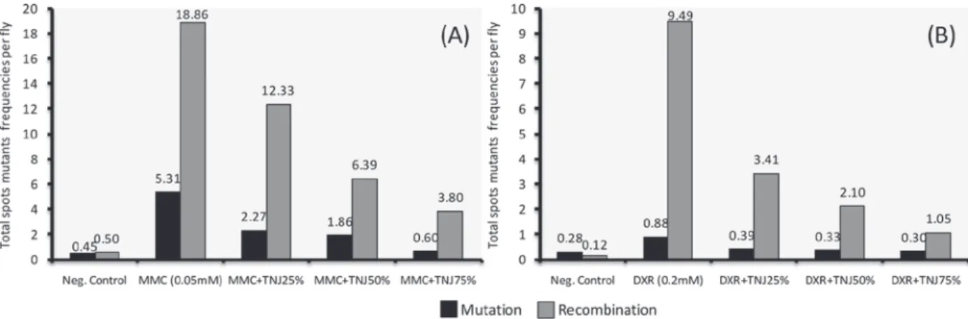
The effect on mutation and recombination induced by both genotoxins, and its modulation by TNJ is shown in Figure 1. MMC (0.05 mM)
induced a total of 24.17 spots per fl y through
a combination of mutation and recombination, while 25, 50 and 75% concentrations of TNJ decreased this frequency to 14.60, 8.25 and 4.40, respectively. MMC alone induced 5.31 spots per
fl y due exclusively to mutation, and 18.86 due
to mitotic recombination. TNJ administered in the culture media reduced the MMC mutational frequency to 2.27, 1.86 and 0.60, while the MMC-recombinational frequency was decreased to 12.33, 6.39 and 3.80 respectively, demonstrating a dose-response effect for both MMC-genotoxic events.
DXR (0.2 mM) induced a total frequency of 10.37 spots, but when TNJ (25, 50 and 75%) were co-administered this frequency dropped to 3.80, 2.43 and 1.35 (Table I). DXR induced 0.85 spots
per fl y due exclusively to mutation and all TNJ
Spots per fl y (nº of spots) statistical diagnosisa
Clone induction frequencies (per 105 cells per
cell division)d (n/NC)ef
Inhibitiong (%) Genotype
and compounds
N. of
fl ies
(N)
Small single spots
(1-2 céls)b
m=2
Large single spots
(>2 céls)b
m=5
Twin spots
m=5
Total spots
m=2
Total
mwh
clonesc (n)
mwh/fl r3
Neg. Control 40 0.73 (29) 0.13 (5) 0.10 (4) 0.95 (38) 38 1.95
MMC
(0.05mM) 40 12.05 (482) 7.90 (316) 4.23 (169) 24.18 (967) 925 47.39 [45.44]
MMC+
TNJ25% 40 6.78 (271) + 5.08 (203) w+ 2.75 (110) w+ 14.60 (584) w+ 562 28.79 [26.84] 40.93
MMC+
TNJ50% 40 3.70 (148) + 2.85 (114) + 1.70 (68) + 8.25 (330) + 319 16.34 [14.40] 68.31
MMC+
TNJ75% 40 1.90 (76) + 1.50 (60) + 1.00 (40) + 4.40 (176) + 171 8.76 [6.81] 85.01
mwh/ TM3,BdS
Neg.
Control 40 0.40 (16) 0.05 (2) h 0.45 (18) 18 0.92
MMC 40 3.78 (151) 1.30 (52) 5.08 (203) 203 10.40 [9.48]
MMC +
TNJ25% 40 1.70 (68) + 0.48 (19) + 2.18 (87) + 87 4.46 [3.53] 62.76
MMC +
TNJ50% 40 1.38 (55) + 0.43 (17) + 1.80 (72) + 72 3.69 [2.77] 70.78
MMC +
TNJ75% 40 0.50 (20) + 0.08 (3) + 0.58 (23) + 23 1.18 [0.26] 97.26
TABLE I
Effect of the co-treatment (TNJ + MMC and TNJ + DXR) in somatic cells of
Drosophila melanogaster using the wing SMART standard cross.
Drosophila melanogaster
DISCUSSION
In a previous report we have demonstrated that TNJ (25, 50, 75 and 100% v/v) has no genetic toxicity effects in somatic cells of Drosophila melanogaster (Franchi et al. 2008), indicating the absence of TNJ mutational and recombinational actions. Previous studies have also shown that TNJ did not induce gene mutations at the HPRT locus (in 0.003 to 3
μL/mL dose range), in presence and absence of S9 mix. In the same study, no mutagenic activity of ethyl-acetate extract from noni juice (100-fold concentrated) was observed in Chinese hamster V79-cell line (Westendorf et al. 2007). There was no increase in micronuclei nor any evidence of
systemic toxicity in mice ascribed to dehydrated TNJ (10 g/kg body weight) when administered via oral gavage (Edwards 2002). Also, no
signifi cant increases were noted in the frequency
of chromosome aberrations in cultured cells from human lymphocytes (625, 1250, 2500, and 5000
μg/mL) in presence or absence of S9 mix (Edwards
2003, Ratanavalachai et al. 2008, West et al. 2006).
Considering these fi ndings, we decided to
evaluate the antimutagenic and/or antirecom-binagenic action of TNJ. In our study, the co-treatment with different concentrations of TNJ plus
DXR or MMC induced a statistically signifi cant
dose-response reduction in the frequencies of
DXR and MMC are two well known antineo-plasic agents used in the treatment of solid tumors (Begleiter 2000, Minotti et al. 2004). DXR inhibits the activity of the enzyme topoisomerase II, inducing DNA strand breaks (Islaih et al. 2005, Resende et al. 2006), while MMC acts primarily
by promoting DNA crosslinkage (Efi mov and
Fedyunin 2010, Riley and Workman 1992, Tomasz et al. 1987). Through these mechanisms, MMC and DXR are able to induce mutations and
chromosomal aberrations in both tumor and non-tumor cells. Moreover, cellular enzymes are capable of converting DXR and MMC into free radical metabolites (Benchekroun et al. 1993, Dusre et al. 1989, Menegola et al. 2001), which in turn induce damage to several molecules, such as DNA.
Many studies have suggested the co-administration of antineoplasic agents and free radical scavengers, such as antioxidants, to reduce the genotoxicity of such treatments in non-tumor
TABLE I (CONTINUATION)
Spots per fl y (nº of spots) statistical diagnosisa
Clone induction frequencies (per 105 cells per
cell division)d (n/NC)ef
Inhibitiong (%) Genotype
and compounds
N. of
fl ies
(N)
Small single spots
(1-2 céls)b
m=2
Large single spots
(>2 céls)b
m=5
Twin spots
m=5
Total spots
m=2
Total
mwh
clonesc (n)
mwh/fl r3
Neg.
Control 40 0.35 (14) 0.03 (1) 0.03 (1) 0.40 (16) 16 0.82
DXR (0.2mM) 40 1.43 (57) 7.03 (281) 1.93 (77) 10.38 (415) 400 20.49 [19.67]
DXR +
TNJ25% 40 1.28 (51) - 1.28 (51) + 1.25 (50) + 3.80 (152) + 147 7.53 [6.71] 65.89
DXR +
TNJ50% 40 1.05 (42) - 0.60 (24) + 0.78 (31) + 2.43 (97) + 97 4.97 [4.15] 78.90 DXR +
TNJ75% 40 0.90 (36) + 0.30 (12) + 0.15 (6) + 1.35 (54) + 54 2.77 [1.95] 90.09
mwh/TM3,BdS
Neg.
Control 40 0.28 (11) 0.00 (0) 0.28 (11) 11 0.56
DXR 40 0.73 (29) 0.13 (5) 0.85 (34) 34 1.74 [1.18]
DXR +
TNJ25% 40 0.35 (14) + 0.03 (1) i h 0.38 (15) + 15 0.77 [0.20] 83.05
DXR +
TNJ50% 40 0.30 (12) + 0.03 (1) i 0.33 (13) + 13 0.67 [0.10] 91.53
DXR +
TNJ75% 40 0.30 (12) + 0.00 (0) + 0.30 (12) + 12 0.61 [0.05] 95.76
aStatistical diagnosis according to Frei and Würgler (1988): +, positive; -, negative; i, inconclusive. m, factor of multiplication for
evaluation of results signifi cantly negatives. Levels of signifi cance α= 0,05. bIncluding rare single spots fl r3. cConsidering clones
mwh for single spots mwh and for twin spots. dCalculated accordance with Frei et al. (1992). eC = 48,800, i. e., approach number
of cells examined for individual. fffNumbers between keys are the corrected frequencies of induction in relation incidence estimate Numbers between keys are the corrected frequencies of induction in relation incidence estimate
of the negative control. gCalculated according to Abraham (1994): (genotoxin alone-genotoxin plus TNJ/genotoxin alone)×100. hJust single spots mwh can be observed in heterozygous individual mwh/TM3,BdBdBdS, since TM3 balancer chromosome does not
Figure 1 - Contribution of mutation and recombination to total mutant spots frequencies per fl y in trans-heterozygous fl ies treated
with MMC (A) or DXR (B) in combination with TNJ (25%, 50%, 75%). The recombinagenic action was calculated according to Graf et al. (1992) and Sinigaglia et al. (2006) as follows: Frequencies (et al. (2006) as follows: Frequencies ( ) = total FFF) = total mwh clones (n) /number of fl ies (N); Mutation
Frequencies (F
Frequencies ( M) = frequencies of FFM) = frequencies of mwh clones in balancer-heterozygous/frequencies of mwh clones in marker trans-heterozygous; Recombination Frequencies (FFFR) = 1-R) = 1-FMFMFM; Frequencies of Total Spots (; Frequencies of Total Spots (FFFT) = total spots in T) = total spots in mwh//fl rfl r3 fl ies (considering mwh and fl r3 spots)/number of fl ies; Mutation = FFFT × T × FFFM; Recombination = FFFT × T × FFFR.
cells (Amara-Mokrane et al. 1996, Antunes et al. 2007, Antunes and Takahashi 1998, Costa and Nepomuceno 2006, Fragiorge et al. 2007, Gentile et al. 1998, Tavares et al. 2006). So, the noni juice may be promising in this scenario.
In this study, TNJ produced a marked decrease in all kinds of MMC- and DXR-induced mutant spots. However, the largest effect observed in the co-administration of TNJ with MMC and DXR was antirecombinagenic, resulting in 82 and 87% reductions, respectively.
The mechanisms by which TNJ exerts its antigenotoxic activity are not clear at the present. However, antioxidant and/or free radical scavenger activities could be suggested. The radical scavenging activity has been measured in vitro using the terazolium nitroblue (TNB) assay, by assessing the juice’s potential capacity to protect cells or lipids from oxidative alteration promoted by a superoxide anion radical (SAR). The SAR scavenging activity of TNJ was 2.8 times higher than that of vitamin C, and 1.4 times higher than that of pycnogenol and grape seed powder (Wang and Su 2001). TNJ’s protective effects were proportional to the concentrations applied, indicating a dose-response correlation.
Iridoids and potent antioxidant phenolic compounds, such as deacetylasperulosidic acid, scopoletin, isoscopoletin, aesculetin, 7-hydroxycoumarin
(7-HC), quercetin, americanin A have been identifi ed in
noni fruits (Dussossoy et al. 2011, Ikeda et al. 2009, Liu et al. 2007, Su et al. 2005, West et al. 2011).
These fi ndings suggest that several compounds,
in particular iridois and phenolic compounds, could contribute separately or synergistically to an antioxidative and antigenotoxic activity in TNJ.
Alternatively, the antigenotoxic activity detected for TNJ could be attributed to the presence of QR inducers. These compounds, such as 2-methoxy-1,3,6-trihydroxyanthraquinone (Pawlus et al. 2005), scopoletin and quercetin (Nitteranon et al. 2011), have been described in noni fruit. QR is a phase II metabolizing enzyme that is induced in conjunction with other protective phase II enzymes. This induction of phase II enzymes, such as QR, is considered as a cancer chemopreventive - since potential oxidative and electrophilic molecules can be more readily metabolized and excreted before its interaction with cellular macromolecules, such as DNA. QR is also responsible for maintaining the
and coenzyme Q10. QR inducers are sometimes referred to as “indirect antioxidants”, and this activity is considered protective at the initiation stage of carcinogenesis (Dinkova-Kostova and Talalay 2000, Kensler 1997).
This study has successfully used the SMART to demonstrate the protective effects of TNJ on the genotoxicity of DXR and MMC. We conclude that TNJ provides greater protection against these drugs, and that antirecombinagenic activity was the predominant effect. A dose-response relationship was also observed and might be attributed mainly to their powerful scavenger ability. Nevertheless, further experiments should be carried out to gain a better understanding of the mechanism of action of noni phytocompounds and to ensure their safe clinical use.
RESUMO
Noni, nome Havaiano da fruta de Morinda citrifolia L., é uma planta medicinal e tradicional da Polinésia
amplamente usada para o tratamento de muitas doenças
entre as quais artrite, diabetes, asma, hipertensão e câncer. Neste trabalho, um suco comercial de noni (TNJ) foi avaliado para suas atividades protetoras
contras as lesões induzidas por mitomicina C (MMC) e doxorrubicina (DXR) usando o teste de mutação e recombinação somática em Drosophila melanogaster. Larvas de terceiro estágio, trans-heterozigotas para dois genes marcadores (mwh e fl r3), foram cotratadas com
TNJ mais MMC ou DXR. Observamos uma redução
dos efeitos genotóxicos de MMC e DXR pelo suco. TNJ
induziu uma forte redução em todos os tipos de manchas
mutantes induzidas por MMC e DXR principalmente devido a uma atividade antirecombinante. Os efeitos
protetotes de TNJ foram dependentes da concentração, indicando uma correlação dose-resposta, os quais podem ser atribuídos a uma potente ação antioxidante
e/ou habilidade de sequestrar radicais livres pelo TNJ.
Palavras-chave: antigenotoxicidade, doxorrubicina, mitomicina C, SMART, suco de noni.
REFERENCES
ABRAHAM SK. 1994. Antigenotoxicity of coffee in the Drosophila assay for somatic mutation and recombination. Mutagenesis 9: 383-386.
AMARA-MOKRANE YA, LEHUCHER-MICHEL MP, BALANSARD
G, DUMÉNIL G AND BOTTA A. 1996. Protective effects of alpha-hederin, chlorophyllin and ascorbic acid towards the induction of micronuclei by doxorubicin in cultured human lymphocytes. Mutagenesis 11: 161-167.
ANDRADE
A
A HHR, RREGULY ML AND LEHMANN M. 2003. Wing Somatic Mutation and Recombination Test. In: HENDERSON DS (Ed), Drosophila Cytogenetics Protocols - Methods in Molecular Biology. Humana Press Inc., Totowa, p. 389-412.
ANTUNES
A
A LMG, BUENO RDE, DIAS FD AND BIANCHI MDP.
2007. Acetylsalicylic acid exhibits anticlastogenic effects on cultured human lymphocytes exposed to doxorubicin. Mutat Res Genet Toxicol Environ Mutagen 626: 155-161. ANTUNES
A
A LMG AND TAKAHASHI CS. 1998. Effects of high doses of vitamins C and E against doxorubicin-induced chromosomal damage in Wistar rat bone marrow cells. Mutat Res Genet Toxicol Environ Mutagen 419: 137-143. BEGLEITER A. 2000. Clinical applications of quinone-containing alkylating agents. Front Biosci 5: E153-E171. BENCHEKROUN MN, SINHA BKBK AND AND ROBERT J. 1993.
Doxorubicin-induced oxygen-free radical formation in sensitive and doxorubicin-resistant variants of rat glioblastoma cell-lines. FEBS Lett 322: 295-298.
CARDON D. 2003. Le Monde des Teintures Naturelles. Belin, Paris, France, 586 p.
CHAN-BLANCO Y, VAILLANT F, PEREZ AM, RRREYNES M,
BRILLOUET JM AND BRAT P. 2006. The noni fruit (Morinda citrifolia L.): A review of agricultural research, nutritional and therapeutic properties. J Food Compos Anal 19: 645-654.
COSTA WF AND NEPOMUCENO JC. 2006. Protective effects of a mixture of antioxidant vitamins and minerals on the genotoxicity of doxorubicin in somatic cells of Drosophila
melanogaster. Environ Mol Mutagen 47: 18-24.
DENG Y, CHIN YW, CHAI H, KKKELLER WJWJ AND AND KINGHORN AD.
2007. Anthraquinones with quinone reductase-inducing activity and Benzophenones from Morinda citrifolia
(Noni) roots. J Nat Prod 70: 2049-2052.
DINKOVA-KOSTOVA AT AND TALALAY P. 2000. Persuasive evidence that quinone reductase type 1 (DT diaphorase) protects cells against the toxicity of electrophiles and reactive forms of oxygen. Free Radic Biol Med 29: 231-240.
DUSRE L, MIMNAUGH EG, MYERS CE AND SINHA BK. 1989.
Potentiation of doxorubicin cytotoxicity by buthionine sulfoximine in multidrug-resistant human breast tumor cells. Cancer Res 49: 511-515.
DUSSOSSOY E, BRAT P, BONY E, BOUDARD F, POUCHERET
EARLE J. 2001. Plantas Medicinales en el Trópico Húmedo. San José, CR, Editorial Guayacán, 246 p.
EDWARDS CN. 2002. Tahitian Noni Juice-mouse micronucleus test. Test Report. Scantox Biologisk Laboratorium, Lille Skensved, DK (Lab no. 47053).
EDWARDS CN. 2003.In vitro mammalian chromosome aberra-tion test performed with human lymphocytes. Test Report. Scantox Biologisk Laboratorium. Lille Skensved, DK (Lab no. 48877).
EFIMOV V AND FEDYUNIN S. 2010. Cross-linked nucleic acids: isolation, structure, and biological role. Biochemistry 75: 1606-1627.
FRAGIORGE EJ, SPANÓ MA AND AAANTUNES LMG. 2007.
Modulatory effects of the antioxidant ascorbic acid on the direct genotoxicity of doxorubicin in somatic cells of Drosophila melanogaster. Genet Mol Biol 30: 449-455. FRANCHI LP, GUIMARÃES NN, LEHMANN M, AAANDRADE
HHR AND
HHR AND CUNHA KS. 2008. Ausência de efeito tóxico-genético de Morinda citrifolia (noni) em células somáticas de Drosophila melanogaster. Rev Elet Farm 5(3): 46-53. FRANCHI LP, PENTIADO NHGR, SILVA RDN, GUIMARÃES NN,
JESUINO RSA, DE ANDRADE HHR, LEHMANN M AND
CUNHA KS. 2009. Mutagenic and recombinagenic effects of lamivudine and stavudine antiretrovirals in somatic cells of Drosophila melanogaster. Food and Chem Toxicol 47: 578-582.
FREI H AND WÜRGLER FE. 1988. Statistical-methods to decide whether mutagenicity test data from drosophila assays indicate a positive, negative, or inconclusive result. Mutat Res 203: 297-308.
FREI H AND WÜRGLER FE. 1995. Optimal experimental design and sample size for the statistical evaluation of data from somatic mutation and recombination tests (SMART) in Drosophila. Mutat Res 334: 247-258.
FURUSAWA E, HIRAZUMI A, STORY S AND JENSEN J. 2003.
Antitumour potential of a polysaccharide-rich substance from the fruit juice of Morinda citrifolia (Noni) on sarcoma 180 ascites tumour in mice. Phytother Res 17: 1158-1164.
GENTILE JM, RRRAHIMI S, ZWIESLER J, GENTILE GJGJ AND AND
FERGUSON LR. 1998. Effect of selected antimutagens on the genotoxicity of antitumor agents. Mutat Res Fundam Mol Mech Mutagen 402: 289-298.
GRAF U AND VANSCHAIK N. 1992. Improved high bioactivation cross for the wing somatic mutation and recombination test in Drosophila melanogaster. Mutat Res 271: 59-67. GRAF U, WURGLER FE, KKKATZ AJ, FREI H, JUON H, HALL CB
AND KALE PG. 1984. Somatic mutation and recombi-nation test in Drosophila melanogaster. Environ Mutagen 6: 153-188.
HARADA S, HAMABI W, KKKAMIYA K, SATAKE T, YAMAMOTO J
AND TOKUYAMA S. 2009. Preventive effect of Morinda citrifolia fruit Juice on neuronal damage induced by focal ischemia. Biol Pharm Bull 32: 405-409.
HIRAZUMI A AND FURUSAWA E. 1999. An immunomodulatory polysaccharide-rich substance from the fruit juice of
Morinda citrifolia (noni) with antitumour activity.
Morinda citrifolia (noni) with antitumour activity.
Phytother Res 13: 380-387.
HIRAZUMI A, FURUSAWA E, CHOU SC AND HOKAMA Y.
1996. Immunomodulation contributes to the anticancer activity of Morinda citrifolia (noni) fruit juice. Western Pharmacology Society - Proceedings of the Thirty-Ninth Annual Meeting 39: 7-9.
IKEDA R, WADA M, NISHIGAKI T AND NAKASHIMA K. 2009.
Quantifi cation of coumarin derivatives in Noni (Morinda citrifolia) and their contribution of quenching effect on reactive oxygen species. Food Chem 113: 1169-1172. ISLAIH M ET AL. 2005. Relationships between genomic, cell
cycle, and mutagenic responses of TK6 cells exposed to DNA damaging chemicals. Mutat Res Fundam Mol Mech Mutagen 578: 100-116.
KASTENBAUM
K
K MA AND BOWMAN KO. 1970. Tables for determining the statistical signifi cance of mutation frequencies. Mutat Res Fundam Mol Mech Mutagen 9: 527-549.
KENSLER
KENSLER
K TW. 1997. Chemoprevention by inducers of carcinogen detoxication enzymes. Environ Health Perspec 105: 965-970.
LIU CH, XUE YR, YE YH, YUAN FF, LIU JY AND SHUANG JL.
2007. Extraction and characterization of antioxidant compositions from fermented fruit juice of Morinda citrifolia (noni). Agric Sci China 6: 1494-1501.
MENEGOLA E, BROCCIA ML AND RENZO FD. 2001. Terato-genic effects of doxorubicin in rats at midgestation and at term. Teratogen Carcinogen Mutagen 21: 283-293. MINOTTI G, MENNA P, SALVATORELLI E, CAIRO G AND
GIANNI L. 2004. Anthracyclines: Molecular advances and pharmacologic developments in antitumor activity and cardiotoxicity. Pharmacol Rev 56: 185-229.
NELSON SC. 2006. Species profi les for Pacifi c Island
agroforestry: Morinda citrifolia (noni). http://www. traditionaltree.org/
NITTERANON V, ZHANG G, DARIEN BJBJ AND AND PARKIN K. 2011.
Isolation and synergism of in vitro anti-infl ammatory and
quinone reductase (QR) inducing agents from the fruits of Morinda citrifolia (noni). Food Res Int 44: 2271-2277. OHSAWA Y AND OHBA S. 1993. Damnacanthal. Acta Crystallographica Section C-Crystal Structure Communi-cations 49: 2149-2151.
PALU AK, KIM AH, WEST BJ, DENG S, JENSEN JJ AND AND WHITE
L. 2008. The effects of Morinda citrifolia L. (noni) on the immune system: Its molecular mechanisms of action. J Ethnopharmacol 115: 502-506.
PAWLUS AD, SU BN, KKELLER WJWJ AND AND KINGHORN AD. 2005.
An anthraquinone with potent quinone reductase-inducing activity and other constituents of the fruits of Morinda citrifolia (Noni). J Nat Prod 68: 1720-1722.
POTTERAT O AND HAMBURGER M. 2007. Morinda citrifolia
(Noni) fruit - Phytochemistry, pharmacology, safety. Planta Med 73: 191-199.
RATANAVALACHAI
R
R T, THITIORUL S AND NANDHASRI P. 2008.
RESENDE
R
R FA, MATTOS DE AAANDRADE BARCALA CA, DA SILVA
FARIA MC, KKKATO ATO FH, CUNHA WRWR AND AND TAVARES DC.
2006. Antimutagenicity of ursolic acid and oleanolic acid against doxorubicin-induced clastogenesis in Balb/c mice. Life Sci 79: 1268-1273.
REZENDE
R
R AAA ET AL. 2011. The effect of the dibenzylbutyro-lactolic lignan (-)-cubebin on doxorubicin mutagenicity and recombinogenicity in wing somatic cells of Drosophila
melanogaster. Food Chem Toxicol 49(6): 1235-1241. RILEY RJRJ AND AND WORKMAN P. 1992. DT-diaphorase and
cancer-chemotherapy. Biochem Pharmacol 43: 1657-1669. SALUDES JP, GARSON MJ, FRANZBLAU SG AND AGUINALDO
AM. 2002. Antitubercular constituents from the hexane fraction of Morinda citrifolia Linn. (Rubiaceae). Phytother Res 16: 683-685.
SANTOS JH, GRAF U, RREGULY ML AND AAANDRADE HHR.
1999. The synergistic effects of vanillin on recombination predominate over its antimutagenic action in relation to MMC-induced lesions in somatic cells of Drosophila
melanogaster. Mutat Res Gen Toxicol Environ Mutagen 444(2): 355-365.
SINIGAGLIA M, LEHMANN M, BAUMGARDT P, AMARAL VS,
DIHL RR, RREGULY ML AND AAANDRADE HHR. 2006. Vanillin as a modulator agent in SMART test: Inhibition in the steps that precede N-methyl-N-nitrosourea-, N-ethyl-N-nitrosourea-, ethylmethanesulphonate- and bleomycin-genotoxicity. Mutat Res 607: 225-230.
SU BN, PAWLUS AD, JUNG HA, KKKELLER WJ, MCLAUGHLIN JL
AND KINGHORN AD. 2005. Chemical constituents of the fruits of Morinda citrifolia (Noni) and their antioxidant activity. J Nat Prod 68: 592-595.
TAVARES DC, BARCELOS GRM, SILVA LF, TONIN CCC AND
BASTOS JK. 2006. Propolis-induced genotoxicity and antigenotoxicity in Chinese hamster ovary cells. Toxicol in Vitro 20: 1154-1158.
TÉLLEZ MGO, RODRÍGUEZ HB, OLIVARES GQ, SORTIBRÁN
ANC, CETTO AA AND RODRÍGUEZ-ARNAIZ R. 2007. A phytotherapeutic extract of Equisetum myriochaetum is not genotoxic either in the in vivo wing somatic test of Drosophila or in the in vitro human micronucleus test. J Ethnopharmacol 111: 182-189.
TOLEDO VM, TÉLLEZ MGO, SORTIBRÁN ANC, AAA
NDRADE-CETTO A AND RODRÍGUEZ-ARNAIZ R. 2008. Genotoxicity testing of Cecropia obtusifolia extracts in two in vivo
assays: The wing somatic mutation and recombination test of Drosophila and the human cytokinesis-block micronucleus test. J Ethnopharmacol 116: 58-63.
TOMASZ M, LIPMAN R, CHOWDARY D, PAWLAK J, VERDINE
GL AND NAKANISHI K. 1987. Isolation and structure of a covalent cross-link adduct between mitomycin-c and DNA. Science 235: 1204-1208.
VOGEL EW, GRAF U, FREI HJHJ AND AND NIVARD MMJ. 1999. The results of assays in Drosophila as indicators of exposure to carcinogens. In: MCGREGOR DB, RICE JM AND VENITT S (Eds), Use of short- and medium-term tests for carcinogens and data on genetic effects in carcinogenic hazard evaluation. IARC Sci Publ 146: 427-470.
WANG MY AND SU C. 2001. Cancer preventive effect of
Morinda citrifolia (Noni). Canc Prev 952: 161-168. WANG MY, WEST BJ, JENSEN CJ, NOWICKI D, SU C, PALU AK
AND AAANDERSON G. 2002. Morinda citrifolia (Noni): A literature review and recent advances in Noni research. Acta Pharmacol Sin 23: 1127-1141.
WEST BJ, DENG S AND JENSEN CJ. 2011. Nutrient and phyto-chemical analyses of processed noni puree. Food Res Int 44: 2295-2301.
WEST BJ, JENSEN CJ, WESTENDORF JJ AND AND WHITE LD. 2006.
A safety review of Noni fruit juice. J Food Sci 71: R100-R106.
WESTENDORF J, EFFENBERGER K, IZNAGUEN H AND BASAR S.
2007. Toxicological and analytical investigations of noni (Morinda citrifolia) fruit juice. J Agric Food Chem 55: 529-537.
YU H, LI S, HUANG MT AND HO CT. 2008. Antiinfl ammatory