(Annals of the Brazilian Academy of Sciences) ISSN 0001-3765
www.scielo.br/aabc
Assessment of sperm production and reproductive organs
of Wistar rats to long-term exposure of
Caesalpinia ferrea
LEDA M.F. LUCINDA1, CAMILA B. ROCHA1, MAYCON M. REBOREDO1, VINÍCIUS C. FARIA1 and RITA C.S. SÁ2
1Universidade Federal de Juiz de Fora, Centro de Biologia da Reprodução, Rua José Lourenço Kelmer, s/n Campus Universitário, Bairro São Pedro, 36036-900 Juiz de Fora, MG, Brasil
2Universidade Federal da Paraíba, Departamento de Fisiologia e Patologia Cidade Universitária, 58059-900 João Pessoa, PB, Brasil
Manuscript received on February 5, 2009; accepted for publication on June 24, 2010
ABSTRACT
Caesalpinia ferreaMart (Leguminosae) is a medicinal plant used to treat diabetes, among other therapeutic properties, but which is also reported to have hepatotoxic effects. Although it contains substances such as flavonoids and coumarin, which are known to have antifertility activity, no studies have apparently been conducted to evaluate the potential adverse side effects of this plant on the function of the reproductive system after a chronic treatment. Therefore, this investigation was carried out to evaluate the effect and safety of the long-term exposure toC. ferreaon male Wistar rats’ vital organs, reproductive system and sperm production. Adult and immature male rats were treated with an aqueous extract ofC. ferreaat a dose level of 300 mg/kg of body weight, administered during one or two spermatogenic cycles of this species.
The reproductive and vital organs were analyzed, and sperm was collected from the epididymal secretion of the right epididymis cauda. The long-term administration ofC. ferreadid not significantly alter the body, vital and reproductive
organs weights. Gamete production was not affected either. The chronic assessment of C. ferreasuggests that this
plant does not affect the normal functioning of the Wistar rat reproductive system.
Key words: Caesalpinia ferrea, Wistar rat, reproductive system, toxicological assessment, chronic and sub-chronic
treatment.
INTRODUCTION
Caesalpinia ferrea Mart (Leguminosae), popularly known in Brazil as “Pau-ferro” or “Juca”, is a plant commonly used by the population for its antiulcerogenic (Bachi et al. 1995), anti-inflammatory (Carvalho et al. 1996) and cancer chemopreventive properties (Naka-mura et al. 2002). This plant is also used for treating respiratory tract diseases, dysentery, diabetes (Bragança 1996) and liver inflammation (Di Stasi et al. 2002), al-though the occurrence of hepatotoxicity has been re-ported in the literature (Di Stasi et al. 2002). Phyto-chemical studies have shown the presence of sitosterol,
Correspondence to: Rita de Cássia S. Sá E-mail: ritacassia.sa@bol.com.br
flavonoids, saponins, tanins, coumarins, steroids and phenolic compounds in the hydroalcoholic extract of the
stem bark and leaves of C. ferrea (Gonzalez et al.
In addition, the administration of an aqueous extract ofC. ferreato Wistar rats submitted to a short-term bioassay
showed a non-significant weight reduction of the seminal vesicle (Reboredo et al. 2006).
Reproductive toxicity is linked to the occurrence of adverse effects expressed as alterations to the reproduc-tive organs, the related endocrine and nervous systems, or pregnancy outcomes. Toxicity for males may result from adverse effects on sexual maturation, gamete pro-duction and transport, sexual behavior and fertility (Kimmel et al. 1995). A variety of chemical and phys-ical agents, including the natural occurring substances found in plants, have been associated with male repro-ductive toxicity (Dixit et al. 1989, Montanari et al. 1998, He et al. 2010, Wang et al. 2010). Folk medicine is traditionally employed by many people throughout the world. However, the clinical knowledge on the effec-tiveness of plants in the treatment of diseases and fertil-ity-related problems, as well as the risks and nature of any adverse side effects, are quite often not clearly de-termined.
Acute, chronic and subchronic tests are among the standard protocols routinely used for toxicity tests of the reproductive system. Short-term tests are important to identify target sites and affected cell types, while term tests provide information on toxic effects of long-term dosing, covering one or more spermatogenic cycles (Zenick et al. 1994). Considering the popular use ofC. ferrea, the seminal vesicle being a possible target organ,
and the presence of substances with antifertility activity among its constituents, this study was designed to inves-tigate the potential reproductive toxicity in male Wistar rats submitted to long-term treatments with an aqueous extract of this plant.
MATERIALS AND METHODS
ANIMALS ANDHOUSING
Immature male Wistar rats (Rattus norvegicus
Berken-hout 1769) (30 days old and weighing about 70 g) and adult male Wistar rats (90 days old and weighing about 250 g) were obtained from the vivarium of the Federal University of Juiz de Fora (UFJF), where they were born and bred. The animals were housed individually un-der standard laboratory conditions, with a 12h light/12h
dark photoperiod. They were fed on rat chow pellets and received waterad libitum. The experimental protocol
was approved by the Ethical Committee of the Repro-duction Biology Center (UFJF) (protocol number 013/ 2004-CEA), which follows the international principles in ethics for animal experimentation.
PLANTMATERIAL
C. ferreaMart (Leguminosa) was collected in the Belém region (Pará State, Brazil) in 2005 and authenticated by Dr. Antonio Barioni Guzman in the Herbarium of the Biology Department of USP in Ribeirão Preto (São Paulo State, Brazil), where a voucher specimen regis-tered under the number 3221 is kept. The air-dried fruits were cut into small pieces and macerated in distilled wa-ter to obtain a crude aqueous extract (w/w yield = 23%). Afterwards, 100 mL of the crude extract was diluted to 300 mL of an aqueous solution to yield 76.6 mg/mL as the final concentration. The extract was, then, frozen for storage. The aqueous extract was subsequently recon-stituted in water at the appropriate concentration for the experiment.
TREATMENTPROCEDURE
Reproductive toxicity was assessed through two chronic test protocols. In the first protocol, 24 adult Wistar rats were selected at random and divided evenly into a treat-ment group (T1) and a control group (C1). In the second protocol, 30 immature Wistar rats were divided evenly into a treatment group (T2) and a control group (C2). The rats in the treatment groups received a single daily dose by gavage of 1 mL ofC. ferreaextract of 300 mg/
kg of body weight, which was administered for 52 days (T1 group) or 104 days (T2 group). The 52 day period corresponds to the spermatogenic cycle of this species (Hilscher 1964). The control groups received 1 mL of distilled water following the same protocol as the treat-ment group.
The animals of the first protocol, T1 and C1, were euthanized by inhalation of an anesthetic (halothane) on the 53rdday, and those of the second protocol, T2 and C2, on the 105th day. Immediately after death, the fol-lowing organs were dissected out and weighed: testes, left epididymis, seminal vesicle, prostate, liver, kidneys, lungs, brain and the pituitary gland. The left testis and epididymis were, then, fixed in Bouin for histological examination. To conduct a light microscope examina-tion, the tissues were dehydrated in a graded series of
ethanol, embedded in paraffin and sectioned at 3 µm
thickness for routine haematoxylin and eosin staining (Humason 1972).
Sperm was collected from the epididymal secre-tion of the right epididymis cauda. The secresecre-tion was placed in a 0.3 mL drop of physiological serum extract and later diluted in distilled water. From this homo-genate, a sample was taken and a sperm count was ob-tained using a hemocytometer with improved double Neubauer ruling (Moraes 1994).
The Student’s t test and the Mann-Whitney test
were applied for statistical comparison of the differ-ences in data between the test groups (α= 0.05) (Sokal and Rohlf 1996) and the results were expressed by mean and standard deviation (SD).
RESULTS
During the treatment period, no deaths and no changes in locomotor activity, piloerection, nor any other clinical signs of toxicity were observed. Body weight did not change significantly, and the treatment did not interfere with food consumption, although the average food intake of the treatment group (T2, 104-day test protocol) was systematically a little below the average consumption of the respective control group.
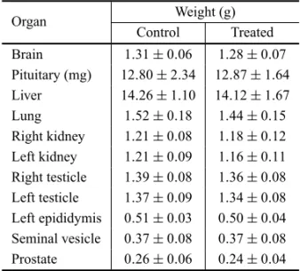
The weight of the reproductive organs, the acces-sory sex glands, the pituitary gland and other analyzed organs were not significantly different between control and treated groups on both toxicity test protocols (Tables I and II).
Sperm concentrations from the secretion of the
right epididymis cauda were: control (C1) = 108.4±
64×106 sperm/mL (mean ±SD) and treated (T1) =
191.4±138.3×10⁶sperm/mL; control (C2) = 1095±
334.8×10⁶sperm/mL (mean±SD) and treated (T2)
TABLE I
Organ weights of control andC. ferreaextract-treated Wistar rats submitted to chronic treatment, with an
exposure of 52 days and death on the 53rdday.
Organ Weight (g)
Control Treated Brain 1.15±0.08 1.16±0.06
Pituitary (mg) 11.33±3.74 13.0±3.64
Liver 12.01±1.60 12.62±1.90
Lung 1.51±0.16 1.53±0.12
Right kidney 1.19±0.13 1.23±0.11
Left kidney 1.16±0.13 1.20±0.11
Right testicle 1.36±0.07 1.41±0.03
Left testicle 1.37±0.07 1.41±0.07
Left epididymis 0.42±0.04 0.45±0.04
Seminal vesicle 0.37±0.04 0.40±0.11 Prostate 0.30±0.05 0.30±0.04
Results expressed in mean±SD. N=12.
TABLE II
Organ weights of control andC. ferreaextract-treated Wistar rats submitted to chronic treatment, with an
exposure of 104 days and death on the 105thday.
Organ Weight (g)
Control Treated Brain 1.31±0.06 1.28±0.07
Pituitary (mg) 12.80±2.34 12.87±1.64 Liver 14.26±1.10 14.12±1.67
Lung 1.52±0.18 1.44±0.15
Right kidney 1.21±0.08 1.18±0.12 Left kidney 1.21±0.09 1.16±0.11
Right testicle 1.39±0.08 1.36±0.08
Left testicle 1.37±0.09 1.34±0.08 Left epididymis 0.51±0.03 0.50±0.04
Seminal vesicle 0.37±0.08 0.37±0.08
Prostate 0.26±0.06 0.24±0.04 Results expressed in mean±SD. N=15.
= 1079±196.6×10⁶ sperm/mL. There were no
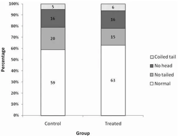
Fig. 1 – Proportion of normal and abnormal spermatozoa of control andC. ferreaextract-treated Wistar rats submitted to chronic treatment, with an exposure of 52 days and death on the 53rdday. N = 12.
Fig. 2 – Proportion of normal and abnormal spermatozoa of control andC. ferreaextract-treated Wistar
The histological examination revealed that there was no structural change in the seminiferous epithelium of the treated groups’ testis when compared to that of the control groups. Similarly, the epididymis of control and treated rats showed a normal epithelium lining and the presence of spermatozoa in all segments (head, body and tail) of this organ.
DISCUSSION
Plants contain many active compounds that are prob-ably responsible for their different therapeutic properties exploited by folk medicine. However, these natural oc-curring compounds may also exert a toxic effect in the development or normal functioning of the reproductive system (Abdel-Magied et al. 2001, Bidwai et al. 1990, Hiremath et al. 1997, Montanari et al. 1998, Rajase-karan et al. 1988). Coumarin, flavonoids and sitosterol are plant substances that have been related to the occur-rence of adverse effects on the reproductive system. Pre-vious studies have shown that coumarin had antifertility activity in female rats (Ulubelen et al. 1994), whereas flavonoids produced antiandrogenic activities in male dogs (Bhargava 1989) and sitosterol reduced sperm pro-duction in rats (Malini and Vanithakumari 1991). These substances have been reported to be among the active compounds of the aerial parts ofC. ferrea, thus
suggest-ing a possible toxic behavior of this plant on the repro-ductive system.
Reproductive tests designed to evaluate the toxic-ity of compounds are important to identify which tar-gets in the reproductive system are affected by the toxin. Toxic agents may reduce male fertility potential by im-pairing sperm production, maturation, function and sur-vival by acting directly on the sperm in the testis mi-lieu or by affecting epididymal function. In the acces-sory sex gland secretions, the toxic agents could affect sperm directly or affect the female genital tract. The chronic reproductive test protocols include organ weight data, measures of sperm production and quality, and histopathologic evaluations as some of their major end points. They are also recommended to assess male re-productive toxicity because the exposure period covers the entire spermatogenic cycle (Zenick et al. 1994).
C. ferreais a plant used by the population for its
therapeutic properties, although very little is known
about its side effects due to the lack of pre-clinic and clinic studies. A preliminary assessment of the toxicity of this plant showed a non significant weight reduction of the seminal vesicle of rats submitted to short-term tests using an aqueous extract ofC. ferrea(Reboredo et al. 2006). Following the acute reproductive screening, the next step involves the investigation of the long-term exposure of the substance on the organism. Therefore, in this work, two long-term treatment protocols were per-formed on an animal experimental model to investigate a possible toxic effect on several organs, with emphasis on the reproductive system.
Since the aqueous extract of C. ferrea has been
used in an acute toxicity assessment on the reproduc-tive system of Wistar rats at the dose of 300 mg/kg of body weight (Reboredo et al. 2006), the same dose was selected in this experiment. This dose level also cor-responds to the dose used on previous studies for the evaluation of the analgesic and anti-inflammatory prop-erties of this plant (Carvalho et al. 1996). The choice of one dose is also in accordance with the guidelines of the Ethical Committee to reduce the number of animals in the experimental procedure. The long-term adminis-tration of the extract did not seem to have caused any toxic effect in the rats from both test protocols, since no deaths nor clinical signs of toxicity, such as locomotor activity changes and piloerection, were detected.
Body weight reduction of animals under treatment is indicative of toxic action of a substance in the organ-ism (Hiremath et al. 1997). In the present work, the treatments did not cause a significant body weight re-duction nor did interfere with food consumption in the treated animals, which presented similar weight gain when compared to the control.
undesirable side effects on the pituitary glands, nor did affect the organs of the reproductive system or the ac-cessory sex glands, as evidenced by their weights that were not changed significantly. At the beginning of the 104-day treatment group, the reproductive organs of the rats were not completely developed, but by the time the treatment was over, the animals were fully grown and sexually mature. Although some sort of damage would be expected to occur because the animals were still undergoing sexual maturation during the experimen-tal procedure, no androgenic or anti-androgenic effects were detected in the treated animals, giving further sup-port to the normal functioning of the reproductive or-gans, including the androgen-dependent accessory sex glands. Apart from the reproductive organs, the treat-ment did not affect the other vital organs such as the liver, whose hepatotoxic effect has been reported in the literature (Di Stasi et al. 2002).
In addition to the organ weight data, a comprehen-sive assessment of the effects of chemicals on male re-productive functioning also requires the study of effects on spermatogenesis and the quality of the spermatozoa (Blazak et al. 1985, Wu et al. 2010). Despite the long duration of the treatments and the presence of active compounds with reported toxic action on the reproduc-tive system, the C. ferrea extract was not toxic to the
testis, the left epididymis, the ventral prostate and the seminal vesicle. No impairment of the spermatogenic cycle was observed, and the sperm concentration from the secretion of the right epididymis cauda did not dif-fer significantly between the control and treated groups of both test protocols. The sperm morphology seemed unaffected by the treatment as the proportion of nor-mal and abnornor-mal spermatozoa was comparatively sim-ilar between the control and treated groups. These ob-servations were corroborated by the histological data that showed no alteration on the histoarchitecture of the testis and epididymis.
In conclusion, the data in this work points to the absence of toxicity of the aqueous extract ofC. ferrea
considering that no toxic effect was observed in the Wistar rat following a long duration exposure. The an-drogen production and gamete secretion were not af-fected, showing a normal functioning of the reproduc-tive system and its related endocrine system.
ACKNOWLEDGMENTS
The authors are grateful to Prof. José Carlos Tavares for supplying the botanical material, to Prof. Henrique Hippert for helping with the statistical analysis, and to Dr. Michael Paluch for reviewing the English version of this manuscript. This work was sponsored by Fun-dação de Amparo à Pesquisa do Estado de Minas Gerais (FAPEMIG).
RESUMO
Caesalpinia ferreaMart (Leguminosae) é uma planta
medi-cinal utilizada principalmente no tratamento do diabetes, den-tre outras propriedades terapêuticas, mas que também apresen-ta relatos de hepatotóxicos. Embora apresente em sua consti-tuição substâncias capazes de interferirem na fertilidade, como flavonóides e cumarina, nenhum estudo foi ainda rea-lizado para avaliar os efeitos adversos dessa planta no fun-cionamento do sistema reprodutor após tratamento de longa duração. Portanto, este trabalho foi desenvolvido com o obje-tivo de avaliar a utilização segura e os efeitos deC. ferreanos
órgãos vitais, no sistema reprodutor e na produção de esperma-tozóides de ratos Wistar submetidos a tratamento crônico. Ani-mais imaturos e adultos foram tratados com o extrato aquoso deC. ferreana dose de 300 mg/kg de peso corporal,
admi-nistrado durante um ou dois ciclos espermatogênicos dessa espécie. Os órgãos reprodutores e vitais foram analisados e os espermatozóides foram coletados na secreção epididimária proveniente da cauda do epidídimo direito. A administração crônica deC. ferreanão alterou significativamente o peso cor-poral e nem o peso dos órgãos reprodutores e vitais. A produção de gametas também não foi afetada. Os dados sugerem que a utilização crônica deC. ferreanão interfere com o
funciona-mento normal do sistema reprodutor do rato Wistar.
Palavras-chave: Caesalpinia ferrea, rato Wistar, sistema reprodutor, avaliação toxicológica, tratamento crônico e sub-crônico.
REFERENCES
ABDEL-MAGIEDEM, ABDEL-RAHMANHAANDHARRAZ FM. 2001. The effect of aqueous extract ofCynomorium coccineum and Withania somniferaon testicular devel-opment in immature Wistar rats. J Ethnopharmacol 75: 1–4.
BACHI EM, SERTIE JAA, VILLA NANDKATZ H. 1995. Antiulcer action and toxicity of Styrax camporum and
BHARGAVASK. 1989. Antiandrogenic effects of a flavonoid rich fraction ofVitex negundoseeds: a histological and biochemical study in dogs. J Ethnopharmacol 27: 327– 339.
BIDWAIPP, WANGOODANDBHULLARN. 1990. Antisper-matogenic action ofCelastrus paniculatusseed extract in
the rat with reversible changes in the liver. J Ethnophar-macol 28: 293–303.
BLAZAKWF, ERNSTTLANDSTEWARTBE. 1985. Poten-tial indicators of reproductive toxicity: testicular sperm production and epididymal sperm number, transit time, and motility in Fischer 344 rats. Fund Appl Toxicol 5: 1097–1103.
BORN SL, CAUDILL D, SMITH BJ AND LEHMAN-MC -KEEMANLD. 2000. In vitrokinetics of coumarin 3,4-epoxidation: application to species differences in toxicity and carcinogenicity. Toxicol Scienc 58: 23–31.
BRAGANÇALAR. 1996. Plantas Medicinais Antidiabéticas. Niterói, RJ, Brasil, EDUFF Press, p. 172.
CARVALHO JCT, TEIXEIRA JRM, SOUZA PJC, BASTOS JK, FILHODSANDSARTISJ. 1996. Preliminary stud-ies of analgesic and anti-inflammatory propertstud-ies of Cae-salpinia ferrea crude extract. J Ethnopharmacol 53:
175–178.
DISTASILC, GUIMARÃESEM, SANTOSCM, HIRUMA -LIMA CA AND SOUZA-BRITO ARM. 2002. Fabales medicinais. In: DISTASILCANDHIRUMA-LIMACA (Eds), Plantas medicinais na Amazônia e na Mata Atlân-tica, São Paulo, SP, Brasil, Editora UNESP, p. 276–320. DIXIT VP, GUPTA RS AND GUPTA S. 1989. Antifertility
plant products: testicular cell population dynamics fol-lowing solasodine (C27H43O2N) administration in Rhe-sus monkey (Macaca mulatta). Androl 21: 542–546. ELBETIEHAA, BATAINEHH, DARMANI HANDAL-HA
-MOODMH. 2001. Effects of long-term exposure to man-ganese chloride on fertility of male and female mice. Tox-icol Lett 119: 193–201.
GONZALEZFG, BARROSSBMANDBACHIEM. 2004. Ati-vidade antioxidante e perfil fitoquímico deCaesalpinia ferreaMart. Braz J Pharmacol Scienc 40 (supl 1), 79.
HEY, ZENGF, LIUQ, JUW, FUH, HAOH, LILANDXIE Y. 2010. Protective effect of magnesium isoglycyrrhiz-inate on ethanol-induced testicular injuries in mice. J Biomed Res 24(2): 153–160.
HILSCHERW. 1964. Beitrage zur orthologre und palhologie des “spermatogoniogenes” der ratte. Beitr Pathol Anat 130: 69-132.
HIREMATHSP, BADAMIS, SWAMYHKS, PATILSBAND LONDONKARRL. 1997. Antiandrogenic effect ofStriga orobanchioides. J Ethnopharmacol 56: 55–60.
HUMASONGL. 1972. Animal Tissue Techniques. San Fran-cisco: WH Freeman, p. 641.
KIMMEL GL, CLEGG E AND CRISP TM. 1995. Repro-ductive toxicity testing: A risk assessment perspective. In: WITORSCHRJ (Ed), Reproductive Toxicology, New York, Raven Press, p. 75–98.
KRITCHEVSKY D AND CHEN SC. 2005. Phytosterols – health benefits and potential concerns: a review. Nut Res 25: 413–428.
MACLATCHY DLANDVANDER KRAAK GJ. 1995. The phytoestrogen β-sitosterol alters the reproductive endo-crine status of gold fish. Toxicol Appl Pharmacol 134: 305–312.
MAHONYMCANDHODGENGD. 1995. Toxic effects on the hypothalamus-anterior pituitary-gonadal axis, control on the male and female reproductive system, and related is-sues. In: WITORSCHRJ (Ed), Reproductive Toxicology, New York, Raven Press, p. 195–213.
MALINITANDVANITHAKUMARIG. 1991. Antifertility ef-fects ofβ-sitosterol in male albino rats. J Ethnopharmacol 35: 149–153.
MANSONJMANDKANGYJ. 1994. Test methods for assess-ing female reproductive and developmental toxicology. In: HAYEAW (Ed), Principle and methods of toxicology, 3rded., Raven Press, New York, p. 980–1037.
MONTANARI T, CARVALHO JE AND DOLDER H. 1998. Antiespermatogenic effect of Achillea millefoliumL. in mice. Contraception 58: 309–313.
MORAESGES. 1994. Espermocitograma. Porto Alegre, RS, Brasil, Editora Médica Missau, p. 61–93.
NAKAMURAES, KUROSAKIF, ARISAWAM, MUKAINAKA T, TAKAYASU J, OKUDAM, TOKUDAH, NISHINOH ANDPASTOREF. 2002. Cancer chemopreventive effects of a Brazilian folk medicine, Juca, on in vivotwo-stage
skin carcinogenesis. J Ethnopharmacol 81: 135–137. RAJASEKARANM, BAPNAJS, LAKSHMANANS, RAMA
-CHANDRANNAIRAG, VELIATHAJANDPANCHANA -DAMM. 1988. Antifertility effect in male rats of oleano-lic acid, a triterpene fromEugenia jambolanaflowers. J Ethnopharmacol 24: 115–121.
repro-dutor e na produção de espermatozóides de ratos Wistar submetidos a tratamento subagudo. Bol Cent Biol Reprod UFJF 25: 17–29.
SOKALRRANDROHLFFJF. 1996. Biometry. The principles and practice of statistics in biological research. New York, WH Freeman and Co., p. 850.
ULUBELEN A, ERTUGRUL L, BIRMAN H, YIGIT R, ER -SEVEN GANDOLGACV. 1994. Antifertility effects of some coumarins isolated fromRuta chalepensis and R. chalepensis var. latifoliain rodents. Phytother Res 8: 233–236.
WANGH, HUANGP, LIET, LIJ, HUTZC RJ, LIKAND SHIF. 2010. Reproductive toxicity of acrylamide-treated male rats. Rep Toxicol 29: 225–230.
WUP-F, CHIANGT-A, CHENM-T, LEEC-P, CHENP-H, KOAM, YANGK-J, CHANGP-Y, KED-SANDKO Y-C. 2010. A characterization of the antioxidant enzyme activity and reproductive toxicity in male rats following sub-chronic exposure to areca nut extracts. J Haz Mat 178: 541–546.