211
Portuguese consensus document statement
in diagnostic and management of atypical
hemolytic uremic syndrome
Ana Azevedo1, Bernardo Faria2, Catarina Teixeira3, Fernanda Carvalho4, Gisela Neto5, Josefina Santos6,7, Maria do Céu Santos8, Nuno Oliveira9,10, Teresa Fidalgo11, Joaquim Calado12,13
1 Nephrology Consultant in Hospital Vila Franca de Xira 2 Nephrology Department, Hospital de Braga
3 Nephrology Department, Hospital Beatriz Ângelo, Loures 4 Nephropathologist in Nefrolab-Clara Saúde, Paço de Arcos
5 Pediatric Nephrology Unit, Department of Women, Children and Adolescents – Hospital de Dona Estefânia – Centro Hospitalar de Lisboa Central
6 Nephrology Department, Hospital de Santo António – Centro Hospitalar do Porto
7 Unit for Multidisciplinary Research in Biomedicine, Instituto de Ciências Biomédicas Abel Salazar (ICBAS) – Universidade do Porto 8 Immunobiologist in Clinical Pathology Department; Hospital de São José – Centro Hospitalar de Lisboa Central
9 Nephrology Department, Centro Hospitalar Universitário de Coimbra
10 University Clinic of Nephrology, Faculty of Medicine – Universidade de Coimbra
11 Molecular Haematology Laboratory, Department of Clinical Hematology, Centro Hospitalar Universitário de Coimbra 12 Nephrology Department, Hospital Curry Cabral – Centro Hospitalar de Lisboa Central
13 Centre for Toxicogenomics and Human Health (ToxOmics), Genetics, Oncology and Human Toxicology, Nova Medical School – Universidade Nova de Lisboa
ABSTRACT
Among thrombotic microangiopathies (TMA), the hemolytic uremic syndrome associated with dysregulation of the alternative complement pathway (aHUS) is one of the most challenging diseases a nephrologist can face. By the end of the XXth century, the complement’s role was unraveled with the discovery that mutations in the factor H coding gene were responsible for aHUS. But it was the acknowledgment that pharmacological C5-9 blockage provided a cure for aHUS that fostered the interest of the nephrology community in the genet-ics, pathophysiology and therapeutics of, not only of aHUS, but TMA in general. The molecular genetics of aHUS is technically demanding and, as such, in Portugal (alike many other European countries) a single labora-tory emerged as a national reference center. The fact that all samples are evaluated in a single center provides a unique opportunity for data collection and a forum for discussion for all those interested in the field: immu-nologists, molecular geneticists, pathologists and nephrologists. The current consensus document emerged from such a discussion forum and was sponsored by the Portuguese Society of Nephrology. The goal is more to portray the Portuguese picture regarding the diagnostic approach and therapeutic options than to exten-sively review the state of the art of the subject. The accompanying documents that are published as supple-mentary data are in line with that goal. They range from the informed consent and clinical form to be sent together with the biological samples for genetic testing, to the appendix regarding the actual sampling and storing conditions. The document is also intended to set an example for future documents and independent discussion forums on other kidney diseases for which emerging diagnostic and/or therapeutic strategies are reaching clinical practice.
Keywords: atypical hemolytic uremic syndrome, diagnosis, kidney transplantation, terminal complement blockage. Received for publication: Sep 19, 2018 Accepted in revised form: Sep 26, 2018
212 Port J Nephrol Hypert 2018; 32(3): 211-232
INTRODUCTION
Thrombotic microangiopathy (TMA), hemolytic ure-mic syndrome in particular, can be a devastating illness, with significant renal and non-renal morbidity and mortality. The awareness of the role played by com-plement in the pathophysiology of atypical hemolytic uremic syndrome (aHUS)1-3 and the introduction, in 2011, of a drug able to block the effects of complement system activation4 has dramatically changed the treat-ment and the prognosis of this entity.
However, its differential diagnosis is still challenging and this is at odds with the need for a fast diagnosis and early initiation of treatment in order to minimize organ damage. Despite the publication of several inter-national consensus documents5-8, a recent survey invol-ving European medical staff, mostly nephrologists with more than 10 years of clinical practice, revealed that approximately half of clinicians stated that making a diagnosis of TMA would take 3 days; similarly, treatment was delayed for at least 3 days in 57% of cases and in 13% of the cases for more than 1 week after presenta-tion9. In this same survey, only 70 and 78% of the enqui-red physicians requested complement evaluation and ADAMTS13 (A Disintegrin and Metalloprotease with ThromboSpondin type 1 motif, member 13) activity in the initial medical workup, respectively9. This reinforces the need to establish clear guidelines that may increase national and regional awareness of TMA, its differential diagnosis, and the proper lab test prioritization accor-ding to the local availability of these tests.
The Portuguese Society of Nephrology and the Socie-ty of Pediatric Nephrology of the Portuguese SocieSocie-ty of Pediatrics working group on aHUS aims at esta-blishing National recommendations for the differential diagnosis and management of aHUS by reviewing cur-rent definition and etiological classification of TMA, aHUS differential diagnosis and physiopathology as well an update on therapeutic management.
OVERVIEW OF THROMBOTIC
MICROANGIOPATHIES
Thrombotic microangiopathy describes the patho-logical findings of microvascular endothelial injury and thrombosis. Originally introduced in 1952 by Sym-mers(10), it described the vascular lesions seen in Thrombotic Thrombocytopenic Purpura (TTP); later, Habib11 included under the same designation the
pathological observations of HUS; since then, it has been recognized that these changes are present in many other conditions (e.g. scleroderma renal crisis, malignant hypertension) and its use has become more widespread11. In the heart of its pathophysiology lies an imbalance between the immune, clotting and com-plement systems, precipitated by environmental factors (prevalent in secondary TMAs) in the proper setting of genetic predisposition (prevalent in aHUS)12. Currently, this strictly pathological definition has evolved to a clinical diagnostic triad of a Coombs negative microan-giopathic hemolytic anemia (MAHA), consumptive thrombocytopenia and platelet-mediated microvascu-lar occlusion, leading to organ failure.
TMA classification can be both challenging and con-fusing. Ancient designations are not always reconcilable with the ever-evolving knowledge on pathogenesis. Traditionally, HUS and TTP have been considered the two main variants of TMA. Around 90% of HUS cases were considered to be associated with infection by Shiga-Toxin producing Escherichia Coli (STEC), with the-se commonly designated as typical or diarrhea (D+) HUS7. The remaining non-STEC cases were referred to as atypical HUS13. Finally, the latter were either consi-dered as secondary aHUS, whenever a cause could be identified, or, if not the case, primary aHUS13. More recently, alternative pathway complement dysregula-tion was found to underlie most aHUS cases and hence the designation of complement-mediated aHUS13. On the other hand, a deficit in the activity of the von Wil-lebrand factor (VWF) cleaving protease ADAMTS13 is the hallmark of TTP; ultra large VWF multimers are formed, leading to platelet aggregation and microvas-cular thrombosis14. These definitions are reflected in the Kidney Disease: Improving Global Outcomes (KDI-GO) consensus document of 20166. In our paper we will designate complement mediated HUS as aHUS. We refrained from using the designation of secondary aHUS for TMA conditions once STEC-HUS, TTP and comple-ment mediated HUS are excluded and chose to desig-nate these conditions as secondary TMA.
Upon presentation, it is critical to identify plausible causes as well precipitating factors, in order to initiate supportive or specific therapy within the first 24-48h after patient admission5. In figure 1 we present the gene-ral outline for the different diagnoses. Although the importance of a detailed clinical background (including family history and trigger events, such as infections and drug exposure) cannot be overemphasized, it is impor-tant to realize that the underlying cause is rarely iden-tifiable on clinical grounds alone5. Hence the reliance
Port J Nephrol Hypert 2018; 32(3): 211-232 213 on multiple laboratory tests; in table 1 we present the
key tests that should be ordered. We also reference Por-tuguese labs where these tests are available.
The key presenting findings that should evoke the differential diagnoses of TMA are:
– MAHA: low hemoglobin, elevated serum lactate dehydrogenase (LDH) level, undetectable (or mark-edly decrease) serum haptoglobin and the pres-ence of schistocytes on a peripheral blood smear (although a non-obligatory criteria) with negative Coombs test.
– Thrombocytopenia;
– Organ injury (kidney disease, neurologic symptoms, gastrointestinal manifestations or other);
– Normal coagulation evaluation.
Etio-pathogenic classification of TMA Atypical HUSAtypical HUS, caused by dysregulation of the alter-native complement, is essentially, but not entirely, a diagnosis of exclusion; i.e. mainly to be considered after ruling out TTP or STEC-HUS by testing ADAMTS13 acti-vity and culturing for STEC, respectively5. But, and as detailed below, although driven by dysregulation of the alternative complement pathway, only a fraction of aHUS cases will display a decrease in serum com-plement components, C3 in particular, and in roughly half of them, mutations in genes encoding for the main regulatory proteins of the complement alternative pathway (or others) will not be identified. So, and although genotype and phenotype characterization of an aberrant alternative pathway complement are cri-tically helpful for the diagnosis of aHUS, they will not be uniformly present in every single patient. In addition, even with the advent of the next generation sequencing technologies, genotype results can take several weeks1. Treatment, therefore, has to be initiated based on a high degree of suspicion; not only excluding TTP and STEC-HUS, but being aware that secondary TMAs do exist and that they clinically and histopathologically may overlap with aHUS. A prompt diagnose of aHUS at presentation is one of the most challenging tasks a nephrologist can face these days. But is also one of the most rewarding. The major focus of the manuscript, aHUS is detailed in section III.
Thrombotic Thrombocytopenic Purpura
Thrombotic Thrombocytopenic Purpura (TTP) has a reported incidence of six cases per million per year in the UK and a mortality rate of 90%, that can be reduced
by the prompt delivery of plasma exchange (PEX) 15. It is probably the main primary TMA to be considered in an adult patient. Though there is a lower incidence in children, we recommend the determination of the activity level of ADAMTS13 and, eventually, anti ADA-MTS13 antibodies, in adolescents13,16. The key lab test is ADAMTS13 activity: levels < 5-10% confirm the diag-nosis; a level > 5-10% rules it out15,17. It is mandatory to collect samples prior to initiating any kind of plasma therapy: donor plasma ADAMTS13 activity may con-found results and plasmapheresis may remove patients’ anti ADAMTS13 antibodies, both instances leading to false-negative results. Although central nervous symp-toms are key to TTP and renal involvement argue against it, one must realize that up to 35% of patients do not have neurological signs at presentation and that modest renal impairment can be present in up to 50% of the patients15. Platelet count can help in discrimi-nation: usually between 10-30.000/µL for PTT while 50-100.000/µL for HUS15.
STEC-HUS
STEC-SHU is the major systemic complication of ente-rohemorrhagic Siga-Toxin producing Escherichia Coli infection, usually serotype O157:H7. Routine investi-gation for STEC-HUS is mandatory whenever diarrhea presents. Around 5% of STEC-HUS do not have a pro-dromal diarrhea and 30% of complement mediated HUS do have diarrhea or gastroenteritis6. HUS compli-cates 6-9% of overall STEC infections and about 15% if children under age 10 are considered18. Around appro-ximately two-thirds of STEC-HUS cases in the United States occurred among children less than five years of age19 and is the most common cause of pediatric HUS, accounting for 90 percent of cases20. But it also needs to be considered in adults: in the O104:H4 outbreak in Germany, 88% of the patients were 18 years of age or older21. Clinical manifestations may include history of bloody diarrhea in the previous 5-10 days, a peripheral white blood cell count above 10.000/µL and abdominal tenderness. Fever is frequently absent18.
Secondary forms of TMA
Beyond the diagnoses discussed above, several other entities may be associated with TMA. We generally define these as secondary TMA (see Figure 1).
One of the most common conditions associated with TMA that needs to be considered is severe
hyperten-sion. The differential diagnosis is sometimes difficult
since TMA caused by hypertension is undistinguishable from all the other entities that cause TMA; also, most patients with other TMAs such as TTP or aHUS also
214 Port J Nephrol Hypert 2018; 32(3): 211-232
present with new-onset severe hypertension. Hyper-tension causing MAHA and thrombocytopenia is typi-cally severe, with systolic pressures over 200 mmHg and diastolic pressures over 100 mmHg, and is asso-ciated with severe kidney injury as well neurological symptoms related to posterior reversible encephalo-pathy syndrome22. The key to the differential diagnosis is the evolution of the clinical condition: TMA secondary to hypertension should improve quickly with blood pressure control, without the need for plasma therapy or terminal complement blockage17.
Systemic infections must also be considered while
evaluating TMA. In the Oklahoma Registry, between 1989 and 2010, 31 (7%) of 415 patients were initially diagnosed as having TTP and therefore treated with PEX, but subsequently, the TMA was found to be secon-dary to a systemic infection17.
Streptococcus pneumoniae TMA needs to be
exclud-ed, particularly in the pediatric population, since 5% of TMA cases in children are associated with Pneumo-coccal invasive infections5. Children under 2 years have
the highest incidence, similarly to other invasive pneu-mococcal infections23. Though invasive Streptococcus
pneumoniae infections are common, these are only
complicated by TMA in 0.4 – 0.6% of patients but are often more serious in terms of morbidity and mortal-ity23. The commonest precipitating illness for TMA is pneumococcal pneumonia with empyema (65–92%)24. Pneumococcal meningitis has been less commonly reported though most of the patients with worse neu-rologic disease also presented with meningitis23. The clinical features can often cause confusion and delay in diagnosis due to a significant cross over with dis-seminated intravascular coaguIation25. Approximately 90% of patients have positive direct Coombs test results7. The epidemiology has changed with the emer-gence of different pneumococcal serotypes as newer pneumococcal vaccines became available. Pneumococ-cal 13-valent conjugate vaccine (Prevenar13®), included in our National Vaccination Program since 2015, is important to help prevent Streptococcus pneumoniae TMA. However, Prevenar13® protects against many but not all HUS-related serotypes. Pneumococcal 23-valent conjugate vaccine covers all strains but is not
Figure 1
TMAs diagnostic diagram
Figure 1 – TMAs diagnostic diagram
Abbreviations: ADAMTS13 - a disintegrin and metalloproteinase with a thrombospondin type 1 motif, member 13; aHUS - Atypical hemolytic uremic syndrome; ANA - antinuclear antibodies; HA - hemolytic anemia; HIV - Human Immunodeficiency Virus; STEC - Shiga toxin-producing Escherichia coli; TMA - thrombotic microangiopathy; TTP - thrombotic thrombocytopenic purpura;
- - - In these cases a full complement evaluation should be performed
Abbreviations: ADAMTS13 – a disintegrin and metalloproteinase with a thrombospondin type 1 motif, member 13; aHUS – Atypical hemolytic uremic syndrome; ANA – antinucle-ar antibodies; HA – hemolytic anemia; HIV – Human Immunodeficiency Virus; STEC – Shiga toxin-producing Escherichia coli; TMA – thrombotic microangiopathy; TTP – thrombotic thrombocytopenic purpura;
Port J Nephrol Hypert 2018; 32(3): 211-232 215 recommended before 2 years of age. Although the
pathogenesis of Streptococcus pneumoniae TMA remains uncertain, the Thomsen–Friedenreich antigen (T antigen) seems to play a central role23. Streptococcus pneumoniae produces neuraminidase, thereby expos-ing the T antigen on the surface of cell membranes, leading to IgM antibody activation via reaction with a complement-fixing antibody, resulting in red cell poly-agglutination and hemolysis26. Ruling out Pneumococ-cal invasive infections is important since therapeutic plasma may aggravate the condition. Early identification of patients with Streptococcus pneumoniae TMA is critical so that fresh frozen plasma or plasma containing products for PEX may be avoided because most healthy donors will have anti–T IgM in their serum23,26. When Streptococcus pneumoniae infection is suspected or proven, it is recommended that washed blood products should be used23.
Autoimmune diseases can also be associated with
TMA. The differential diagnosis may sometimes be diffi-cult and there is a considerable overlap between enti-ties12. Indeed, mutations of the complement system may be present in up to 33% of the patients that present with HUS associated with autoimmune diseases12.
Clinically evident TMA is rare in Systemic Lupus Erythematosus (SLE), but occasionally superimposes in the anti-phospholipid syndrome and scleroderma. In the case of SLE, TMA is mostly of histopathological nature. In a series of 149 SLE patients TMA was present in 36 (24.3%), but only 7 patients (4.7%) had associated clinical features: in the remaining 29 patients TMA was exclusively a pathological finding in kidney biopsies, but one associated with severity and worse renal prog-nosis, as assessed by higher proteinuria, serum creati-nine and total activity scores27. The pathogenesis of renal TMA in lupus nephritis remains unclear. SLE is an immune-complex mediated disease, so one can assume that the classical pathway drives TMA in lupus nephritis. Nevertheless, several studies suggest that the dysre-gulation of the alternative complement pathway may play a role as well. For instances, nearly half of the patients with TMA features had decreased serum com-plement factor H levels and this was related to higher activity indexes and poorer renal prognosis27. Antiphos-pholipid syndrome (aPLS) can also manifest through TMA, particularly in patients with a catastrophic pre-sentation, and successful use of eculizumab has been reported28. Finally Scleroderma Renal Crisis (SRC) may clinically and pathologically overlap with TMA, since, in addition to its clinical features of new onset severe hypertension and rising serum creatinine levels, MAHA
is apparent in up to 50% of patients29. SRC is more likely to occur in patients with diffuse cutaneous sys-temic sclerosis, usually within the first 3–5 years of onset29. Contrary to SLE or aPLS, in SRC, complement dysregulation does not appear to be involved in the pathogenesis, and treatment rests on blood pressure control through blockage of the renin angiotensin system29.
Drug induced TMA may be mediated by
drug-depen-dent antibodies reacting with platelets, neutrophils, and other cells, the best-documented case being qui-nine17. Drugs may also cause TMA in a dose-dependent way17. Examples are mitomycin C, gemcitabine, calci-neurin inhibitors (cyclosporine, tacrolimus [CNI]), mTOR inhibitors and vascular endothelial growth factor inhi-bitors (bevacizumab).
Disseminated malignancies may also be an
occasio-nal cause of TMA. Oncology literature series suggest that probably any metastatic malignancy may cause TMA 30. In most reports, the systemic micro vascular tumor emboli is the proposed mechanism of TMA 17. Clues that may suggest the presence of systemic malig-nancy include older age, weight loss, gradual onset of symptoms and peripheral leukoerythroblastosis17.
Among the cobalamin (Cbl) deficient inborn
metabo-lism errors, the combined methylmalonic acidemia and
homocystinuria (also termed CblC type), is the most frequent. Although an extremely rare cause of TMA, CblC should always be considered, particularly in the pediatric ages31-33. Failure to thrive, poor feeding, lethargy and neurologic abnormalities are usually pre-sent in infantile CblC-associated TMA31,32. However, late-onset in childhood and adulthood can occur. Par-ticularly challenging is the late onset CblC presenting as isolated TMA in the absence of neurological involve-ment34. Since serum total homocysteine determination is inexpensive and available in most hospital labs, we recommend that it should be performed at the initial workup of every TMA case, regardless of age or neuro-logical involvement. If appropriate, the search for urine organic acids, serum methylmalonic acid (MMA) and plasma amino acids (low methionine levels) should follow. Molecular diagnosis can be accomplished by
MMACHC gene sequencing. In Portugal, the MMACHC
c.271dupA (p.Arg91Lysfs*14) allele is the most preva-lent, either in homozygosity or compound heterozygo-sity35. The treatment goal is to normalize serum methio-nine and to lower homocysteine and MMA as soon as possible, which can be achieved through the adminis-tration of hydroxocobalamin and betaine31,32.
216 Port J Nephrol Hypert 2018; 32(3): 211-232
Pregnancy is associated with various forms of TMA,
accruing significant perinatal and maternal morbidity and mortality36. The differential diagnosis of TMA in pregnancy primarily concerns aHUS and TTP, as well as the TMA occurring in the syndrome of hemolysis, ele-vated liver enzymes, and low platelets (HELLP syndro-me)37, which is part of the clinical spectrum of pree-clampsia38. TMA occurring late in the third trimester or postpartum is usually caused by aHUS39-41. A high proportion (>50%) of these women will have identifia-ble complement mutations, and pregnancy acts as a trigger in those with an underlying genetic predispo-sition42. This provides the rationale for complement inhibition therapy, and increasing data suggests that eculizumab is safe during pregnancy43,44. According to KDIGO recommendations all patients with pregnancy--associated HUS should have a full complement eva-luation. On the other hand, TMA that occur during pregnancy, particularly in the second and third trimes-ters and resolve following pregnancy, are usually TTP. This might be explained by the physiologic increase in von Willebrand factor during pregnancy, which consu-mes ADAMTS1342. Accordingly, on the basis of obser-vational data and knowledge of pathogenesis, PEX is recommended45.
Although the etiology of HELLP and preeclampsia is not fully understood, it has been linked to elevated circulating antiangiogenic factors soluble Flt1 (sFlt1) and endogline. sFlt 1 reduces the concentration and activity of vascular endothelial growth factor (VEGF), leading to endothelial dysfunction, hypertension, and
proteinuria37. There is some evidence that complement is activated in preeclampsia and HELLP, although it is unclear whether it plays a role in pathogenesis, and seems to be one of many predisposing factors rather than the main triggering event42 in contrast to preg-nancy-associated aHUS. Management should be sup-portive, and the definitive treatment of HELLP is expe-dited delivery.
Histopathological Features of Thrombotic MicroangiopathiesEndothelial cells are crucial players in TMA. Their damage is the most important event in the pathoge-nesis of the disease and lesions are predominant in vessels, consisting in microthrombi with red blood cells fragmentation in arterioles, capillaries and arteries46. It is also important to realize that severity and duration of the disease do influence the development of these features, either in glomeruli or vessels. The endothelial cell aggression commands almost all the morphological aspects in the early stages of the disease.
Light microscopy findings in glomerular early lesion consists of endotheliosis, the swelling of the glomerular endothelium, with subendothelial widening that, depen-ding of the intensity of the aggression, may occlude the capillary lumina. Eventually, bloodless glomeruli (closure of capillary lumina) may be observed (Figure 2a). The separation of the endothelial cell from the basement membrane with production of a new basement
Figure 2a
Closure of lumina and trombosis of afferent arteriole with wall invasion. HE x200
Figure 2a
Closure of lumina and
trombosis of afferent arteriole
with wall invasion. HE x200
Figure 2b
Chronic ischemic glomerulus.
Vessel with a thrombus and
“mucoid” aspect of the intima.
Tricrome x 200.
Figure 2b
Chronic ischemic glomerulus. Vessel with a thrombus and “mucoid” aspect of the intima. Tricrome x 200
Figure 2a
Closure of lumina and
trombosis of afferent arteriole
with wall invasion. HE x200
Figure 2b
Chronic ischemic glomerulus.
Vessel with a thrombus and
“mucoid” aspect of the intima.
Tricrome x 200.
Port J Nephrol Hypert 2018; 32(3): 211-232 217 membrane gives rise to double contours, best observed
by PAS or Silver stains. Congested glomeruli, or
glome-rular paralysis, is due to the presence of red blood cells
in the glomeruli, in cases where there is severe invol-vement of the efferent arteriole. Fibrin can be detected within thrombus or in cases where there is fibrinoid necrosis of the afferent arteriole. Exceptionally, cres-cents may develop in some cases, secondarily to necro-sis. Additional findings include glomerular capillary infiltration by large number of neutrophils, a mesangial fibrillary appearance due to edema and mesangiolisis, the dissolution of mesangial matrix. The glomeruli in biopsies performed later along the natural history of the disease (Figure 2b) will display a solid aspect, reflec-ting a collapse of the glomerular tuft, with thickening and wrinkling of capillary walls, as a consequence of severe vascular lesions and associated ischemia. The migration of mesangial cells to the sub-endothelial space will consolidate the double contour pattern, sometimes mimicking a membranoproliferative aspect. Also, a continuous augmentation of matrix due to ische-mia will lead to mesangial sclerosis. In far advanced disease, the ischemic aspects of glomeruli are more frequent: in the chronic ischemic glomeruli there is a simplification of the glomerular tuft, which comes smal-ler, with the resulting widening of the Bowman space being filled by collagen.
Various changes may occur in arteries and arterioles in TMA. Some cases have mild aggression to the vessels, but whenever it happens, it is the severity of the lesions of vessels that dictates prognosis. In arterioles, once again, endotheliosis induces narrowing of the capillary lumen, thrombi may develop, fibrin may invade the arteriolar wall and necrosis tend to occur at the hilum of the glomerulus. In arteries, swelling of the intima is responsible for the intimal mucoid appearance, thrombi are present, as is fibrin in the lumen, sometimes inva-ding the wall. Infiltration of the intima by cells may induce a pattern of “onion skin”, the hallmark of evo-lution towards fibrosis11.
Immunofluorescence findings tend to be scarce and inconstant. The majority of biopsies will not display deposits. Fibrin in thrombi or areas of fibrinoid necrosis of glomerular capillaries and vessels is expected. IgM may be seen in parietal vessel walls. In some cases, C3 or C5b-C9 have been reported in glomeruli, not only in aHUS but also in TMAs associated with drug toxicity or after hematopoietic stem cell transplantation. It is striking why complement is so rarely seen in aHUS biopsies, a disease driven by the alternative pathway dysregulation. There are two hypotheses to justify this
point: one is technical; the actual immunofluorescence method may not be sufficiently sensitive to label them; another hypothesis relates to the time lag between the acute phase of the disease and time of biopsy, usually a few weeks after presentation and once treat-ment-induced hematological remission has occurred: by then, complement components may have already been degraded6,11,47.
Ultrastructural studies of biopsy specimen are rather non-specific and affect mainly the endothelial cell, with sub-endothelial expansion, swelling and loss of fenestration.
Similarly to what was already stated for the clinical findings, the reported morphological aspects are sha-red among TMAs in general, and cannot be used to differentiate them. However, for an experience nephro-pathologist, subtle changes may suggest a specific diagnosis. For instance, in aHUS the thrombi are richer in fibrin, whereas in TTP the thrombi are full of platelets.
Renal biopsy remains an important procedure in the evaluation of TMA. As abovementioned, per se it will not establish the etiology of any given TMA, nor is it, with the current awareness of TMA (aHUS in particular), expected to provide the first clue to a TMA or aHUS diagnosis, with the occasional exception of TMA occur-ring in renal allografts. Still, it is critically important in ruling out secondary forms, such as TMA in lupus nephritis, and establishing a reliable renal prognosis.
ATYPICAL HUS
General Overview: Epidemiology, Clinical Presen-tation and PhysiopathologyAtypical hemolytic uremic syndrome is an ultra-rare disease. In Portugal, the true incidence and prevalence is unknown. In Europe aHUS has an estimated preva-lence of 2/1,000,000 adults and 3/1,000,000 children. This disease can manifest in any age, but usually affects predominantly children and young adults. Gender dis-tribution appears to be similar48,49.
The diagnosis of aHUS is mainly clinical, because as previously mentioned, complement abnormalities are not uniformly present and secondary TMA forms are essentially ruled out on clinical grounds alone. There-fore, the diagnosis is based on the combination of
218 Port J Nephrol Hypert 2018; 32(3): 211-232
clinical evaluation and biochemical findings. Additio-nally, results of genetic testing are not immediately available.
The classical presentation is laboratory evidence of the classical triad of hemolytic anemia (decreased hemoglobin, elevated LDH, decreased haptoglobin, schistocytes on blood smear) with Coombs test nega-tive, thrombocytopenia and acute kidney injury. Howe-ver, a subacute presentation can occur with proteinu-ria, acute kidney injury, arterial hypertension with signs of TMA on renal biopsy with or without thrombocyto-penia and hemolysis. Therefore, in every patient pre-senting with renal insufficiency and low-grade hemoly-sis the differential diagnohemoly-sis with TMA needs to be considered50.
Extra-renal findings are present in up to 20% of the patients. Gastrointestinal involvement (diarrhea, colitis, abdominal pain, and pancreatitis) was documented in one-fourth of aHUS patients. Also, some of these patients have bloody diarrhea. Neurological symptoms were also found, as cardiac (acute myocardial infarction, cardiomyopathy, heart failure) and pulmonary49,51.
The complement system (Figure 3) is an essential part of innate immunity and provides a first-line defense against invading pathogens infections and non microbial forms of stress. Three distinct activation pathways belong to the complement system: the classical, lectin, and alternative pathways, all resulting in the formation of C3 convertases. The C3 convertases (C4b2a and C3bBb) continuously cleaves C3 in an amplification loop. The terminal complement cascade is initiated by the C5 convertase and generates the membrane attack complex inducing cell lysis. The C3 convertase ampli-fication loop requires rigorous control to prevent unin-tended tissue inflammation and damage52.
In aHUS, the underlying pathogenesis lies behind the alternative pathway dysregulation on host cell surface, secondary to complement gene mutations or presence of antibodies against complement factors. The dysregulation of the alternative complement path-way can result from either a loss-of-function mutation in a regulatory gene (CFH, CFI, MCP and THBD [throm-bomodulin coding gene]) or a gain-of-function muta-tion in an effector gene (CFB and C3). Penetrance of
Figure 3
The complement cascade
Factor I Factor H MCP
iC3b
Alternative pathwayC3b
Factor H C3bBb Factor D Factor B ProperdinC5
C9
CD55
CD59
Terminal pathway Classical pathway C1q, C1r, C1rs C4 C2 C4b2a MBL, MASP Lectin pathwayAbbreviations: MASP – mannose-binding lectin-associated serine protease; MBL – mannose-binding lectin; MCP – membrane cofactor protein
Port J Nephrol Hypert 2018; 32(3): 211-232 219 aHUS in carriers of mutations in some genes is around
50%53-57.
Also, the presence of autoantibodies against factor H (fH)58,59 that interfere with the alternative pathway regulation in a similar way to mutations, has been reported in aHUS children. Usually the presence of antibodies against (anti-fH) is associated with a homo-zygous deletion of the genes for complement factor H-related (CFHR) proteins (CFRH1 and CFRH3)60.
In addition to the mutations in complement proteins, some patients with aHUs present mutations in other molecules not directly linked to complement system, such as mutations in diacylglycerol kinase ε, plasminogen, and factor XII (although only in the presence of anti-factor H autoantibodies). Some patients have mutations in the thrombomodulin coding gene, which has a role in both coagulation and complement regulation61,62.
Diagnostic of atypical Hemolytic Uremic Syndrome General evaluationThe diagnosis of aHUS relies, firstly, on the proper characterization TMA as above mentioned (section II.a).
Kidney injury is identified as creatinine elevation and hematuria or proteinuria. Figure 1 summarizes the diagnostic work-up to perform in these patients.
As a priority, TTP needs to be ruled out. An ADAMTS 13 activity <10% confirms the diagnosis of TTP (see Table 1 for sample conditions). Excluding STEC-HUS comes next. Screening specifically for E. coli O157:H7 serotype in stool with sorbitol-MacConkey (SMAC) agar is often considered. However, other serotypes of STEC have been reported to account for a significant fraction of outbreaks21. Therefore, whenever diarrhea is present, the direct detection in stool specimens of one or two phage-encoded Shiga toxins, Stx1 and Stx2, by an enzyme-linked immunosorbent assays (ELISA) is preferred as the initial approach. Once stool cultures are positive and available, detection of the responsible genes, stx1 and stx2, by PCR amplification techniques (real-time PCR) using commercial kits, is another pos-sibility, depending on local availability. In the event of a highly suggestive STEC-SHU case but with negative ELISA and/or real-time PCR assays for the Shiga toxins, bacteria isolated from stool cultures should be refer-red to the Instituto Nacional de Saúde Doutor Ricardo Jorge, Lisboa, for further molecular bacterial DNA genotyping63.
Table 1
Information for sample collection for analyses in thrombotic microangiopathies
Analyses Sample Transport conditions Aliquots Lab
• C3; C4
• serum • frozen sample (sample should arrive frozen at the Lab) • All labs perform these tests • Factor H, B, I • serum • frozen sample
(sample should arrive frozen at the Lab) 2 (10µl each)
• CHLC – Immunology lab – Clinical Pathology
• CH50; AH 50 • serum • frozen sample
(sample should arrive frozen at the Lab) 2 (10µl each)
• CHP; CHLC, HBA, HSM; Terceira; • Antibodies
• Anti factor H • serum
• frozen sample
(sample should arrive frozen at the Lab) 2 (10µl each)
• CHLC; CHP • MCP (CD46)
• Flow cytometry
• all blood • sample with EDTA
• Sample with EDTA
(should arrive in the same day)
• No lab performs this test in Portugal • ADAMTS13 Activity and
Antigen
• Antibodies Anti ADAMTS13
• Plasma • Sample with citrate (sample should arrive frozen at the Lab) 3 (20µl each)
• CHUC – Haemostasis and Molecular Haematology Lab
• CLHC – Immunology lab • Shiga Toxin detection
• ELISA
• Real-time PCR
• Sorbitol-MacKonkey (SMAC)
• Fresh stool sample
• Standard stool culture enrichment • Stool culture with selective media
• Sterile container
• For solid phase immunodetection of shiga toxins in diluted fecal samples (without the need of stool cultures); • Requires stool culture enrichment
before bacterial DNA extraction and PCR amplification;
• Requires stool culture enrichment with SMAC: solely identifies the E. coli O157:H7 serotype.
• Local microbiology lab (see text for indications for sending samples to Instituto Nacional de Saúde Doutor Ricardo Jorge, Lisboa)
220 Port J Nephrol Hypert 2018; 32(3): 211-232
After the exclusion of these two conditions, second-ary causes of TMA, which will eventually require specific treatment, need also to be ruled out (see Figure 1). Phenotype evaluation of the complement
The levels of complement proteins should be eva-luated in all patients with TMA and clinical suspicion of aHUS, prior to plasma therapy6. In Table 1 we present the complement studies which must be carried out in these patients.
It should be noticed that in general, low C3 and nor-mal C4 concentrations in serum suggests complement alternative pathway activation. But one must be aware that decreased serum C3 level is not specific for aHUS and also that normal C3 and C4 concentrations do not exclude the diagnosis of aHUS47.
A large decrease in C3 is usually observed with muta-tions in C3, CFI and CFH genes. Factor B (fB) is a protein unique to alternative pathway, and a decrease in levels of fB is an indication of activation of alterative pathway. Factor I and fH should also be evaluated, since low levels of these tend be associated with low C3 levels. Quantification of MCP factor (CD46), found to be redu-ced in 10% of patients with aHUS, can be performed by flow cytometry and, importantly, the levels of MCP factor are not influenced by plasma therapy47. Recent guidelines recommend the evaluation of CD46 surface expression by flow cytometry6 but, in Portugal, no lab is currently performing this test.
Many complement functional assays and activation markers were, in the meantime, developed. CH50 is the most used assay to screen complement abnorma-lities. A low CH50 can result from congenital ment deficiencies, increased consumption of comple-ment factors or insufficient syntheses of complecomple-ment factors. AH50 is used to evaluate the alternative path-way activity. If the test is performed during the acute phase of the disease, the AH50 activity is low because of consumption of complement cascade components 47. The KDIGO recommendations are to evaluate CH50 and AH50 in all patients with aHUS suspicion6.
Additionally, complement activation markers are being evaluated, including C3 decay products (C3a, C3b), C4 decay products (C4d), C5 decay products (C5a) and fB decay products (Ba, Bb). The most interesting of these products is the fB decay products, in particular Bb. The fB is specific for alternative pathway and ele-vated levels of Bb fragment with decreased levels of fB were reported in patients with aHUS(47, 64).
However, the results obtained with complement acti-vation markers are conflicting and not recommended in evaluation of these patients, according to interna-tional guidelines6. These tests are not available in Por-tugal and there is no evidence to recommend its use. Human molecular genetics of the complement
Atypical HUS is characterized by dysregulation of the alternative complement pathway resulting from either a loss-of-function mutation in a regulatory gene (CFH,
CFI, MCP or THBD) or a gain-of-function mutation in
an effector gene (CFB or C3)(65, 66). The mutations were mainly found in the heterozygous state, and approximately 5% of patients have combined muta-tions, usually in CFH with either CD46 or CFI. Homo-zygosity for risk haplotypes of CFH (rs3753394, c.1--332C>T and rs1065489, c.2808G>T, p.Glu936Asp) that tag the disease risk haplotype CFH-H3 and one poly-morphism in MCP (rs7144, c.*897T>C) that tags the
MCPggaac risk haplotype have been shown to
signifi-cantly increase disease penetrance and severity(67-69). Additional genetic risk factors include a deficiency of CFH–related proteins 1 (CFHR1) and 3 (CFHR3), due to
CFHR1–3 deletion in homozygous state caused by
non--allelic homologous recombination of CFHR3 and
CFHR1(70, 71). Finally, recessive mutations in DGKE,
which encodes diacylglycerol kinase-ε and is expressed in endothelial cells, platelets, and podocytes, were identified in children with the onset of aHUS in the first year of life16.
However, detectable complement abnormalities have been described in only approximately 50%–60% of patients; therefore, the aHUS diagnosis is based on clinical criteria and the exclusion of a severe ADAMTS13 deficiency (<10%) and STEC-HUS 72.
The advent of next-generation sequencing (NGS) allows simultaneously sequencing of large gene panels and generates competitive results at a lower cost and in a shorter amount of time.
Therefore, the strategy for TMA diagnosis should be based on a workflow that includes the measure of ADAMTS13 activity and screening of ADAMTS13 and complement genes using a NGS gene panel –
ADA-MTS13, CFH, CFHR1, CFHR3, CFHR4, CFHR5, CFI, CFB, C3, THBD and DGKE (see Annex 1 – Human molecular genetics: information for sampling and where to send
and Annex 2 – Informed consent for genetic testing). The NGS-targeted gene panels have changed the paradigm of routine molecular studies. In the face of
Port J Nephrol Hypert 2018; 32(3): 211-232 221 the multiple genetic changes found in every patient,
the critical challenge was discriminating disease-asso-ciated variants (historically referred as mutations) from the broader background of variants present in all patients’ genomes(73-78). The prediction of pathoge-nicity of the genetic variation has become crucial for understanding the great inter-individual variability of these patients.
According to the practice guidelines for the evalua-tion of pathogenicity recently published by the Ameri-can College of Medical Genetics and Genomics and the Association for Molecular Pathology77 the following criteria must be evaluated: 1) whether the variant was a stop/frameshift variant, which was considered to most likely be disease causing, 2) co-segregation in the family, 3) whether the variation had been previously identified in international databases, 4) in silico evalu-ation and 5) presence of the second mutant allele in the case of autosomal recessive inheritance. Thus, the variants will be classified as pathogenic, likely patho-genic, uncertain significance, likely benign or benign based on the available evidence.
This approach provides an accurate molecular analy-sis; however, its interplay with a detailed clinical data registry sustained by a multidisciplinary team is crucial for a correct TMA differential diagnosis (see Annex 3 – Clinical information).
OutcomeThe availability of eculizumab has significantly modi-fied the prognosis for patients with aHUS. With PEX alone, the mortality was about 10% in the first episode and more than 50% of the patients needed dialysis during the first year of the disease48,49.
Clinical outcomes remain critically dependent on the genotype. Table 2 summarizes patient survival as well both native and graft kidney survivals according to genotype, based on which we can stratify the risk of death or End-Stage Renal Disease (ESRD), recurrence and transplant relapse. This information was compiled from several published aHUS cohorts48,49,69. Geno-phenotype correlations in a cohort of 273 aHUS patients revealed49 that complete remissions were more com-mon with THBD and MCP mutations (62% and 90% respectively), and that patients displaying MCP muta-tions also spontaneously remitted more frequently. Poor responses were observed in patients with CFH and CFI mutations, with a complete remission rate of only 5% and 12.5%, respectively. Additionally, partial remission was observed in more than 50% of treated patients with CFH mutations or in the presence of anti-fH antibodies. Overall, better treatment responses were observed in children than adults (78% vs 53%) and mortality or ESRD after plasma therapy was high, espe-cially with CFH (77%) and CFI (67%) mutations49. In another retrospective analysis of a cohort of 214 patients with aHUS, more than half of adult patients with CFH mutations developed ESRD after the first epi-sode, despite treatment with high volume PEX48.
Similarly, the outcome of kidney transplantation is influenced by genetics: 60% of patients had recurrent disease, with an 80–90% of graft loss, depending on the causative mutation(79, 80). Once again, mutations in CFH are associated with a higher risk of recurrence or graft loss after transplantation (75-90%), particularly those related to abnormalities in terminal 3’ or gene conversions between CFH and CFHR1 (hybrid gene CFH/
CFHR1)81. In two French case series(51, 82) the rate of post-transplant aHUS recurrence in patients with CFH mutations was around 75–80% in 5 children and 16
Table 2
Clinical outcomes of patients with aHUS (before eculizumab)
Gene Risk of death or ESRD in 1st year Risk of relapse Death risk or ESRD 3-5 years Risk of recurrenceKidney Transplant
CFH 50-70% 50% 75% 75-90% CFI 50% 10-30% 50-60% 45-80% MCP 0-6% 70-90% 6-38% <20% C3 60% 50% 75% 40-70% CFB 50% 3/3 75% 100% THBD 50% 30% 54% a) Anti-FH 30-40% 40-60% 35-60% 30% a) Insufficient data
Abbreviations: Anti-FH – anti-complement factor H antibodies; CFB – complement factor B gene; CFH – complement factor H gene; CFI – complement factor I gene; ESRD – end-stage renal disease; MCP – membrane cofactor protein gene; THBD – thrombomodulin gene.
222 Port J Nephrol Hypert 2018; 32(3): 211-232
adults who received 6 and 17 renal transplants, res-pectively. The same recurrence rate was noted in a 2006 meta-analysis of 36 renal transplantations in 27 patients with aHUS associated with CFH mutations, leading to a graft loss in 93% of patients79. In aHUS patients with CFI mutations, the post-transplant recur-rence rate is also high and associated with poor prog-nosis56,83,84. More than half of cases with mutations in CFI had an additional genetic susceptibility for aHUS, which negatively impacted kidney survival85. Patients with MCP mutations are expected to have a low risk of post-transplant aHUS recurrence (<20%) because MCP is a membrane-bound protein, expressed on endo-thelial cells and, therefore, transplantation will restore normal renal endothelial MCP function83. However, a recent large study69 revealed an unexpected recurrence of disease after transplantation in some carriers of MCP mutations. This is probably explained by the fact that a significant proportion of patients with mutations in
MCP also carried mutations in genes encoding
circula-ting complement factors69. Because mutations in other genes are rare, less data is available about the risk of recurrent disease. Nevertheless, existing evidence suggests significant risk of recurrent associated with mutations in C3 and CFB, 60% and 100%, respectively, with a high risk of graft failure on recurrence(79, 86). The risk of post-transplant recurrence in patients with
THBD mutations is not well-established, because
throm-bomodulin exists both in membrane bound and soluble forms, hence, it is not possible to reliably predict the risk of post-transplant recurrence in carriers of these mutations. Considering the available studies(87-89)
THBD mutations may favor recurrence after kidney
transplantation. Concerning patients with fH anti-bodies, the risk of post-transplant aHUS recurrence seems to be related to high and persistent titers of antibodies(59, 82). No posttransplantation recurrence has been observed to date in patients with DGKE muta-tions16. Finally, data on the rate of recurrent aHUS after transplant in patients with no identifiable pathogenic alleles is sparse. Overall, the risk seems to be lower (30%)82.Therefore these patients have to be considered at medium risk of recurrence.
Treatment of atypical HUSAdministration of eculizumab is recommended as first line treatment in paediatric patients with suspected aHUS. Taking into account the technical difficulties of PEX in paediatric patients and potential complications, in addition to the superiority of eculizumab for the recov-ery of renal function, early first-line treatment with
eculizumab is recommended in this population and the use of plasma exchange should be completely avoided.
For adult patients, in the presence of an unequivocal diagnosis of aHUS we recommend the use of eculizumab as first line therapy. Patients presenting with a TMA of uncertain etiological diagnosis, we recommend starting PEX. Once secondary causes of TMA, STEC-TMA and PTT have been excluded, a presumptive diagnosis of aHUS can be assumed in these cases, and if the patient remains plasma exchange dependent or displays plasma resistance, eculizumab is to be initiated.
Plasma therapies in aHUS
Both plasma exchange and plasma infusion (PI), by the administration of large volumes of fresh frozen plasma, can provide functional complement-regulating proteins. PEX has the advantage of removing dysfunc-tional complement factors, as well as anti-fH antibodies and potential inflammatory molecules that might result from endothelial injury, while reducing the risk of volu-me overload5,90-92.
The use of PEX in the setting of aHUS was attempted for the first time in 197993. In the next years, other case reports and small series of patients successfully managed with PEX have been described90. However, some of those reports included TMA secondary to other conditions. Furthermore, the role of complement in the pathogenesis of aHUS was not yet established, consequently no genetic information was available, and we can only assume the presence of aHUS based on the exclusion of other known causes of TMA. Still, some degree of improvement was observed in about 87% of the patients in a disease with a previous high morbidity and mortality rates90. Over the next few years, early and intensive, with high volume PEX was empirically used in the treatment of aHUS92,94,95. Potential com-plications associated with PI and PEX are more common in children than in adults and include hypotension, allergic reactions, nausea and vomiting, hypocalcaemia and catheter-related complications96,97.
Though actual recommendations advocate first line therapy with eculizumab particularly in children5,6,16, PEX still has its role in the management of aHUS patients. Eculizumab is not available (or promptly available) in every center. When a delay in eculizumab administration is expected, PEX should be initiated within the first 24 hours, considering the poor prognosis associated with late treatment6,16,94. Patients with complete hemato-logical and renal recovery after PEX may not require switching to eculizumab5,16. PEX is also recommended
Port J Nephrol Hypert 2018; 32(3): 211-232 223 in the presence of anti-fH antibodies in combination with
immunosuppression given the favorable outcome on renal function and mortality16,91,98-102. In the presence of severe disease, with coma or seizures, and while TTP is not yet ruled out by the presence of ADMTS13 activity > 10%, PEX should also be considered6.
When PEX is the selected therapy option, the repla-cement fluid should be fresh frozen plasma in order to provide functional complement factors and the volume should be 1.5 of plasma volume (60-75ml/Kg). Different treatment schedules have been reported in the litera-ture and cannot be compared. It is recommended to maintain treatments until normal platelet count is obtained, hemolysis stops and sustained improvement in renal function is observed. Protocols of 5 daily treat-ments initially, followed by 5 treattreat-ments per week for 2 weeks and then 3 treatments per week for the next 2 weeks have been proposed. Subsequent treatment should be assessed on individual basis5,16.
Terminal complement blockage
The complement system offers multiple potential therapeutic targets, and several drugs acting on the activation pathways, the anaphylatoxins, the amplifi-cation loop and the terminal pathway, have been deve-loped and entered preclinical and clinical trials103. Blocking the terminal pathway offers the possibility of having a potent inhibitory effect on the cascade, while at the same time allowing the upstream components to retain some of their physiological functions, such as the removal of apoptotic and necrotic cells and solu-bilization of immune-complexes104.
Eculizumab4 is a humanized monoclonal IgG2/4 kappa antibody against C5 that blocks cleavage of the terminal complement protein C5 into the proinflam-matory C5a and lytic C5b-9. After being developed in the 1990s, it was tested in a range of inflammatory conditions in early clinical trials. The most impressive efficacy was found for Paroxysmal Nocturnal Hemoglo-binuria (PNH), and it was for this condition that it was firstly approved for clinical use in 2007 by the FDA (Food and Drug Administration) and EMA (European Medical Agency). As another prototypical disease resulting from complement dysregulation, aHUS was the next obvious candidate for clinical use of eculizumab. After successful case reports105,106, and the preliminary reports of two prospective clinical trials107,108 of primarily adult patients, either with evidence of progressing TMA or with long disease duration, demonstrating efficacy and safety of eculizumab in aHUS, it became in 2011, the first and only approved treatment for aHUS.
In 2016, the first and largest study of eculizumab to treat severe aHUS in an exclusively adult population was published. An open-label single-arm phase 2 trial, with 41 aHUS adult patients receiving eculizumab, highlighted its benefits in improving hematological, renal and quality of life paramaters, dialysis disconti-nuation and transplant protection, after a 26-week treatment period109.
Although there has never been a randomized con-trolled trial on eculizumab in aHUS, the mechanistic rationale and the high morbidity and mortality asso-ciated to this disease, justified that comparison with historical controls is sufficient enough for the recom-mendation of its use. And clearly, the prognosis of aHUS has been transformed: full recovery of renal function is now expected, other than in those who present late in the course of disease. KDIGO guidelines recommends that all patients with a clinical diagnosis of aHUS are eligible for treatment with a complement inhibitor6.
The recommended dosing schedule for eculizumab in aHUS is the one reported in the trials (Annex 4 – Eculizumab dosing schedule), and although options for altered dosing have been considered such as the mini-mal dose required to achieve complement blockade or a discontinuation dosing schedule, there is still not enough data to support either option108,110.
In patients with ant-fH mediated aHUS, eculizumab should be used in patients with severe injury of vital organs16, in association with immunosuppression. Ecu-lizumab discontinuation should be guided by antibodies titers6,16.
Prophylaxis of meningococcal infections
All patients should be immunized against meningo-coccal infection (including type B) given the risk inher-ent to the treatminher-ent with eculizumab. Ideally, the vac-cination should be given at least two weeks before treatment or getting clearance for transplantation. However, eculizumab initiation should not be delayed because lack of previous immunization and this circum-stance antibiotic prophylaxis is mandatory up two weeks after vaccination6. Currently, in Portugal, the following vaccines are available with the preconized schedules for the general population111:
a) Conjugated tetravalent meningococcal vaccine – MenACW135Y (Nimenrix®, Menveo®)
b) Meningococcal B vaccine – MenB (Bexsero®, Trumemba®)
224 Port J Nephrol Hypert 2018; 32(3): 211-232
In addition to invasive meningococcal disease, trea-ted individuals are at increased risk of serious infection by other capsulated bacteria namely Haemophilus influenza type b and Streptococcus pneumoniae. Patient’s vaccination status must be confirmed in accor-dance with the recommendations of the Direção Geral da Saúde (DGS)112.
The working group therefore recommends the fol-lowing scheme:
– Meningococcal vaccination (mandatory): Men-ACW135Y and MenB
– Others (recommended): Pn13, Pn23, Hib, Influenza Since the carriage rate of meningococci is the highest in adolescents and young adults (up to 30% between age 16 and 24) vaccination of susceptible household close contacts may be also considerer (siblings and parents)16.
Antibiotic prophylaxis
The current vaccination scheme is not effective in protecting against all meningococcal serotypes. Also, it is unknown if anti-meningococcal antibodies are protec-tive in the setting of complement blockade. Therefore, giving prophylactic antibiotic treatment to all patients under eculizumab therapy is recommended113.
Prophylactic treatment for adults and children > 12 years old is to be performed with amoxicillin 500 mg orally twice daily; for children two-12 years of age, 250 mg orally twice daily; and < two years old 10 mg/kg twice daily. In the presence of penicillin allergy, a macrolide can be used: for children and adolescents, we recommended azi-thromycin at a dose of 5 mg/kg/day. The antibiotic pro-phylaxis should be used during throughout the treatment and until 60 days after eculizumab treatment has ended6.
Neither vaccines nor antibiotic prophylaxis guaran-tee full protection114, hence the importance of patient/ family educations about signs of meningococcal infec-tion and the availability of an informainfec-tion card to be carried by patients or their care-giver16.
Pregnancy
Recent studies, including those on PNH patients, showed acceptable outcomes with a high rate of fetal survival and a low rate of maternal complications, although preeclampsia and HELLP syndrome have not been prevented in 2 and 1 reported cases respectively, and higher doses of eculizumab than generally recom-mended had to be used during pregnancy43,44.
Monitoring disease activity and therapeutic efficacy
Complement blockade is obtained within 1h of the first dose. With the recommended treatment scheme, most patients will have full complement blockade in between consecutive doses. Special attention should be paid to children and pregnant women who could need an adjustment to the usual dose5,16. Also, patients with massive proteinuria may need higher doses of eculizumab.
During the treatment, monitoring relies on regular measurement of CH506, consisting in the use of acti-vated sheep erythrocytes which are mixed with dilu-tions of patients’ serum to identify the dilution requi-red to lyse 50% of available sheep erythrocytes. Eculizumab treatment suppresses CH50 activity, so in these patients is expected to obtain a CH50 < 10 % of normal value, using this technique47,115. Easier hemolytic or enzyme-linked immunosorbent assays are currently commercially available, and normal range is assay dependent. A limitation of this test is that CH50 cannot be used in patients with complete fH deficiency (e.g. of homozygous CFH mutation) in whom CH50 levels are permanently undetectable regardless treatment status115.
Eculizumab trough levels could be measured in patients under this treatment and is expected to have full com-plement blockade with trough levels above > 100 μg/ mL6,16. In Portugal, no lab yet performs this technique.
Eculizumab discontinuation
One of the most controversial issues concerning the use of eculizumab in aHUS, is the duration of treatment. Some current recommendations suggest that eculi-zumab should be prescribed indefinitely5. Supporting this notion is the fact that aHUS is a chronic disease and TMA manifestations are unpredictable and can lead to irreversible and potentially life-threatening complica-tions116. On the other hand, continued therapy with eculizumab carries an increased risk of meningococcal infection, the need for prolonged prophylactic antibio-therapy and is very costly. In fact the optimal duration of eculizumab treatment is presently unknown6. In the original eculizumab trials108 a continued and time dependent improvement of kidney function was observed up to 12 months (quick increase in estimated glomerular filtration rate (eGFR) in the first 26 weeks and a trend towards stabilisation thereafter). In one of the trials (trial 1) 4 out of 5 patients that were dialysis dependent recovered autonomous kidney function. Based on these observations we follow other consensus boards5,6,117 and recommend that eculizumab should
Port J Nephrol Hypert 2018; 32(3): 211-232 225 be maintained for a minimum of 6-12 months; also, in
patients with acute renal failure in need of renal replacement therapy, eculizumab treatment is recom-mended for at least 3 months before establishing the final diagnosis of end stage renal disease.
If suspension of eculizumab therapy is being consi-dered, the risk of relapse should be discussed with the patient. A recent analysis of discontinuation of eculi-zumab therapy in patients with aHUS revealed that treatment was discontinued in 47% of the patients included in the original clinical trials and in 26% of the patients included in aHUS registries116. Relapse rates ranged from 20% to 31% and in 5%, organ losses were reported116. This analysis was unable to establish any definitive predictor of relapse, although patients with relapses seemed to have a higher proportion of CFH gene pathogenic variants116. The authors consider that until novel tools enabling a more robust risk stratifica-tion and adequate monitoring of complement activa-tion and disease activity are made available, the opactiva-tion to discontinue eculizumab therapy will not be an evi-dence-based decision116.
However the analysis of the French aHUS Registry, including 108 patients, identified 38 in which eculizu-mab was suspended (35%); of these, 12 (32%) relapsed with a median time to relapse of 7,5 months (total follow up time was 22 months)109. Of the 12 patients that relapsed, 8 (67%) had CFH gene pathogenic variants (compared with only 3 nonrelapsers with CFH genic variants, p=0.002) and 4 (33%) had MCP patho-genic variants (compared with 4 nonrelapsers with MCP pathogenic variants, p=0,2). Importantly, all patients that relapsed had an identified pathogenic variant in one of the complement genes (p=<0.001)109.
A key aspect of this analysis was that eculizumab was resumed in all relapsing patients within 48 hours, leading to a good therapeutic response: within the follow-up period no changes in eGFR or relapses were reported109.
In conclusion, the optimal duration of eculizumab treatment in patients with aHUS is currently unknown. If suspension is being considered, a risk analysis of relap-se should be made. Prerelap-sently, the best available data supporting any decision resides on the published analysis of the French aHUS Registry: in patients with no muta-tions or MCP mutamuta-tions eculizumab discontinuation can be considered; in patients with CFH pathogenic variants any decision must take in consideration the high risk of relapse; and in patients with anti-fH antibodies,
eculizumab discontinuation can be considered only when titers have been significantly reduced109.
In all cases of discontinuation of therapy patients must be followed closely. Regular blood and urine tests, including serum creatinine, haemoglobin, platelet count, schizocytes, lactate dehydrogenase, haptoglobin and proteinuria-to-creatinuria ratio, should be per-formed every week for first month, subsequently every two weeks for 3 months and monthly thereafter109; regular blood pressure monitoring is also suggested. Patients should consult their physician in case of poorly controlled blood pressure or any intercurrence that might trigger the disease (e.g. infection, surgery, preg-nancy). Importantly, in case of a relapse, eculizumab therapy should be quickly resumed.
Immunosuppressive therapy
The use of immunosuppression has shown benefit in children with anti-fH antibodies. Steroids in combina-tion with cyclophosphamide or rituximab following PEX reduce antibody titers, prevent disease relapses and improve renal survival(69, 85-87). In a cohort of 138 Indian children with anti-fH antibodies, early combined therapy with PEX and induction immunosuppression was significantly associated with better outcomes in the multivariate analysis (OR 0,22, 95% CI 0,07–0,76; P = 0,016). One case report of an adult successfully treated with PEX followed by steroids and rituximab has also been described88.
Kidney transplantation
Kidney transplantation should be delayed until at least 6 to 12 months after the start of dialysis because renal recovery may occur several months after starting eculizumab118,119. The resolution of hematological and other extrarenal TMA manifestations is a prerequisite for transplantation.
Recurrence of aHUS following renal transplantation
Post-transplant aHUS recurrence is usually an early event, with 60% of cases occurring within the first month after transplantation79. This higher risk of early recur-rence (Figure 4) may relate to endothelial activation by ischemia-reperfusion injury, drugs (e.g. CNI and mTOR inhibitors), infections and alloimmune responses (e.g. donor specific antibodies). Although early recurrence is most frequently seen, late recurrence, up-to several years after transplantation, has also been reported120.
More important, the risk for posttransplantation recurrence is dictated by the underlying genetic defects (see above, section c. Outcome) and the recurrence
226 Port J Nephrol Hypert 2018; 32(3): 211-232
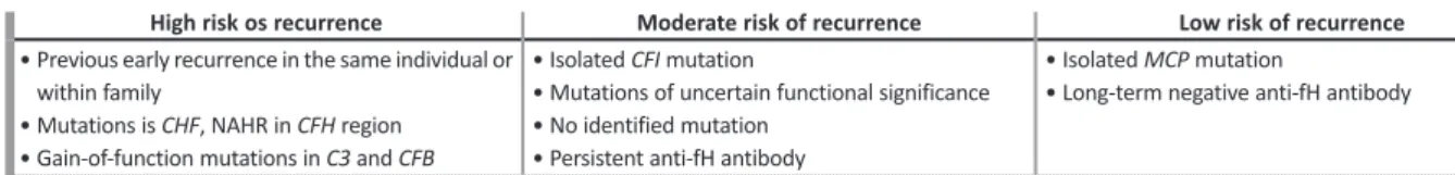
in previous grafts117. According to this information, the risk of recurrence of aHUS can be stratified for patients who are candidates for renal transplantation (Table 3).
De novo TMA following renal transplantation
De novo post-transplant TMA (Figure 5) can develop in
the absence of previous aHUS or any known susceptibility factor for aHUS. However, approximately 30% of patients who develop de novo post-transplant TMA carry mutations in complement regulatory proteins encoding genes82.
Post-transplant immunosuppression regimens
Several strategies related to immunosuppression regimens have been attempted to prevent recurrence of aHUS in the kidney grafts.
Treatment with CNI, cyclosporine (CsA) or tacroli-mus, are reported to be associated with post-transplant
TMA121,122, the risk being apparently higher for CsA
(up to 14% of patients in one series)121. The use of mTOR inhibitors bears a greater risk for post-transplant
TMA and it was shown that early use of mTOR inhibitors is an independent risk factor for the development of TMA123, with CNI having a synergic effect124.
Data on the post-transplant immunosuppressive regimen and the risk of recurrent aHUS is controversial. A small study suggested that the early use of CsA increa-sed the risk of aHUS recurrent in adults125, but analysis of a larger pediatric cohort failed to confirm it126. The-refore, avoidance of CNI treatment is generally not believed to reduce the risk of recurrent aHUS79.
Immunosuppression based on belatacept could be followed depending on the immunological risk of each patient, although, until now, no conclusive data are available in the literature.
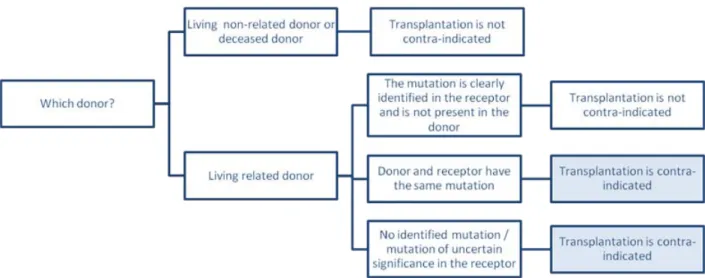
Living kidney donation
Living related donor transplantation has traditionally been contraindicated for patients with aHUS, because it associates with a risk for recurrence in the recipient
Figure 4
Strategies for prevention of recurrence of aHUS in kidney transplantation
Figure 4 – Strategies for prevention of recurrence of aHUS in kidney transplantation Abbreviations
CNIs: Calcineurin inhibitors ; CMV: cytomegalovirus; DSA: donor-specific antibodies; mTORi: mammalian target of rapamycin inhibitors;
Abbreviations: CNIs – Calcineurin inhibitors; CMV – cytomegalovirus; DSA – donor-specific antibodies; mTORi – mammalian target of rapamycin inhibitors
Table 3
Recipient risk assessment of aHUS recurrence in post-transplant
High risk os recurrence Moderate risk of recurrence Low risk of recurrence • Previous early recurrence in the same individual or
within family
• Mutations is CHF, NAHR in CFH region • Gain-of-function mutations in C3 and CFB
• Isolated CFI mutation
• Mutations of uncertain functional significance • No identified mutation
• Persistent anti-fH antibody
• Isolated MCP mutation
• Long-term negative anti-fH antibody
Abbreviations: C3 – complement component 3 gene; CFB – complement factor B gene; CFH – complement factor H gene; CFI – complement factor I gene; fH – factor H; MCP – membrane cofactor protein gene; NAHR – non-allelic homologous recombination.