Purpose: To review the effectiveness of amniotic membrane transplantation (AMT) in severe ocular surface pathologies (OSP).
Introduction: The AMT is performed in severe OSP as support for proliferation of stem cells and reepithelialisation of the cornea, permitting a return to anatomic integrity, a reduction in symptoms and an improvement in best corrected visual acuity (BCVA).
Methods: Retrospective analysis of 32 patients who underwent 37 AMT procedures between 2008-2017 at the Department of Ophthalmology of Hospital Espírito Santo de Évora for treatment of several OSP. Average age was 71.97 years (+-5.82). Grafts were applied through suture in 21 and fibrin glue in 16 cases. The average follow-up was 2.96 years (+-0.86). Failure, retransplantation, relapse, change in anatomic integrity, variation of symptoms and variation in BCVA rates were evaluated.
Results: 37 procedures were performed in 32 eyes, 9 failed, 3 of these were retransplanted and 3 relapsed. Failure was lower in corneal suture than fibrin glue (OR=0.278+-0.221, p =0.136). In 25 cases (78.13%, p<0.001) anatomic integrity was achieved. Regarding symptoms (p<0.01), 19 cases referred improvement, 8 cases no change and 5 cases worsening. Regarding BCVA, average change was irrelevant (p=0.587). In subgroup analysis, 10 cases registered an increase (average change of -0.50393, +- 0.51513 logMAR, p=0.0542), 19 cases remained stable and 3 cases progressed to a decrease (average change of +1.06804, +-1.00248 logMAR, p=0.0444).
Conclusions: AMT is, indeed, an effective and safe option when conservative treatment of OSP fails, being useful to return to anatomic integrity and to reduce OSP symptoms.
Keywords: Amniotic membrane transplantation, Ocular surface pathologies, Anatomic integrity, Best Corrected Visual Acuity, Fibrin Glue
RESUMO
Objectivos: Rever a eficácia da transplantação de membrana amniótica (TMA) nas patologias de superfície ocular (PSO) graves.
Introdução: A TMA é realizada na PSO como suporte para proliferação de células estaminais e para reepitelização da córnea, permitindo um retorno a integridade anatómica, uma redução em sintomas e uma melhoria da melhor acuidade visual corrigida (MAVC).
Métodos: Análise retrospectiva de 32 doentes submetidos a 37 procedimentos de TMA entre 2008-2017 no Serviço de Oftalmologia do Hospital Espírito Santo de Évora para tratamento de diversas PSO. Idade média foi 71.97 anos (+-5.82). Enxertos foram aplicados por sutura em 21 e cola de fibrina em 16 casos. Seguimento médio foi 2.96 anos (+-0.86). Frequência de falência, retransplantação, recidiva, mudança na integridade anatómica, variação de sintomas e variação na MAVC foram avaliadas.
Resultados: 37 procedimentos realizados em 32 olhos, 9 falharam, 3 destes foram retransplantados e 3 recidivaram. Falência foi inferior com sutura corneana do que com cola de fibrina (OR=0.278+-0.221, p =0.136). Em 25 casos (78.13%, p<0.001) antingiu-se integridade anatómica. Quanto à sintomatologia (p<0.01), 19 casos demonstraram melhoria, 8 casos estabilização e 5 casos agravamento. Quanto à MAVC, variação média foi irrelevante
(p=0.587). Em análise de subgrupos, 10 casos registeram aumento (variação media de -0.50393, +- 0.51513 logMAR, p=0.0542), 19 casos estabilizaram e 3 casos progrediram para
diminuição (variação média de +1.06804, +-1.00248 logMAR, p=0.0444).
Conclusões: TMA é, comprovadamente, uma opção segura e eficaz quando a terapêutica conservadora de PSO falha, sendo útil no reestabelecimento da integridade anatómica e na redução de sintomas.
Palavras-chave: Transplantação de Membrana Amniótica, Patologias de Superfície Ocular, Integridade Anatómica, Melhor Acuidade Visual Corrigida
INTRODUCTION
The management of severe ocular superficial disease has changed greatly over the past 25 years. All patients received a poor prognosis and the available tools were just ocular lubrification, keratoplasty and tarsorrhaphy. Currently, recent concepts and treatments are emerging due to better understanding of the ocular surface.
Advances in microsurgical techniques and the
comprehension of limbal stem cells role have taken us to great improvements not only in best corrected visual acuity (BCVA) but also in patients’ life quality.1
The eye surface plays an essential role in vision. Not only is the corneal smooth wet superficial layer the highest refractive interface in the visual system, but also the corneal transparency is essential for the propagation of light through the lens and, finally, onto the retina to activate the photoreceptors. The ocular surface remains the only wet-surfaced epithelium directly exposed to the outside environment, being especially liable to desiccation, injury and pathogens. This smooth, wet, refractive interface pivotal for vision does come with a price, being maintained and protected by the Ocular Surface System.2
The term “Ocular Surface System” is applied due to its synergistic function and due to its continuity. While the cumulative function of the system being, first and foremost, to provide, protect and maintain a smooth refractive superficial interface on the cornea accounts for the “surface” designation, the continuity of all epithelia at the ocular interface, without any breaks between regions and all deriving from the surface ectoderm, accounts for the “system” designation.2
Ocular superficial diseases lead to loss of vision, discomfort, infection, erosion, ulceration and destruction with scarring of the eye surface.3
However, various disorders of the ocular surface, including persistent epithelial defects of the cornea, acute chemical burns with long-term loss of integrity of the ocular surface epithelium or conjunctival scarring as a result of the healing of mucous membrane disorders still pose a clinical challenge in ophthalmic surgery.4
The past 20 years have witnessed the resurgence of
amniotic membrane transplantation (AMT) in
ophthalmology. Amniotic membrane (AM) can function in the eye as a basement membrane substitute or as a temporary graft.5
Several previous studies have reported several beneficial AM properties. AM promotes epithelisation and epithelial differentiation. AM produces growth factors promoters of wound healing (epidermal growth factor and keratocyte growth factor) and structural proteins (laminin and type VII collagen) promoters of epithelial migration. Furthermore, other inherent neurotropic substances enable AM to be the ideal substrate for the reepithelisation process. AM is still responsible for stopping TGF-β transduction in corneal and conjunctival fibroblasts which explains its anti-scarring effect in several ocular surface pathologies. Having said that, AM is yet responsible for the down-regulation of diverse growth factors and pro-inflammatory cytokines expression and for the up-regulation of anti-inflammatory cytokines (IL-10 and IL-1 receptor antagonist) with an overall anti-inflammatory and immunomodulatory effect, with rare tissue rejection in AM clinical application. So AM is a repository of growth factors, mitogenic factors, angiogenic factors, anti-inflammatory proteins and other natural protease inhibitors with anti-scarring properties.4,6
Its importance is therefore due to its ability to reduce scarring and inflammation, enhance wound healing and epithelisation and due to its anti-microbial properties. It
has also been employed as a substrate for cultured limbal stem cell transplantation. AM has been widely used in corneal disorders like neurotrophic ulcers, persistent epithelial defects, shield ulcers, microbial keratitis,
band keratopathy, bullous keratopathy, following
photorefractivekeratectomy, and chemical injury.4,5 Furthermore, AMT has been found to be a good alternative for corneal and conjunctival reconstruction in many clinical situations, including acute burns, persistent epithelial defects of the cornea, and diseases that cause conjunctival scarring.4
Currently, the most important indications for AMT in reconstructive surgery of the ocular surface remain the persistent epithelial defects of the cornea with corneal ulceration of varying etiology, covering defects after surgical removal of large conjunctival lesions, acute chemical burns, symblepharon and fornix reconstruction, in healing conjunctival disorders, and in limbal stem cell deficiency of the cornea with simultaneous stem cell grafting.4
Concerning the surgical principles and methods of implantation, the AMT can be applied through 3 surgical techniques.7
One is the Graft or Inlay technique being the AM placed with the epithelial or basement membrane externally and posteriorly trimmed to match the underlying epithelial or stromal defect, usually with a corneal suture with 10-0 nylon or with a episcleral or conjunctival suture with 9-0 ou 10-0 vycril (preceded by a host corneal epithelial debridement of 1-2mm in the periphery of the lesion). There is the option to use this technique as a single layer inlay (only a single layer of AM) or as a multilayer inlay (multiple layers of AM, with a filling of the defect with successive layers non-sutured with a final superficial layer is sutured to the periphery of the lesion, posterior to a debridement of a ring-shaped area around the corneal lesion).7
Other technique is the Patch or Overlay technique with a larger AM graft (than the underlying lesion) being sutured to the ocular surface so that the host epithelium remains present underneath. The AM is sutured to the surrounding conjunctiva or episcleral using 9-0 vicryl suture with either the epithelial or the stromal side superficially, being expected that the epithelium will proliferate below the graft which acts only as a “biological bandage contact lens”. Posteriorly, the AM graft is expected to fall off or be removed.7
The final technique is the Combined (inlay or overlay) technique in which 2 or more layers of AM are used with two functions: the inner smaller layer(s) act as a graft and the outer larger layer acts as a patch, being also called as the “sandwich technique”. The epithelium grows not only under the patch but also over the uppermost inlay graft.7
The availability of fibrin glue for ophthalmic use has in many cases supplanted the use of sutures with the possibility of the AM being adhered to the ocular surface using the recombinant fibrin glue.7
METHODS
Study design and participants
This study was approved by the Ethics Committee of Hospital Espírito Santo de Évora (HESE) and was conducted in accordance with the Helsinki Declaration and with the European Union’s Good Clinical Practice Directive (Directive 2005/28/EC of 8 April 2005 of the European Parliament).
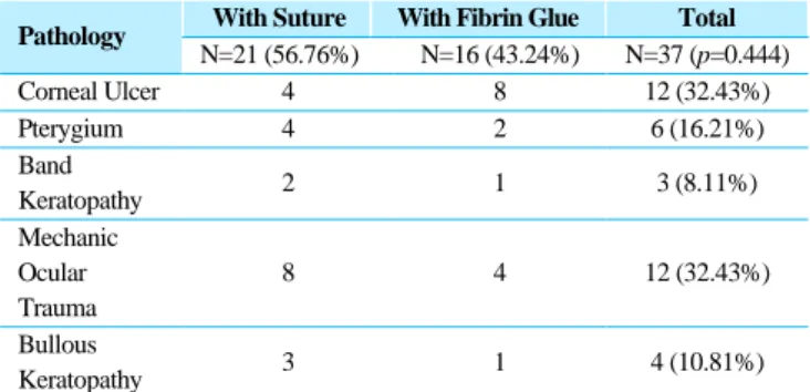
In this retrospective study, we reviewed the medical records of all patients who underwent any AMT procedure from September 2008 to January 2017 at the Department of Ophthalmology of HESE, in Évora, Portugal. A total of 32 patients were identified during this period. Furthermore, all the 32 patients had severe ocular surface pathologies which did not resolve with conservative treatment. Tables 1-4 summarise the data collected regarding demographic characteristics, pathology and surgical technique. While the average patient age submitted to the AMT procedure with recombinant fibrin glue was significantly lower in the fibrin glue when compared with the average patient age submitted to the procedure with suture (p<0.01), the pathology frequencies between the two subgroups were not statistically significant (p=0.444). The foremost common pathologies leading to AMT were corneal ulcers (31.25%) and ocular trauma (31.25%).
Regarding this study outcomes, the primary endpoint is the rate of long-term anatomic integrity, achieved with the total reepithelisation of the ocular surface. The secondary outcomes were the variation in symptoms and BCVA.
Table 3 - Study Population Baseline Pathologies
Pathology Female Male Total
N=15 (46.88%) N=17 (53.13%) N=32 (100%) Corneal Ulcer 4 6 10 (31.25%) Pterygium 1 5 6 (18.75%) Band Keratopathy 1 1 2 (6.25%) Ocular Trauma 7 3 10 (31.25%) Bullous Keratopathy 2 2 4 (12.5%)
AM origin and preservation
The AM was provided to the department by the Centro de Sangue e Transplantação de Lisboa – Área de Transplantação (regional branch of the national blood and transplant centre) and was stored at -80ºC in a temperature-controlled environment within the hospital pharmacy.
Surgical Technique
All pathologies with ocular surface inflammation, dryness or exposure were all refractory to the conservative treatments offered before surgery. Oxiboprucainehydrochloride 4 mg/ml (Anestocil©) was administered as preoperative topical anaesthesia. The eyebrows, eyelids and the eye surface were always disinfected with 5% iodopovidone solution. The surgical technique included the application of the AMT through corneal suture in 21 procedures (56.76%) while in 16 procedures (43.24%) the AMT was applied through recombinant fibrin glue (Table 4). Concerning the AMT technique employed, this was a decision made by the surgeon based upon their surgical expertise and experience. Overall, AMT procedures was applied through the Patch or Overlay technique.
Table 4 - Study Population Baseline Pathologies subgroup analysis
Pathology With Suture With Fibrin Glue Total
N=21 (56.76%) N=16 (43.24%) N=37 (p=0.444) Corneal Ulcer 4 8 12 (32.43%) Pterygium 4 2 6 (16.21%) Band Keratopathy 2 1 3 (8.11%) Mechanic Ocular Trauma 8 4 12 (32.43%) Bullous Keratopathy 3 1 4 (10.81%)
Table 5 - Corneal Ulcer causes Corneal Ulcer Infectious 1 Asseptic 1 Graft rejection 1 Post-endophthalmitis 2 Pemphigus 1 Metaherpetic 2 Neurotrophic 2 Corneal melting 2 Total 12
Post-Operative Care
Post-operative care included a follow-up period which averaged 2.96 (+-0.86) years. Moreover, 83.78% of AMT procedures were followed beyond the first 6 months and 78.83% of AMT procedures were followed beyond the first 12 months (Table 5). Failure was defined as peeling of the amniotic membrane graft during the first 7 days post-surgery. Relapses were defined as resurgence of the pathology posterior to an existing period of remission.
Anatomic integrity, achieved with the total
reepithelisation of the ocular surface, was assessed by slit--lamp examination with fluorescein dye.
Prior to surgery, the three most commonly reported symptoms were ocular pain, burning and dryness. The post-surgical evaluation of change in these symptoms was done by the patients themselves and registered by the physician upon the post-surgical appointment.
BCVA was registered before and after the AMT procedure in decimal, counting fingers and hand motion scale. Conversion from decimal, counting fingers and handmotion to logMAR scale was achieved through Holladay table. Furthermore, patients were also classified as low BCVA recovery potential candidates and high BCVA recovery potential candidates according to prior BCVA being higher or lower than 1 logMAR, respectively.8
Statistical Analysis
All data calculations were obtained using IBM SPSS Statistics 25©. To analise the data, the One-Sample T test, the Independent-Samples T test, Paired- Samples T test, the Chi-square test and the Fisher Exact Test were used to obtain data and respective significance levels. All data
value intervals refer to a 95% Confidence Interval (CI 95%). The data was considered statistically significant at a significance value of 5% (α<0.05 or p <0.05).
RESULTS
Over the 37 AMT procedures performed and during the average follow-up period of 2.96 years (+-0.86), 9 cases failed (24.32%) with a recombinant fibrin glue failure rate of 37.5% (6 cases, p=0.136, OR=0.278+-0.221) and corneal suture failure rate of 14.29% (3 cases, p=0.136 OR=0.278+-0.221). It was still reported a total relapse rate of 8.11% (3 cases), with a corneal suture relapse rate of 14.29% (3 cases, p=0.243) and a fibrin glue relapse rate of 0% (p=0.243), as documented in Table 6.
Table 6 - AMT Procedures with recombinant fibrin glue or corneal suture
Procedure With Suture With Fibrin Glue Total
Initial Procedure N=21 (56.76%) N=16 (43.24%) N=37 (100%) Failures (% of initial procedures) N=3 (14.29%) OR=0.278 (+-0.221) N=6 (37.5%) OR=1.00 N=9 (24.32%) p=0.243 Retransplantation (% of failures) N=1 (33.33%) N=2 (33.33%) N=3 (33.33%) Relapses (% of procedures) N=3 (14.29%) N=0 (0.00%) N=3 (8.11%) p=0.243
Table 7 - AMT Procedures Failure rates according to pathology
Failures
Pathology With suture With Fibrin glue Total
N=3 (14.29%) N=6 (43.24%) N=8 Corneal Ulcer 3 (75%) 4 (50%) 7/12 (58,33%) Pterygium - - - Band Keratopathy - 1/1 1/4 (25%) Ocular Trauma - - - Bullous keratopathy - 1/1 1/1
Regarding anatomic outcomes, 25 cases achieved final anatomic integrity (78.13%, p <0.001), as shown in Figure 1.
Figure 1 - Anatomic integrity Long-term outcome (LTO, p<0.001)
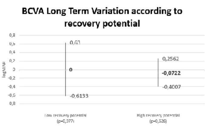
Regarding symptom variability (p<0.01), 19 cases (59.38%) reported improvement, 8 cases (25%) reported no change and 5 cases (15.63%) reported worsening of symptoms. Regarding BCVA, the total average change was of +0.05735 logMAR (+-0.21329, p=0.587). Regarding the subgroup analysis, 10 cases (31.25%, p<0.01) registered an improvement with a BCVA average change of -0.50393 (+- 0.51513, p=0.0542) logMAR, 19 cases (59.38%, p<0.01) registered no change of BCVA and 3 cases (9.38%, p<0.01) registered worsening of BCVA with an average variation of +1.06804 (+-1.00248, p=0.0444) logMAR (Figures 2 and 3).
Figure 2 - Symptoms and BCVA Long-term outcomes (LTOs, p<0.01)
Figure 3 - BCVA Long Term Variation Subgroup Analysis
Figure 4 - BCVA Long Term Variation according to recovery potential
DISCUSSION
Our results possess long-term validity since the registered follow-up periods were superior to 6 months in 83.78% of cases, superior to 1 year in 78.83% of cases and superior to 3 years in 35.13% of cases with an average follow-up period of 2.96 years (+-0.86).
According to our statistically significant results, AMT
procedures in severe ocular surface disorders
unmanageable with conservative treatment have proved their positive value not only in the achievement of anatomic integrity (78.13%, p <0.001) but also in the reduction and stabilisation of symptoms (84.38%, p <0.01), which is similar to the results of previous studies.3,9
Despite clear beneficial results of transplantation, retransplantation rate remained only in 33% due to two main reasons. Firstly, 4 cases were cases were referred to specialised tertiary care centres. Secondly, 2 cases progressed too quickly and required evisceration and enucleation.
However, despite the promising results mentioned above, the mostly looked for “holy grail” of improvement of BCVA through the AMT procedure remains mainly elusive (an improvement rate of 31.25% against a 59.38% stabilisation rate and a 9.38% worsening rate). Moreover, only in the BCVA worsening subgroup was the average change of BCVA statistically significant (p <0.05). While these rates are disappointing when compared to the rates in other trials,3,9 it remains important to emphasise that symptoms and BCVA worsening rates are mainly due to the evolution and progression of the underlying
pathologies and not to a negative effect of the AMT procedure.
The failure rates allow us to confirm the higher short-term effectiveness of the corneal suture procedure group when compared to the recombinant fibrin glue procedure group (14.29% versus 37.5%, respectively, OR=0.278+-0.221, p=0.136). Furthermore, the long-term effectiveness was higher in the corneal suture procedure group than in the recombinant fibrin glue procedure group when comparing the incidence rate of failure and relapse between procedures (28.57% versus 37.5%, respectively). Despite these rates, it is recognised that statistical significance was not achieved (p>0.05).
The small size of the sample studied did not permit to obtain significant conclusions from the comparison between BCVA low recovery potential and BCVA high recovery potential cases concerning long-term BCVA variation (p>0.05).
Recently, researchers have started to study the use of autologous, noncorneal epithelial cells as a tissue source. Future studies will focus on the further development of cellular expansion and/or the establishment of new alternative sources for replacing limbal epithelial stem cells.10
CONCLUSIONS
Amniotic membrane transplantation procedure is, indeed, an effective and safe therapeutic option when conservative treatment of ocular surface disorders fails, being extremely useful in the return to anatomic integrity and in the reduction of ocular surface pathology symptoms.
REFERENCES
1. Holland EJ, Schwartz GS. Changing concepts in the management of severe ocular surface disease over
twenty-five years. Cornea. 2000;19(5):688-698.
doi:10.1097/00003226-200009000-00014
2. Gipson IK. NIH Public Access. Ocul Surf.
2010;48(10):4390-4398. doi:10.1167/iovs.07-0770.The 3. Gheorghe A, Pop M, Burcea M, Serban M. New clinical
application of amniotic membrane transplant for ocular surface disease. J Med Life. 2016;9(2):177-179.
https://www.ncbi.nlm.nih.gov/pmc/articles/PMC4863510 /pdf/JMedLife-09-177.pdf.
4. Meller D, Pauklin M, Thomasen H, Westekemper H, Steuhl K-P. Amniotic Membrane Transplantation in the Human Eye. Dtsch Aerzteblatt Online. 2011;108(14):243-249. doi:10.3238/arztebl.2011.0243
5. Maharajan VS, Shanmuganathan V, Currie A,
Hopkinson A, Powell-Richards A, Dua HS. Amniotic
membrane transplantation for ocular surface
reconstruction: Indications and outcomes. Clin Exp
Ophthalmol. 2007;35(2):140-147. doi:10.1111/
j.14429071.2006.01408.x
6. Röck T, Bartz-Schmidt KU, Landenberger J, Bramkamp M, Röck D. Amniotic membrane transplantation in reconstructive and regenerative ophthalmology. Ann Transplant. 2018;23:160-165. doi:10.12659/AOT.906856 7. Malhotra C, Jain AK. Human amniotic membrane
transplantation: Different modalities of its use in ophthalmology. World J Transplant. 2014;4(2):111. doi:10.5500/wjt.v4.i2.111
8. Holladay J. Proper method for calculating average visual acuity. J Refract. 1997;13(4):388-391.
9. Ashraf NN, Adhi MI. Outcome of application of amniotic membrane graft in ocular surface disorders. J Pak Med
Assoc. 2017;67(7):1045-1049. doi:DOI
10.1093/schbul/sbq085
10. Rosen R. Amniotic membrane grafts to reduce pterygium
recurrence. Cornea. 2018;37(2):189-193.
doi:10.1097/ICO.0000000000001407
CONTACTO
Tiago Morais-Sarmento Department of Ophthalmology
Hospital do Espírito Santo de Évora E.P.E. Largo Senhor da Pobreza
7000-811, Évora, Portugal
E-mail: [email protected]
The authors have no financial interests in the topic of this manuscript. No conflicting relationship exists for any author.