Expression of epidermal growth factor receptor (EGFR) in
cholangiocarcinomas: predictive factors and survival
Expressão do receptor do fator de crescimento epitelial (EGFR) em
colangiocarcinomas: fatores preditivos e sobrevida
RodRigo VieiRa gomes1; michele Ângela RodRigues2; João BeRnaRdo sancio Rocha RodRigues, acBc-mg1; Paula TeixeiRa Vidigal3;
KaRine aRaúJo damasceno2; henRique aRaúJo lima1; dawidson assis gomes4; caRla JoRge machado5; ViVian Resende, TcBc-mg1
INTRODUCTION
C
holangiocarcinoma (CCA) is the second mostfrequent primary malignant neoplasm in the liver and accounts for about 3% of all gastrointestinal cancers1. It originates from the epithelium of intra
or extrahepatic bile ducts2. It has been described by
Durand-Fardel for the first time in 1840, and can be divided according to its location into intrahepatic, peri-hilar (Klatskin tumor) and distal3. Histologically, it is
usually an adenocarcinoma and displays varying degrees of differentiation and phenotypes4. Some risk factors for
cholangiocarcinoma have already been established, such
as parasitic infections, primary sclerosing cholangitis, bile duct cysts, hepatolithiasis, and some toxins. Other potential risk factors include inflammatory bowel disease, hepatitis B and C virus, liver cirrhosis, diabetes
mellitus, obesity, alcoholism, smoking, and genetic mutations5. The incidence of CCA has been increasing
in recent years6.
More attention has been devoted to CCA due to poor prognosis, coupled with the poor efficacy of therapeutic modalities, especially for patients with
advanced disease7. Chemotherapy has been widely
used, though with a low response rate7,8. Staging,
size and degree of tumor differentiation, resection
1 - Faculty of Medicine, Surgery Department, Federal University of Minas Gerais, Belo Horizonte, MG, Brazil. 2 - Institute of Biological Sciences, Ge-neral Pathology, Federal University of Minas Gerais, Belo Horizonte, MG, Brazil. 3 - Faculty of Medicine, Pathological Anatomy and Legal Medicine, Federal University of Minas Gerais, Belo Horizonte, MG, Brazil. 4 - Institute of Biological Sciences, Biochemistry and Immunology, Federal University of Minas Gerais, Belo Horizonte, MG, Brazil. 5 - Faculty of Medicine, Preventive and Social Medicine, Federal University of Minas Gerais, Belo Horizonte, MG, Brazil.
A B S T R A C T
Objective: to evaluate the expression of the epithelial growth factor receptor (EGFR) by immunohistochemistry, and to verify its association with prognostic factors and survival of patients operated by cholangiocarcinoma. Methods: we verified the immunohistochemical expression of EGFR in 35 surgical specimens of cholangiocarcinoma (CCA). We obtained survival curves with the Kaplan-Meier method.
Results: we found significant EGFR expression in ten (28.6%) of the 35 CCAs, eight with score 3 and two with score 2. Advanced stages (III and IV) presented higher EGFR expression (p=0.07). The clinical characteristics that were most associated with positive EGFR expression were female gender (p=0.06) and absence of comorbidities (p=0.06). Overall survival at 12, 24, 36 and 48 months was 100%, 82.5%, 59% and 44.2%, respectively. The survival of EGFR positive patients at 12, 24, 36 and 48 months was 100%, 75%, 50% and 0%, whereas for negative EGFR patients it was 100%, 87.5%, 65.6% and 65.6%, respectively. Conclusion: EGFR expression occurred in 28.6% of the cases studied and was associated with lower survival.
margins, and perineural and angiolymphatic invasion are described as prognostic factors9. Among the factors
that contribute to poor prognosis, the advanced stage of the disease is highlighted, which decreases the chance of cure by surgical treatment10. More than 50% of the
cases are diagnosed at an advanced stage, leading to a general average survival of approximately seven to eight months10. Patients who undergo surgical resection have
a better prognosis, with a five-year survival rate of 30% and mean survival of 30 months11-13.
Epidermal growth factor receptor (EGFR) is a cell membrane receptor tyrosine kinase encoded by proto-oncogenes. Different ligands, such as epidermal growth factor (EGF), heparin binding factor (HB-EGF), and amphiregulin, couple in the extracellular domains of these receptors and initiate an intracellular signaling cascade, increasing the potential for cell proliferation, angiogenesis and resistance to apoptosis. EGFR activation also impairs cell-cell adhesion through the destabilization of junctional complexes (E-cadherin, beta-catenin), which contributes to the acquisition of motile and invasive phenotype14,15.
Epidermal growth factor inhibitors, antibodies and other molecules, such as erlotinib, gefitinib, cetuximab, panitumumab, trastuzumab and lapatinib, have been used in the treatment of colorectal, breast, lung and head and neck cancers, with promising results, having as targets the EGFR or the human epidermal growth factor receptor 2 (HER2)11,16. The overexpression
of EGFR in tumor cells was associated with a worse prognosis in some studies, being found in up to 80% of the cholangiocarcinomas, making this receptor a potential therapeutic target in patients with this type of neoplasia16-19.
The role of EGFR in malignant liver diseases is still poorly understood. The identification of groups of patients with more aggressive subtypes and risk of lower survival could contribute to the development of more effective therapeutic options. To better understand the clinical significance of EGFR in cholangiocarcinoma, the main objectives of the present study were to evaluate
the degree of immunohistochemical expression of EGFR and to verify its association with clinical-pathological factors and survival of patients submitted to surgical treatment.
METHODS
This research was approved by the Institutional Ethics Committee with the following reference number: CAAE - 37156714.6.0000.5149. From January 2008 to December 2015, we selected patients undergoing surgical treatment and histological diagnosis of cholangiocarcinoma. We collected data regarding gender, age, previous history, history of smoking or alcoholism, presence of gallstones, family history for neoplasia, histological type, tumor staging and survival after treatment. The laboratory tests analyzed were bilirubins and Ca 19-9. The classification of the anatomical location of the tumors and the staging were based on the description of the radiological studies (Computed Tomography, Magnetic Resonance and/ or Magnetic Resonance Cholangiopancreatography) associated with the findings of the surgical description and anatomopathological study. We revised the hematoxylin and eosin (HE) stained slides to classify tumors as polypoid and non-polypoid. Classification and staging followed the TNM pattern of the American Joint Cancer Committee/Union for International Cancer Control - 7th edition20,21.
We included patients whose tissue samples were obtained from surgical resection specimens or surgical biopsies of cholangiocarcinomas. Samples of gallbladder cancer and the hepatopancreatic (Vater’s) ampulla and cases of biopsies in which the material was insufficient for analysis (six cases) were considered exclusion criteria. All samples were fixed in 10% formaldehyde, embedded in paraffin, cut into fragments with a thickness of four micrometers and stained with hematoxylin and eosin.
In cases with variable intensity, the predominant intensity was selected for the final score. Scores 2+ and 3+ were defined as positive for protein expression.
We performed statistical analysis using the statistical package Stata for Mac 2016. To compare the distribution of the categories of the study variables according to EGFR expression (negative and positive), we performed the Student’s t-test for the continuous variables and Chi-square or Fisher’s test for categorical ones. Survival analysis had as the dependent variable the occurrence of death in relation to time, which was measured in months. We excluded cases of death of patients occurring within 30 postoperative days. We constructed survival curves for the variables using the Kaplan-Meier method. In the comparison between groups for each variable, we used the Logrank test. In all analyzes, the level of significance considered was 5% (p<0.05).
RESULTS
There was no significant difference in the proportion between men (n=17) and women (n=18). The mean patients’ age was 58 years, ranging from 28 to 83. There were lower proportions of smokers, alcohol users and gallstones (Table 1).
The following procedures with curative intent were performed in 30 patients: eight (26.66%) right hepatectomies, two (6.66%) right trisegmentectomies and nine (30%) left hepatectomies, associated with resection of segment one, four (13.33%) bisegmentectomies (three including segments two and three and one including segments six and seven). Six (20%) pancreatoduodenectomies and one (3.33%) resection of the extrahepatic biliary tract were also performed.
of the primary tumor site, consisting of 30 specimens of surgical resection with curative intent and five biopsies.
The slides were initially processed with pepsin and hydrochloric acid at 37°C for 30 minutes for antigen retrieval and, thereafter, blocking of endogenous peroxidase and proteins. Next, incubation with the primary antibody (EGFR - clone 3G7 - Invitrogen - 1:100 dilution) was performed for 16 to 18 hours (overnight incubation). Thereafter, the antibody was
removed, the polymer was placed and the slides were incubated in the humid chamber for 30 minutes at room temperature. After removal of the polymer, the chromogen 3,3’ Diaminobenzidine tetrahydrochloride (DAB), which was withdrawn after the reaction. When the reaction was positive, brown staining was observed at the antigen site. Then, we performed HE
counter-staining and assembled the slides with transparent medium.
All slides were evaluated by a pathologist with experience in immunohistochemical analysis, without prior knowledge of the clinical and pathological information of the patients. The intensity of EGFR
expression was defined according to the previously described score, based on the work of Yoshikawa et al.22:
• 0: no staining of the cell membrane or staining of the membrane in 10% or less of the tumor cells;
• 1+: weak and partial staining of the
membrane in more than 10% of tumor cells;
• 2+: moderate and complete staining of the membrane in more than 10% of tumor cells;
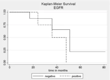
Table 1. Expression of EGFR according to the clinicopathological variables.
Variables
EGFR Total Valor p
Negative (n=25; 71.4%)
Positive
(n=10; 28.6%) 35 (100.0) 0.017
Age
(mean; SD) (n=35) 59.5 (12.6) 56.2 (13.7) 0.496
Gender (n; %) Male Female
15 (88.2) 10 (55.6)
2 ( 11.8) 8 (44.4)
17 (100.0) 18 (100.0)
0.060
Smoking (n; %) No Yes 14 (63.6) 8 (80.0) 8 (36.4) 2 (20.0) 22 (100.0) 10 (100.0) 0.440
Alcohol use (n; %) No Yes 17 (65.4) 5 (83.3) 9 (34.6) 1 (16.7) 26 (100.0) 6 (100.0) 0.637
Comorbidities (n; %) No Yes 10 (55.5) 13 (86.7) 8 (44.5) 2 (13.3) 18 (100.0) 15 (100.0) 0.070
Family history (n; %) No Yes 10 (71.4) 7 (58.3) 4 (28.6) 5 (41.7) 14 (100.0) 12 (100.0) 0.683
Biliary lithiasis (n; %) No Yes 17 (65.4) 5 (83.3) 9 (34.6) 1 (16.7) 26 (100.0) 6 (100.0) 0.637
Classification (n; %) Intra-hepatic Peri-hilar Distal 6 (60.0) 15 (83.3) 4 (57.1) 4 (40.0) 3 (16.7) 3 (42.9) 10 (100.0) 18 (100.0) 7 (100.0) 0.185
CA 19-9 (n; %) >37U/ml <37U/ml 5 (55.5) 8 (61.5) 4 (44.4) 5 (38.5) 9 (100.0) 13 100.00) 0.999
Bilirubin (n; %) 0-4 mg/dl 4-10 mg/dl >10mg/dl 8 (57.1) 5 (71.4) 8 (88.8) 6 (42.9) 2 (28.6) 1 (11.1) 14 (100.0) 7 (100.0) 9 (100.0)
0.151
Treatment (n; %) Palliative Resection 4 (80.0) 21 (70.0) 1 (20.0) 9 (30.0) 5 (100.0) 30 (100.0) .999
T (n; %) 1-2 3-4 15 (83.3) 10 (58.8) 3 (16.7) 7 (41.2) 18 (100.0) 17 (100.0) 0.146 N Negative Positive 15 (75) 6 (60) 5 (25) 4 (40) 20 (100) 10 (100) 0.331
Staging (n; %) I-II III-IV 14 (87.5) 11 (57.9) 2 (12.5) 8 (42.1) 16 (100.0) 19 (100.0) 0.071
Table 1 shows the EGFR expression in relation to the variables of interest in the 35 CCA cases. There was significant EGFR expression in ten (28.6%) of the 35 lesions, being score 3+ in eight patients and 2+ in two patients. In 25 patients (71.4%), EGFR expression was negative, being 1+ in five patients and 0 in 20 patients.
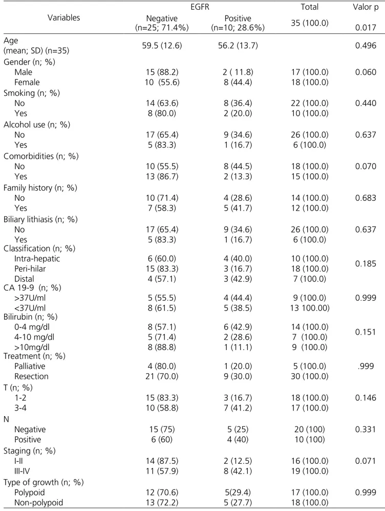
The clinical characteristics of patients who had the greatest association with positive expression were female (p=0.06) and absence of comorbidities (p=0.07). Regarding TNM staging, those with the more advanced stages III and IV had greater EGFR expression (p=0.071). Figure 1 shows the intensities and scores attributed to the cholangiocarcinomas.
Figure 1. Intensity of EGFR in cholangiocarcinomas. HE staining on the left (a, c, e, g) and EGFR immunohistochemistry on the right (b, d, f, h). (b) Complete membrane, strong staining, score 3+. (d) Complete membrane, moderate staining, score 2+. (f) Incomplete membrane, weak staining, score 1+. (h) Negative, score 0.
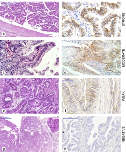
The overall survival at 12, 24, 36 and 48 months was 100%, 82.5% (CI: 45.1-95.5), 59% (CI: 22.9-82.8) and 44.2% (CI: 11.9-73.2), respectively . The survival of patients with EGFR-positive tumors at 12, 24, 36 and 48
patients with EGFR-positive tumors was lower than that of negative EGFR patients, with no statistical significance (p=0.328) (Figure 2).
Figure 2. Survival curves obtained by the Kaplan-Meier method (p=0.328) in patients with positive and negative EGFR cholangiocarci-nomas.
DISCUSSION
The expression of ErbB family growth factor receptors, EGFR and HER2 has been studied in different tumor types and has led to the use of targeted therapies with inhibitors or antibodies specific for these receptors in colorectal, breast, lung and head and neck neoplasias16.
So far, the use of monoclonal antibodies and tyrosine kinase inhibitors, such as cetuximab, trastuzumab, erlotinib, gefitinib, is still being investigated in other types of cancer. In biliary tract cancer, EGFR expression usually occurs in small groups of patients. Yoshikawa et al. recently described the expression of these receptors in malignant tumors of the biliary tract22. Harder et al.
studied the expression of EGFR and HER2 in a group of patients with advanced and unresectable biliary tract tumors. The results of these authors suggest that routine testing and therapeutic targeting of HER2 does not appear to be useful in patients with bile duct cancer, whereas EGFR may be promising as a target11. In a phase
II study, Chiorean et al. tested Erlotinib and Docetaxel in hepatocellular carcinoma and advanced cancer of the biliary tract and concluded that for a significant
improvement in survival, treatment should probably be individualized according to the tumors’ molecular and genetic profiles23.
The expression of EGFR in CCA in previous studies occurred with a variability of 8.1% to 81%, and has been associated with tumors’ greater aggressiveness16-19.
In our study, EGFR expression was considered positive in 28.6% of the cases and, although there was no statistical significance, we found a higher rate of expression in tumors in advanced stages, occurring in 42.1% of stages III/IV versus 12.5% of stages I/II (p=0.071), translating characteristics of aggressive phenotype in the presence of invasive disease. One hypothesis is that the tumor will express more EGFR upon reaching advanced stages. Survival analysis also corroborated the hypothesis that EGFR expression is associated with a worse prognosis, since patients considered positive had lower survival rates than negative ones.
These findings, previously mentioned, can
be explained by the work of Clapéron et al., who
demonstrated that the EGF/EGFR axis is a potent inducer of the epithelial-mesenchymal transition, one of the main molecular factors associated with the progression of cholangiocarcinoma24. It is known that
EGFR activation interferes with intercellular adhesion, destabilizing the E-cadherin/beta-catenin complex, promoting the epithelial-mesenchymal transition, which contributes to increase in the capacity of migration, invasion and resistance to apoptosis24,25. Similarly,
Hoffmann et al. found a worse prognosis in patients with EGFR expression, with an average survival of only 8.5 months, while patients with low expression had a mean survival rate of more than three years17. The
significant association of EGFR with survival may help to select more effective treatment strategies for patients with CCA.
Our EGFR staining score readings were based on the work of Yoshikawa et al.22, who considered
of EGFR was absent in 39.3% (22/56) of patients, whilst they observed weak expression (1+) in 21.5% (12/56), moderate (2+) expression in 23.2% (13/56) and intense (3+) in 16% (9/56). The fact that we used a different way to obtain the score may be the reason we obtained results (28.6%) inferior to those authors’. In addition, Harder et al.11 performed biopsy investigations of
advanced and unresectable cases of biliary carcinoma without distinguishing between gallbladder carcinoma and bile duct cholangiocarcinoma, whereas we only studied patients with carcinoma of the biliary ducts, and in different stages.
Yoshikawa et al. showed an overexpression in 26.4% of extrahepatic CCAs and in 17.7% of intrahepatic CCAs. These authors considered only two anatomic sites, including peri-hilar carcinomas in the extrahepatic CCA group22. In our study, we considered peri-hilar carcinoma
separately and EGFR expression was only 16.7% in that group, smaller than in the others. Harder et al. found an expression rate of 57.9% in the extrahepatic lesions and 25% in the intrahepatic ones11. In our study, expression
in extrahepatic CABG was 42.9%. These divergences can be explained by the different ways of anatomical classification adopted, which are still controversial in the literature, especially in relation to extrahepatic cholangiocarcinoma.
Although the World Health Organization (WHO) includes peri-hilar and distal CCAs in the extrahepatic modlity20,21, they are known to have
different biological behaviors and respond differently to chemotherapy. In a systematic review, Wiggers et al. showed significant differences in the expression
of biomarkers between intrahepatic and extrahepatic CCAs, being more relevant in the markers that are potential targets of systemic therapies (EGFR, c-erB-2 and VEGF-A), suggesting that they should be treated as distinct tumors26. Ercolani et al. have already classified
CCAs into three subgroups (intrahepatic, peri-hilar and extrahepatic) and concluded that CCAs appear to be the same type of tumor, but depending on the location, have different invasion properties of adjacent structures, which may also influence prognosis27. Our results corroborate
this conclusion, since the anatomical location implied different survival outcomes, although the correlation with EGFR expression displayed no significant difference between the three locations.
Challenges for the development of targeted therapies for CCAs include not only the heterogeneity of these tumors but also the rarity of the cases when compared to other solid tumors and the lack of interest of the pharmaceutical industry19. Some limitations
may be noted in our study. Conducting the research in a single center limited the size of the sample, since it is a relatively rare disease. This fact may have been responsible for the lack of statistical significance in some of our findings.
REFERENCES
1. Khan SA, Thomas HC, Davidson BR, Taylor-Robinson SD. Cholangiocarcinoma. Lancet. 2005;366(9493):1303-14. Erratum in: Lancet. 2006;367(9523):1656.
2. Resende V, Santos JP, Gomes RV, Vidigal PV, Pedrosa MS. Papillary neoplasias of the biliary tract. Rev Col Bras Cir. 2014;41(6):445-50.
3. Castellano-Megías VM, Ibarrola-de Andrés C, Colina-Ruizdelgado F. Pathological aspects of so called “hilar cholangiocarcinoma”. World J Gastrointest Oncol. 2013;5(7):159-70.
4. Nakanuma Y, Kakuda Y. Pathologic classification of cholangiocarcinoma: new concepts. Best Pract Res Clin Gastroenterol. 2015;29(2):277-93.
5. Tyson GL, El-Serag HB. Risk factors for cholangiocarcinoma. Hepatology. 2011;54(1):173-84. 6. Singal AK, Vauthey JN, Grady JJ, Stroehlein JR.
Intra-hepatic cholangiocarcinoma--frequency and demographic patterns: thirty-year data from the M.D. Anderson Cancer Center. J Cancer Res Clin Oncol. 2011; 137(7):1071-78.
7. Huang Y, Li X, Zhao Y. Progression of targeted therapy in advanced cholangiocarcinoma. Chin J Cancer Res. 2015;27(2):122-7.
8. Thomas MB. Biological characteristics of cancers in the gallbladder and biliary tract and targeted therapy. Crit Rev Oncol Hematol. 2007;61(1):44-51.
9. Ruys AT, Groot Koerkamp B, Wiggers JK, Klümpen HJ, ten Kate FJ, van Gulik TM. Prognostic biomarkers in patients with resected cholangiocarcinoma: a
systematic review and meta-analysis. Ann Surg Oncol. 2014;21(2):487-500.
10. Esnaola NF, Meyer JE, Karachristos A, Maranki JL, Camp ER, Denlinger CS. Evaluation and management of intrahepatic and extrahepatic cholangiocarcinoma. Cancer. 2016;122(9):1349-69. 11. Harder J, Waiz O, Otto F, Geissler M, Olschewski
M, Weinhold B, et al. EGFR and HER2 expression in advanced biliary tract cancer. World J Gastroenterol. 2009;15(36):4511-7.
12. DeOliveira ML, Cunningham SC, Cameron JL, Kamangar F, Winter JM, Lillemoe KD, et al. Cholangiocarcinoma: thirty-one-year experience with 564 patients at a single institution. Ann Surg.2007;245(5):755-62.
13. Tsukahara T, Shimoyama Y, Ebata T, Yokoyama Y, Igami T, Sugawara G, et al. Cholangiocarcinoma with intraductal tubular growth pattern versus intraductal papillary growth pattern. Mod Pathol. 2016;29(3):293-301.
14. Faria JAQA, Andrade C, Goes AM, Rodrigues MA, Gomes DA. Effects of different ligands on epidermal growth factor receptor (EGFR) nuclear translocation. Biochem Biophys Res Commun. 2016;478(1):39-45. 15. Sohal DP, Shrotriya S, Abazeed M, Cruise M,
Khorana A. Molecular characteristics of biliary tract cancer. Crit Rev Oncol Hematol. 2016;107:111-8. 16. Leone F, Cavalloni G, Pignochino Y, Sarotto I, Ferraris
R, Piacibello W, et al. Somatic mutations of epidermal growth factor receptor in bile duct and gallbladder carcinoma. Clin Cancer Res. 2006;12(6):1680-5. Objetivo: avaliar a expressão do receptor do fator de crescimento epitelial (EGFR) por meio de imuno-histoquímica, e verificar sua associação com fatores prognósticos e com a sobrevida dos pacientes operados por colangiocarcinoma. Métodos: a expressão imuno-histoquímica de EGFR foi verificada em 35 peças cirúrgicas de colangiocarcinomas (CCA). Curvas de sobrevida foram obtidas pelo método de Kaplan-Meier. Resultados: expressão significativa de EGFR foi encontrada em dez (28,6%) de 35 CCA, oito com escore 3 e dois com escore 2. Estágios avançados (III e IV) apresentaram maior expressão de EGFR (p=0,07). As características clínicas que mais estiveram associadas com a expressão positiva de EGFR foram o sexo feminino (p=0,06) e ausência de comorbidades (p=0,06). A sobrevida global aos 12, 24, 36 e 48 meses foi de 100%, 82,5%, 59% e 44,2%, respectivamente. A sobrevida de pacientes EGFR positivos aos 12, 24, 36 e 48 meses foi de 100%, 75%, 50% e 0%, enquanto que para EGFR negativos foi de 100%, 87,5%, 65,6% e 65,6%, respectivamente. Conclusão: a expressão do EGFR ocorreu em 28,6% dos casos estudados e esteve associada a menor sobrevida.
Descritores: Colangiocarcinoma. Genes erbB-1. Prognóstico. Análise de Sobrevida.
17. Hoffmann AC, Goekkurt E, Danenberg PV, Lehmann S, Ehninger G, Aust DE, et al. EGFR, FLT1 and heparanase as markers identifying patients at risk of short survival in cholangiocarcinoma. PLoS One. 2013;8(5):e64186.
18. Yang X, Wang W, Wang C, Wang L, Yang M, Qi M, et al. Characterization of EGFR family gene aberrations in cholangiocarcinoma. Oncol Rep. 2014;32(2):700-8.
19. Voss JS, Holtegaard LM, Kerr SE, Fritcher EG, Roberts LR, Gores GJ, et al. Molecular profiling of cholangiocarcinoma shows potential for targeted therapy treatment decisions. Hum Pathol. 2013;44(7):1216-22.
20. Edge SB, Compton CC. The American Joint Committee on Cancer: the 7th edition of the AJCC cancer staging manual and the future of TNM. Ann Surg Oncol. 2010;17(6):1471-4.
21. Blechacz B, Komuta M, Roskams T, Gores GJ. Clinical diagnosis and staging of cholangiocarcinoma. Nat Rev Gastroenterol Hepatol. 2011;8(9):512-22. 22. Yoshikawa D, Ojima H, Iwasaki M, Hiraoka N, Kosuge
T, Kasai S, et al. Clinicopathological and prognostic significance of EGFR, VEGF, and HER2 expression in cholangiocarcinoma. Br J Cancer. 2008;98(2):418-25.
23. Chiorean EG, Ramasubbaiah R, Yu M, Picus J, Bufill JA, Tong Y, et al. Phase II trial of erlotinib and docetaxel in advanced and refractory hepatocellular and biliary cancers: Hoosier Oncology Group GI06-101. Oncologist. 2012;17(1):13.
24. Clapéron A, Mergey M, Nguyen Ho-Bouldoires TH, Vignjevic D, Wendum D, Chretien Y, et al. EGF/EGFR axis contributes to the progression of cholangiocarcinoma through the induction of
an epithelial-mesenchymal transition. J Hepatol. 2014;61(2):325-32.
25. Brivio S, Cadamuro M, Fabris L, Strazzabosco. Epithelial-to-mesenchymal transition and cancer invasiveness: what can we learn from cholangiocarcinoma? J Clin Med. 2015;4(12):2028-41.
26. Wiggers JK, Ruys AT, Groot Koerkamp B, Beuers U, ten Kate FJ, van Gulik TM. Differences in immunohistochemical biomarkers between intra- and extrahepatic cholangiocarcinoma: a systematic review and meta-analysis. J Gastroenterol Hepatol. 2014;29(8):1582-94.
27. Ercolani G, Dazzi A, Giovinazzo F, Ruzzenente A, Bassi C, Guglielmi A, et al. Intrahepatic, peri-hilar and distal cholangiocarcinoma: three different locations of the same tumor or three different tumors? Eur J Surg Oncol. 2015;41(9):1162-9.
Received in: 19/02/2018
Accepted for publication: 22/03/2018 Conflict of interest: none.
Source of funding: Foundation for Research Support of the State of Minas Gerais (FAPEMIG), Coordination for Improvement of Higher Education Personnel (CAPES) and National Council for Scientific and Technological Development (CNPQ).
Mailing address:
Vivian Resende