A R T I G O D E R E V I S Ã O
S P O R A D I C I N C L U S I O N B O D Y M Y O S I T I S
:
A N U N S O L V E D M Y S T E R Y
Pedro Machado,
*,**Adrian Miller,
**Janice Holton,
**Michael Hanna
**são pistas importantes para um diagnóstico preco-ce. A disfagia é um sintoma frequente no decurso da doença. A biópsia muscular pode apresentar al-terações miopáticas crónicas, infiltrados linfocíti-cos envolvendo fibras não-necróticas, «rimmed va-cuoles» e acumulação de proteínas amilóides. Per-manece a dúvida sobre se a MCIe é primariamen-te uma miopatia inflamatória imuno-mediada ou se se trata de uma miopatia degenerativa com um componente inflamatório associado. Esta revisão descreve a epidemiologia e as características clíni-cas da doença, bem como a sua abordagem tera-pêutica e conceitos genéticos e etiopatogénicos actuais. Apesar das mais recentes descobertas, em muitos aspectos, a MCIe permanece um mistério por resolver.
Palavras-chave: Miosite de Corpos de Inclusão; Miosite; Miopatia; Inflamação; Degeneração.
Introduction
Sporadic IBM is traditionally classified as an idio-pathic inflammatory myopathy, along with poly-myositis (PM) and dermatopoly-myositis (DM).1The his-topathologic changes in sIBM were first described in the mid-1960s,2,3although the disorder was not distinguished from PM and named until 1971.4
sIBM is now considered the commonest acqui-red muscle disease among those aged over 50 years. It is a severely disabling disorder, still without effec-tive treatment.5Characteristically, sIBM causes a selective pattern of muscle weakness, predomi-nantly involving the forearm flexor and quadriceps
femoris muscles early in the disease course.6This leads to loss of manual control, impaired mobility and a propensity to fall. There is often subsequent involvement of the distal leg, proximal arm, and pharyngeal muscles resulting in dysphagia.6-10 Be-cause of the limited awareness among medical practitioners of its existence, the protracted clini-cal course, and histologiclini-cal similarity with other *Department of Rheumatology, Coimbra University Hospital,
Coimbra, Portugal
**MRC Centre for Neuromuscular Diseases, UCL Institute of Neurology, National Hospital for Neurology and Neurosurgery, Queen Square, London, United Kingdom
Abstract
Sporadic inclusion body myositis (sIBM) is consi-dered to be the most common acquired muscle di-sease associated with aging. It is a disabling disor-der still without effective treatment. sIBM causes weakness and atrophy of the distal and proximal muscles. Involvement of quadriceps and deep fin-ger flexors are clues to early diagnosis. Dysphagia in the course of the disease is common. Muscle biopsy shows chronic myopathic features, lym-phocytic infiltration invading non-necrotic fibbers, rimmed vacuoles and accumulation of amyloid-re-lated proteins. It remains uncertain whether sIBM is primarily an immune-mediated inflammatory myopathy or a degenerative myopathy with an as-sociated inflammatory component. This review describes the epidemiology and clinical features of the disease as well as the current genetic and pa-thogenic concepts and therapeutic approaches. Despite recent clues, in many respects sIBM re-mains an unsolved mystery.
Keywords: Inclusion Body Myositis; Myositis; Myo-pathy; Inflammation; Degeneration.
Resumo
A forma esporádica da miosite de corpos de inclu-são (MCIe) é considerada a mais frequente doença muscular adquirida associada com o envelheci-mento. É uma doença incapacitante ainda sem tra-tamento eficaz. A MCIe provoca atrofia e fraqueza tanto de grupos musculares proximais como dis-tais. O envolvimento selectivo dos músculos qua-drícipete femoral e flexores profundos dos dedos
S P O R A D I C I N C L U S I O N B O D Y M Y O S I T I S: A N U N S O LV E D M Y S T E R Y
myopathies, the diagnosis of sIBM is commonly delayed and initially inaccurate.11,12
The primary cause of sIBM is unknown, but is thought to involve a complex interplay between environmental factors, genetic susceptibility and aging. The recognition of proteinaceous deposits in sIBM triggered an evolving body of evidence distin-guishing this disorder from the idiopathic inflam-matory myopathies.13sIBM is enigmatic in its com-bination of inflammatory and degenerative featu-res that persist from the early stages of the disease to its most advanced phase. The inflammatory changes include upregulation of proinflammatory chemokines14and cytokines15in an inflammatory environment that attracts clonally expanded, cyto-toxic CD8+ T-cells. These attack myofibres that ove-rexpress major histocompatibility complex (MHC) class I, which is not constitutively expressed by ske-letal muscle.16,17The second hallmark of sIBM is the accumulation of aberrant molecules, notably β-amyloid, within the myofibres.18Other molecules associated with cellular stress and degeneration are also overexpressed in sIBM.19Cytochrome c oxida-se (COX) deficient fibres and ragged-red fibres re-flect mitochondrial impairment.20The relationship between T-cell invasion and other histopathologi-cal changes in muscle fibres is not known, and it re-mains uncertain whether sIBM is primarily an im-mune-mediated inflammatory myopathy or a de-generative myopathy with an associated inflam-matory component.21
This review describes the epidemiology and cli-nical features of the disease as well as the current genetic and pathogenic concepts and therapeutic approaches. Despite recent clues, in many respects sIBM remains an unsolved mystery.
Epidemiology
It has been estimated that sIBM represents 16-28% of patients with idiopathic inflammatory myosi-tis.22,23However, the true frequency may be higher, and apparently exceeds that of PM, frequently ini-tially misdiagnosed in sIBM patients.24
The prevalence of sIBM differs between diffe-rent populations and ethnic groups.25It has been estimated to be 4.9 per million population in the Netherlands11(adjusted to 16.0 per million above 50 years of age), 9.3 per million in Western Australia12 (adjusted to 35.5 per million above 50 years of age) and 10.7 per million in Connecticut, United States
of America (USA) (adjusted to 28.9 per million abo-ve 45 years of age).8These figures are almost cer-tainly an underestimate and the true prevalence of sIBM may be substantially higher than previously thought.
In a recent population-based study in Minneso-ta, USA,26nine patients with sIBM provided an in-cidence estimate of 7.9 per million per year and a prevalence rate of 70.6 per million. These are the highest rates reported for sIBM to date. The inci-dence and prevalence of sIBM exceeded figures for PM, for which the same authors reported an inci-dence of 4.1 per million per year and a prevalence of 34.5 per million, while previous studies in PM es-timated an incidence from 5.5 to 10 per million.27,28 A follow-up survey in Western Australia29has re-cently been published, and the previously repor-ted prevalence of 9.3 per million12has been upda-ted to 14.9 per million (adjusupda-ted to 51.3 per milli-on above 50 years of age). The authors also report a high rate of initial misdiagnosis and a large mean time to diagnosis (5.2 years), again suggesting that even the latest prevalence figures may be an unde-restimate and emphasising the need to increase the level of awareness about the condition among the medical community.
Larger, multicentre trials are needed to define the epidemiology of sIBM among different geo-graphic regions and ethnic groups and to determi-ne the contribution of different gedetermi-netic or environ-mental factors to these variations.
Genetics
Existing evidence for genetic susceptibility in sIBM has been based on candidate gene studies. The ra-rity of the condition has precluded the use of more robust genetic methods such as twin studies, who-le genome screening, and transmission disequili-brium testing. More recently, microarray techno-logy has provided interesting data on sIBM.
So far, MHC associations provide the strongest evidence for a genetic component in sIBM. The strong association of HLA-DR3 and the extended 8.1 ancestral haplotype (AH) (characterised by HLA-A*01, -B*0801, -DRB1*0301, -DQB1*0201, -DQA1*05) with sIBM was first reported in Austra-lian patients30and then confirmed in Dutch, Ger-man and North American cohorts.31-34The suscep-tibility region has been confined to between PBX2 and HLA-DRB.33 A few genes within this region
P E D R O M A C H A D O E C O L.
have been suggested as candidates for further study, including BTNL2 (butyrophilin-like MHC
class II-associated gene, which is expressed in ske-letal muscle), TSBP (testis-specific basic protein), NOTCH4 (a transmembrane receptor that
regula-tes cell fate decisions), GPSM3 (a predicted gene
with an unknown function previously known as G18), and AGER (advanced glycosylation end
pro-duct-specific receptor, a member of the immuno-globulin superfamily of cell-surface molecules pre-viously known as RAGE).33,35
In addition to the 8.1 AH, other AH alleles have been associated with sIBM. Association with HLA--DR52 in a North American population36probably reflected the association with the 8.1 AH, although DR52 is also part of a number of other AHs. AH 35.2 (defined by DR1, BTNL2(E6)*2, PBX2*T, AGER*T, and B35) has been suggested to confer susceptibility to sIBM in Caucasians33and AH 52.1 (defined as HLA-A*2402, Cw*1202, B*5201, DRB1*1502, DQA1*0103, DQB1*0601) has been suggested to confer susceptibility to sIBM in Japa-nese37 (particularly HLA-B*5201 and HLA DRB1*1502). In contrast, the DRB1*04-DQA1*03 and DQA1*0201 alleles have been reported as pro-tective in a North American population38and the HLA-DR53 allele in a Dutch population.34
In addition to MHC associations, polymor-phisms and mutations in genes encoding the de-posited proteins have also been investigated, but results have been inconclusive. These studies have included β-amyloid precursor protein (βAPP),39 prion protein,40-42apolipoprotein E (ApoE)43,44and α-1-antichymotrypsin (α1ACT).43
Mitochondrial mutations45-47have also been in-vestigated. Multiple mitochondrial deoxyribonu-cleic acid (mtDNA) deletions have been demons-trated in different muscle fibres in sIBM,46and can differ even between different segments of the same fibre.45,47In addition, segmental duplications and depletion of mtDNA also occur.47Oldfors et al20 in-vestigated the POLG1, ANT1 and C10orf2
mito-chondrial encoded genes, as variants of these ge-nes had previously been associated with multiple mtDNA mutations, and did not find any mutations in five sIBM patients.
In sIBM and PM muscle, recent microarray stu-dies demonstrated high immunoglobulin gene ex-pression.48As immunoglobulin genes are only transcribed in B cells and their progeny, this fin-ding appeared paradoxical given that sIBM mus-cle has been known to contain abundant cytotoxic
T cells and reported to contain few or no B cells.49,50 This discrepancy was clarified with further immu-nohistochemical studies that showed few B cells as defined by the expression of CD20, but abundant CD138+ plasma cells, effector antibody-secreting cells derived from B cells after antigen stimulation and differentiation.
It was shown that the B-cell immunoglobulin repertoire in sIBM, and also in PM and DM, has de-veloped as a consequence of antigen stimulation.51 As plasma cells produce antigen-specific antibo-dies in sIBM, these autoantiboantibo-dies may be used as reagents to identify the muscle antigens to which they are directed. Using mass spectrometry-based proteomic methods this approach has led to the suggestion of muscle αB-crystallin as an autoanti-gen in sIBM.52The significance and generalization of this finding, published in abstract format, re-mains to be determined.
Microarray studies of sIBM and PM also predic-ted the presence of myeloid dendritic cells, the im-mune system’s professional antigen-presenting cell central to the development of adaptive immu-ne responses. Bioinformatic pathway analyses sug-gested that local intramuscular antigen presenta-tion by myeloid dendritic cells was occurring.53 These cells were subsequently confirmed by im-munohistochemistry in large numbers in most sIBM and PM muscle biopsy samples studied,54 ha-ving been recognized previously in PM55but not in sIBM.
Clues for further candidate gene studies are ari-sing from the progress that has been made over the last decade in identifying genes associated with hereditary inclusion body myopathies and other vacuolar myopathies that have features similar to sIBM, as well as gene expression profiling studies. The identification of susceptibility genes is funda-mental to elucidating the pathogenesis of sIBM and may provide clues to the development of tar-geted therapies.
Familial Inclusion Body Myositis
Multiple case reports of two or more siblings being affected in the same family,56-60and rare re-ports of affected twins61suggest a familial predis-position for developing sIBM.
The familial occurrence of such a rare disease highlights the importance of genetic predisposi-tion in the aetiopathogenesis of sIBM. These cases have been named familial inclusion body myosi-tis (fIBM) because of the similarities with sIBM.
The only exception is the family reported by Nau-mann,56where onset was earlier and with promi-nent weakness in finger and arm extensors. fIBM has been associated with DR3 (DRB1*0301/ /0302)57,60and DR15(2)/DR4 (DRB1*1502/0405).59
In conclusion, sIBM and fIBM share the same clinical, biological, magnetic resonance imaging (MRI) and histological features, and possibly gene-tic markers (DR3).60They differ from the hereditary inclusion body myopathies, which will be discus-sed in the next section of this review.
Hereditary Inclusion Body Myopathies
Askanas and Engel introduced the term “hereditary inclusion body myopathies” (hIBM) in 199362in or-der to specify hereditary muscle diseases with pa-thologic features that strikingly resemble those of sIBM, including rimmed vacuoles and intracyto-plasmic and intranuclear tubulofilamentous in-clusions. They differ from sIBM with an earlier age of onset, negative MHC class I staining and the ra-rity of lymphocytic inflammation, hence the term “myopathy” instead of “myositis”. The hIBM en-compass several autosomal-recessive and autoso-mal-dominant syndromes of progressive muscle weakness, with various clinical presentations. The hIBM can be grouped by their mode of inheritan-ce and genetic mutation.63They may provide im-portant clues for the aetiopathogenesis of sIBM and will be briefly reviewed.
Hereditary Inclusion Body Myopathy 2 (hIBM2; Nonaka; DMRV): Recessive
Hereditary inclusion body myopathy 2 (hIBM2; OMIM 600737) was likely first recognized in Japan. In 1981, Nonaka et al 64,65described an autosomal recessive “distal myopathy with rimmed vacuoles” (DMRV) in the Western literature. Argov et al 66 pu-blished nine cases from four Jewish families of Ira-nian descent of autosomal recessive “rimmed va-cuole myopathy” sparing the quadriceps. A larger series of Iranian Jews with the same disorder was subsequently published by a group from Tel Aviv67 and found in other ethnic groups.68With the iden-tification of the causative gene, it became apparent that Nonaka/DMRV and the “rimmed vacuole myopathy” described by Argov et al 66were the same disease.69
The disorder is characterised by progressive dis-tal and proximal weakness that starts in young adulthood, usually in the second half of the third decade, with a predilection for distal limb muscles.
S P O R A D I C I N C L U S I O N B O D Y M Y O S I T I S: A N U N S O LV E D M Y S T E R Y
The striking feature is quadriceps femoris sparing even at advanced stages of the disease. However, based on results of molecular genetic testing, it is now recognized that quadriceps sparing is not a constant feature since some individuals without this finding have been identified.70Affected indivi-duals are usually wheelchair bound about 20 years after onset.
GNE, encoding UDP-N-acetylglucosamine
2-epimerase/N-acetylmannosamine kinase (chromosomal locus 9p12-p11), is the only gene known to be associated with hIBM2.71The enzyme is best characterised for regulation of the rate-li-miting epimerase step in sialic acid biosynthe-sis.72,73hIBM2 is inherited in an autosomal recessi-ve manner.
Hereditary inclusion body myopathy with joint contractures and ophthalmoplegia (hIBM3): Dominant
Hereditary inclusion body myopathy with joint contractures and ophthalmoplegia (hIBM3; OMIM 605637) was first reported in 1998.74In a large fami-ly with 19 affected cases with autosomal dominant inheritance, the characteristic clinical features were congenital joint contractures, which norma-lized during early childhood, external ophthalmo-plegia and proximal muscle weakness. The clinical course was non-progressive in childhood, but most adult cases experienced deterioration of muscle function, starting from 30 to 50 years of age.
The disease was subsequently mapped to a 2-Mb chromosomal region in 17p13.1,75and then found to be due to a mutation in the myosin heavy chain IIa (MHCIIa) gene (Glu706-Lys).76Myosin type IIa is the main myosin isoform in type 2A fi-bres, the type of fibres frequently abnormal in hIBM3, whereas other fibre types usually appear normal in this disease.
Inclusion body myopathy with dementia and Paget disease of bone (IBMPFD): Dominant Inclusion body myopathy (IBM) with early-onset Paget’s disease of the bone (PDB) and frontotem-poral dementia (FTD) is a rare autosomal domi-nant multisystem disorder, first described in 1982,77 characterised by a variable expression of the three main features and thus designated as IBMPFD (OMIM 167320). The mean age of onset of IBM, PDB and FTD is around 44, 42 and 54 years, respec-tively.78The age-related, incomplete penetrance of the three major signs, as well as the variable
invol-vement of other systems, allow framing a wide spectrum of illnesses in the IBMPFD presentation. After linkage mapping of the IBMPFD locus on chromosome 9p13.3-p12,78missense mutations in the gene encoding for valosin-containing protein (VCP) were found in linked families originating from either Europe or North America.79VCP is an ubiquitous member of the AAA+ (ATPase associa-ted with a variety of cellular activities) family, a group of enzymatic chaperones involved in seve-ral cellular processes such as membrane fusion/ /transport, stress response, reconstitution of endo-plasmic reticulum/Golgi, protein degradation and protein folding, DNA replication, apoptosis and cell cycle control.80
IBMPFD is inherited in an autosomal recessive manner. Nevertheless, despite the simple mode of inheritance, counselling is difficult because of the variable involvement of multiple systems, varia-ble age of onset of the cardinal signs and possivaria-ble occurrence of cognitive decline.
Hereditary Inclusion Body Myopathy 1 (hIBM1): Dominant
Hereditary Inclusion Body Myopathy 1 (hIBM1) is no longer considered to be a distinct entity. Pati-ents who were considered to have hIBM1 are now included in the myofibrillar myopathy group. Distal myopathies and Myofibrillar myopathies The imprecise term “distal myopathy”81is undesi-rable. Some previously designated distal myopa-thies are indeed hIBM. Myofibrillar myopamyopa-thies82 (OMIM 601419) are characterised by slowly pro-gressive weakness that can involve both proximal and distal muscles. Both are groups of genetically determined myopathies that can be part of the dif-ferential diagnosis of sIBM/hIBM but because con-sidered out of the scope of this review, they will not be discussed in detail.
Aetiopathogenesis
Pathologically, sIBM is characterised by an intra-muscular inflammatory component of variable se-verity, with a predominance of clonally expanded83 CD8+ T-cells and upregulation of MHC class I an-tigen, including in non-necrotic muscle fibres.84 The degenerative component is characterised by rimmed vacuole formation, and intracellular pro-teinaceous deposition as tubulofilamentous and
P E D R O M A C H A D O E C O L.
eosinophilic inclusions.85The protein inclusions comprise a number of proteins related to neurode-generative diseases, including β-amyloid and βAPP,86phosphorylated tau,87α1ACT,88 α-synu-clein,89 prion protein90 and ApoE.91Ubiquitins,92 aβ-crystallin,19parkin,93copper zinc superoxide dis-mutase,94manganese superoxide dismutase,95 apoptotic regulators (Bcl-2, Bcl-x and BAX)96and li-poprotein receptors97have also been described as overexpressed in sIBM. Finally, mitochondrial in-volvement is evidenced by non-necrotic COX defi-cient fibres and ragged-red fibres.20Histopathology features will be detailed in the next section of this review.
Recent research has highlighted the importance of both the inflammatory and the degenerative pro-cesses in the pathogenesis of sIBM, but the man-ner of interaction of these pathological mecha-nisms remains uncertain. On the one hand, local expression of proinflammatory chemokines14such as CC- or CXC-chemokine ligands (CXCL)-9,98,99 CXCL-10,98,99CCL-2,100CCL-3101,102and CCL-4,102and cytokines15such as interleukin-1β103,104(IL-1β), tumor necrosis factor alpha103-105(TNF-α), interfe-ron gamma98(IFN-γ) and transforming growth fac-tor-β104(TGF-β) could be an early upstream patho-genic event linking the inflammatory and degene-rative component of sIBM, as hypothesized by Da-lakas.5Proinflammatory cytokines are very effective inducers of MHC class I expression in human myo-tubes.106It has been suggested that MHC class I ex-pression exerts a stressor effect on the endoplasmic reticulum causing NFκB upregulation,107leading to further enhancement of MHC class I antigen as-sembly and cell membrane expression,107which may in turn lead to a self-sustaining T-cell res-ponse.108Proinflammatory cytokines (particularly IL-1),109,110as well as NFκB111have been shown to in-crease βAPP transcription, which results in increa-sed β-amyloid production. This could trigger a cas-cade of endoplasmic reticulum stress, proteasome dysfunction, and protein accumulation, particu-larly in an aged cellular environment which might include mitochondrial DNA mutations, proteaso-mal impairment and an attenuated “Heat Shock Response”.112A relationship between the inflamma-tory and degenerative processes is supported by recent observations of Schmidt et al 113in which hu-man myotubes in vitro demonstrated accumulati-on of βAPP in respaccumulati-onse to exposure to the inflam-matory mediators IL-1β and TNF-α. Schmidt et al113 also found that in sIBM muscles there was a linear
relationship between the mRNA level of cytokines and chemokines, and that of βAPP, tau, and ubiqui-tin. Supporting this interplay between inflamma-tory and degenerative molecules, Kitazawa et al,114 using an sIBM-transgenic mouse model, found that acute and chronic inflammation induced by lipopolysaccharide increased the steady-state level of βAPP and phosphorylated tau in skeletal muscle by inducing glycogen synthase kinase-3β (GSK-3β), a tau kinase. The cytokines IL-1β, IL-6 and TNF-α upregulated GSK-3β, whereas antibodies against them effectively attenuated the inflammation-in-duced tau phosphorylation. The GSK inhibitor, li-thium, had a similar effect and the authors propo-sed that suppression of inflammation in sIBM may slow disease progression. However, evidence against this theory includes previous unsuccessful clinical trials of immunotherapies. Even where his-tological evidence of inflammation was reduced, this was not accompanied by clinical improve-ment. Furthermore, transgenic mice overexpres-sing MHC Class I apparently lack intracellular de-generative protein deposition.
Epiphenomenal inflammation is demonstrated by other diseases of skeletal muscle, notably fas-cioscapulohumeral dystrophy and it is possible that the degenerative aspects of sIBM, notably β-amyloid accumulation (due to overproduction or abnormalities in processing βAPP) are early up-stream events as proposed by Askanas and Engel.18 Cell injury then results from direct toxicity of pro-teins including β-amyloid, as well as from endo-plasmic reticulum stress, oxidative stress, and a se-condary T-cell response to peptides derived from the accumulating proteins, supported by the acti-vation of the proinflammatory transcription factor NFkB. Indeed, overexpression of βAPP in the ske-letal muscle of transgenic mice, causes muscle we-akness and atrophy.115-118This is accompanied by an inflammatory infiltrate which, where β-amyloid42 (an isoform of β-amyloid) is selectively augmented, is by CD8+ lymphocytes.117In vitro, overexpression of βAPP and accumulation of β-amyloid and/or tau protein functionally impairs muscle cells, their contractility and induces features similar to myo-fibres in sIBM, including formation of vacuoles, intracellular protein aggregates, mitochondrial dysfunction and proteasomal inhibition.119,120 Al-terations in sarcoplasmic reticulum Ca2+release and in skeletal muscle contractility have also been associated with a deleterious β-amyloid modula-tion of the ryanodine receptor Ca2+release
chan-nels in sIBM mice.121Since Ca2+handling is a ma-jor determinant of force generation in skeletal muscle, amyloid-mediated changes in Ca2+ ho-meostasis may also have a role in sIBM. Accumu-lations of β-amyloid and even βAPP are toxic to the muscle as well as other cells in vivo and in vi-tro.122,123Interestingly, type 2 myofibres (fast, anae-robic fibres) appear particularly susceptible to their detrimental effects and deposition. It has been postulated that in sIBM muscle fibres, amy-loid toxicity may be less attributable to insoluble aggregates of β-amyloid, but rather to an intracel-lular toxicity of its soluble oligomers and protofi-brils.18
The overexpression of other cell stress and de-generation-associated molecules such as the small heat-shock molecule aβ-crystallin and ubiquitin,19 a tagging molecule for proteasomal degradation of abnormal proteins, would accompany the dege-nerative process. Crucially, since intracellular accumulation of aberrant protein appears to be both a consequence and a trigger of proteasomal dysfunction, this process is likely to be self-sustai-ning.
Regarding the mitochondrial changes, it has been proposed that mutations are likely to occur during the repair of mtDNA damage induced by oxidative stress. The mitochondrial changes could also be related to abnormal βAPP processing, as mitochondrial abnormalities have been demons-trated in muscle cultures overexpressing βAPP,119or to the effects of pro-inflammatory cytokines, as muscle cultures treated with IL-1β also demonstra-te mitochondrial abnormalities.124The clinical sig-nificance of the mitochondrial abnormalities in sIBM is still unclear, particularly given that in vivo 31P magnetic resonance spectroscopy studies have not shown any evidence of impaired muscle oxi-dative metabolism.125,126However, the numbers of fibres showing these changes in muscle biopsies are usually in excess of what would be expected for the patient’s age,45,127 and in some instances are more numerous than in cases of mitochondrial myopathy where they would be considered to be pathogenic. Moreover, in normal aging mtDNA mutations have been associated with muscle fibre atrophy and breakage, and are thought to be an important factor in the sarcopenia of aging.128As suggested by Oldfors et al 20it is therefore possible that the mtDNA mutations and associated respi-ratory deficiency may contribute to the atrophy of muscle fibres and muscle weakness in sIBM.
restingly, the protein DJ-1, proposed to act as an antioxidant and to be an important mitochondri-al protective agent, has been shown to be increa-sed and highly oxidized in sIBM patients.129
The ultimate cause of the postulated proinflam-matory cytokine expression or β-amyloid overpro-duction is still unknown and multiple genetic fac-tors may contribute to the development and pro-gression of sIBM. Although the cytoplasmic and nuclear tubulofilamentous inclusions in muscle fi-bres were first thought to be viral in origin, and subsequent immunohistochemical studies sug-gested the possibility of an aberrant mumps vi-rus,130this hypothesis was not supported by sub-sequent studies.131,132However it does not preclu-de the possibility of a transient viral infection ini-tiating an autoimmune response by inducing transient muscle injury, MHC expression and pre-sentation of auto-antigens by myofibres, or on the basis of molecular mimicry.133Evidence that viral infections may trigger sIBM comes from the repor-ted development of IBM-like phenotypes in cases of retroviral infections including HTLV134and HIV.135 In conclusion, the aetiopathogenesis of sIBM is still an unsolved mystery and there is a need to de-velop better animal models of sIBM in which the relationship between the inflammatory, degene-rative and mitochondrial components of the dise-ase, as well as the differential vulnerability of dif-ferent muscle groups and the interaction with ge-netic and environmental factors can be more cri-tically investigated.
Histopathology
Reflecting its aetiopathogenesis, sIBM is characte-rised by the combination of several histologic pat-terns (Figure 1). Firstly the inflammatory compo-nent that largely mimicks the tissue pattern in PM, which includes upregulation of MHC class I, infil-trates of predominantly CD8+ cytotoxic T-cells in-vading non-necrotic muscle fibres, and the upre-gulation of T-cell specific metalloproteinases-di-sintegrins (ADAMs) proteins, namely ADAMs 17 and 19.136Because of the high similarity in immu-ne related compoimmu-nents between PM and sIBM, their histological differentiation may be challen-ging. Moreover there seems to be no major diffe-rences in the expression of subtypes of macropha-ges between sIBM and PM137and inflammation can be a myopathologic feature not only of sIBM, PM
and DM, but also of other muscle diseases, such as toxic myopathies or limb girdle muscular dystrop-hies (Table I).138
Mitochondrial abnormalities are also a myopa-thologic feature of sIBM. These may include rag-ged-red fibres (abnormal fibres showing a periphe-ral rim of red material when stained with trichro-me, caused by the subsarcolemmal aggregation of mitochondria), dense peripheral staining for the activity of succinate dehydrogenase (SDH) (a mi-tochondrial enzyme involved in the tricarboxylic acid cycle) and, more often, myofibres devoid of COX activity or with partial COX deficiency. Howe-ver, even some of these features may be rarely found in PM.139
From the outset, there may be signs of chroni-city characterised by hypertrophic, atrophic and split fibres with internal nuclei and increased con-nective tissue, indicating that the disease process has begun long before the patient seeks medical attention. Myopathic features such as variation in fibre diameters, necrosis and regeneration of mus-cle fibres are all non-specific findings.
Finally, the myopathologic degenerative featu-res are defined by autophagic/rimmed vacuoles and aggregates of proteins termed “inclusions”. Rimmed vacuoles contain basophilic granular de-posits, consisting of membranous whorls, around the edges, and may show activation of the lysoso-mal marker enzyme acid phosphatase. The vacuo-les themselves usually do not contain the sIBM characteristic inclusions18,85but, rather, membra-nous debris. They are lysosomal and an end-result of muscle-fibre destruction. Recently it was repor-ted that sIBM vacuolarepor-ted muscle fibres, and other vacuolar myopathies, contain a marker of autop-hagosomes (autophagy protein LC3), but only in sIBM is it colocalized with βAPP,140suggesting that sIBM muscle fibres may be attempting through the autophagosome to degrade βAPP, perhaps bound to other simple or complex proteins.
The two major types of proteinaceous inclusions present in sIBM muscle fibres are, first, the roun-ded, plaque-like aggregates comprising predomi-nantly β-amyloid and, second, the “squiggly”, line-ar deposits of vline-arious sizes, comprising phosphorylated tau. In a given section of a sIBM muscle biopsy, the aggregates are present mainly in the vacuole-free cytoplasmic regions of vacuolated muscle-fibres and in the cytoplasm of “nonvacuo-lated” fibres. Phosphorylated tau-containing paired helical filaments (that can be immunostained
S P O R A D I C I N C L U S I O N B O D Y M Y O S I T I S: A N U N S O LV E D M Y S T E R Y
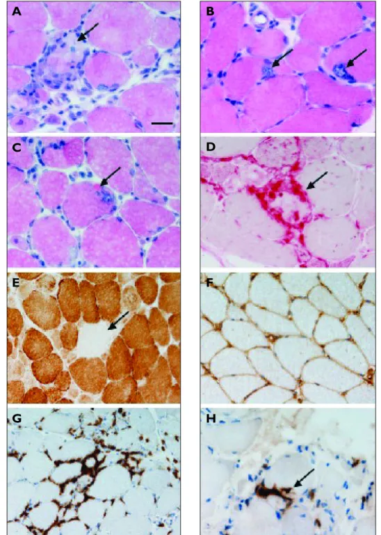
Figure 1. Histological examination of muscle biopsies from patients with inclusion body myositis reveals abnormalities
of varying severity. In haematoxylin and eosin stained sections there may be variation in fibre diameter (A) and fibre necrosis (arrow in A).Vacuoles rimmed by basophilic granular material are seen (arrows in B) and these are sometimes associated with hyalinised eosinophilic inclusions (arrow in C). Fibre necrosis can be confirmed using the acid phosphatase histochemical preparation (D). Fibres lacking cytochrome oxidase activity may be present (arrow in E).There is
widespread expression of MHC Class I at the sarcolemma (F). Lymphocytic infiltrates are largely endomysial and are composed predominantly of CD8 expressing cells (G) which infiltrate into intact myofibres (arrow in H).
A-C: haematoxylin and eosin; D: acid phosphatase histochemistry; E: cytochrome oxidase histochemistry; F: MHC class I immunohistochemistry; G-H CD8 immunohistochemistry. Bar in A represents 25µm in A-D & H, and 50µm in E-G.
A
B
C
D
E
F
P E D R O M A C H A D O E C O L.
ing the SMI-31 antibody141,142) may be seen within nuclei as loosely arranged aggregates of tubulofila-ments, and, more frequently, similar and more den-sely packed aggregates of tubulofilaments within the sarcoplasm, often in the vicinity of autophagic vacuoles. The most prominent protein accumula-ting in muscle fibres in sIBM, β-amyloid, is recog-nizable as small haphazardly deposited filaments which, when forming aggregates, display congop-hilia enhanced by Texas red-type fluorescence mi-croscopy when using the Congo red stain, but also stain with crystal violet and Thioflavin S.
Temiz et al143have recently compared muscle biopsy features of sIBM, polymyositis with mito-chondrial pathology (which can be hypothesised as a variant belonging to the same disease spec-trum as sIBM) and steroid-responsive polymyosi-tis. Interestingly they found that αB-crystallin and the above referred marker of autophagy LC3 were common in sIBM and polymyositis with mito-chondrial pathology (but not in steroid-responsi-ve polymyositis), and that SMI-31 and TDP-43 po-sitive aggregates were common in sIBM (but not in polymyositis with mitochondrial pathology or ste-roid-responsive polymyositis). β-amyloid showed no differences in aggregates among the three groups and, among patients with polymyositis with mitochondrial pathology, the ones with more rapidly progressive weakness also had more COX--negative muscle fibres. TDP-43 (TAR DNA bin-ding protein-43) inclusions in sIBM have also been described as being usually ubiquitin negative and co-localized with T-cells at sites of inflammatory infiltrates.144
As previously discussed, a great number of pro-teins aggregate in sIBM muscle fibres. Some of
them are common to myofibrillar myopathies and the same methods can be used for immunocyto-chemical staining.145,146 Similarly, proteins of the ubiquitin proteasome pathway of extralysosomal protein degradation are upregulated147and ubiqui-tin staining is also a sensitive method for showing the muscle fibre inclusions.92Finally, small angu-lated fibres are often encountered in sIBM muscle specimens, suggesting a subtle neurogenic com-ponent of denervation, but large group atrophy and fibre type grouping (features of reinnervation) are absent. Such angulated atrophic muscle fibres display increased histochemical activity of acid phosphatase and of the oxidative enzymes NADH and MAG. Matrix metalloproteinases (MMP) 2, 7 and 9 have been shown in muscle fibres, inflam-matory cells, and vessel walls.148-150
Electron microscopy (Figure 2) reveals accumu-lation of 15-21 nm tubulofilamentous inclusions and cytoplasmic collections of 6-10 nm amyloid--like filaments that immunoreact with various amy-loid protein related antibodies.151Abnormal myonu-clei with intranuclear 7 nm-wide filaments are also detected in up to 3.5% of the nuclei, but their signi-ficance in vacuolar formation remains unclear.
It should be highlighted that a vacuolar myo-pathy displaying similarities with sIBM can be found in other diseases, namely the above menti-oned hIBM, some of the distal myopathies, oculo-pharyngeal muscular dystrophy, Emery-Dreifuss muscular dystrophy, and even in chronic neuroge-nic conditions such as old poliomyelitis or chroneuroge-nic spinal muscular atrophy.81,82,138,152,153
Clinical manifestations and investigations
Clinical features
The hallmark of idiopathic inflammatory myopathi-es is a progrmyopathi-essive muscle weaknmyopathi-ess, with retained reflexes and without sensory disturbances. sIBM, however, is characterised by male predominance11,12 and causes weakness and atrophy of the distal and proximal muscles. Involvement ofquadriceps femo-ris and deep finger flexors are clues to early
diagno-sis.154Patients often present with falls because their knees collapse owing to quadriceps muscle we-akness, or with difficulty performing certain tasks, such as turning keys, tying knots and holding golf clubs, owing to weakness of finger flexors. Weakness in sIBM may be accompanied by myalgia in up to 40% of cases155and swallowing difficulties in the Table I. Muscle diseases with potential
myopathologic inflammatory features138
• Polymyositis • Dermatomyositis • Inclusion body myositis • Statin myopathy
• Facio-scapulo-humeral muscular dystrophy • Dystrophinopathy
• Dysferlinopathy • Caveolinopathy • Calpainopathy • Merosinopathy
S P O R A D I C I N C L U S I O N B O D Y M Y O S I T I S: A N U N S O LV E D M Y S T E R Y
course of the disease are common, having been re-ported in up to 60% of patients.10
Twelve papers have described the clinical featu-res of between 15 and 78 sIBM patients. Nine of these studies were retrospective and based on re-view of the medical records6,8,22,143,155-159and three were cross-sectional in design.9,10,160
Weakness at the time of diagnosis was reported to be more severe in the lower than in the upper ex-tremities,155,157and to be more or equally severe in proximal muscles compared with distal.155,156,158If weakness was described for specific muscle groups, a different distribution emerged: the knee extensors were considered more affected than the hip flexors and the wrist and finger flexors were more affected than the shoulder abductors.6This pattern has been confirmed by several studies, re-vealing the finger flexors to be most severely affec-ted, along with the knee extensors and foot
dorsi-flexors.8,9,158,160With regard to the least affected mus-cles each study showed a different pattern.9,160 Pro-minent side-to-side differences have been noted, particularly in the distal muscle groups.160
The rate of progression, the mean decrease in muscle strength corrected for observed time, vari-ed from 3.5%8to 15.6 % per year158in retrospective studies and was found to be 7.8% per year in a small prospective study.161In one of the larger studies,10 time of onset of symptoms was generally after the age of 40 (although 20% before the age of 50 years). Patients with sIBM usually present after several years of gradually worsening muscle weakness and those who are untreated or who do not respond to treatment become gradually weaker over a period of years. Peng et al 159assessed diseased progressi-on in 78 patients and found that the older the age at onset of the disease, the more rapid is the loss of strength and function. Patients presenting before age 60 progress to the use of a walker after an ave-rage of 10.2 years and those presenting after age 60 require a walker after only 5.7 years of disease.159By 15 years, most patients require assistance with ba-sic daily activities, and some become wheelchair bound or bedridden.
sIBM can be an indirect cause of death due to respiratory failure or infection, particularly respi-ratory tract infections. Subacute respirespi-ratory failu-re failu-requiring mechanical ventilation was failu-recently reported in one patient with sIBM.162Although most patients have a progressive loss of strength, approximately one-third remain stable or impro-ve when obserimpro-ved for a period of six months.161 Laboratory abnormalities
Muscle enzymes are typically normal or mildly ele-vated in sIBM, with creatine kinase (CK) levels ge-nerally being less than 10-12 times normal.10,22 Markers of systemic inflammation, such as eleva-tion of C-reactive protein, erythrocyte sedimenta-tion rate and anaemia, are usually absent. There are no sIBM specific autoantibodies, although nonspecific positive serum serologies are often present (44% of a total of 99 patients in a study by Koffman et al,163and 32% of a total of 38 patients
in a study by Brower et al,16318% of which were
myositis specific autoantibodies). Electromyography
Electromyography (EMG) in sIBM reveals myopa-thic patterns with increased insertional activity, fi-brillations, and polyphasic potentials. These
fin-Figure 2. Ultrastructural examination of muscle fibres
demonstrates whorled membranous debris (arrow in A) and associated randomly orientated filaments (B). Bar in A represents 700nm in A and 200nm in B.
A
P E D R O M A C H A D O E C O L.
dings are not specific for sIBM and are present in other inflammatory myopathies. In some cases, however, a mixed pattern of myopathic and neu-rogenic changes is seen and that has been descri-bed as more typical of sIBM than PM.22,155,157,164 Ner-ve conduction studies are usually normal. Magnetic resonance imaging
There have been reports of MRI use to characteri-se inflammation in cacharacteri-ses of sIBM,68,165-169which have concentrated mostly on imaging the thigh muscles. Papers have emphasised the sensitivity of MRI when using fat-suppressed imaging techni-ques to detect inflammation, and similarly the va-lue of recognizing the distribution of changes in helping to predict the cause of myopathy or to identify sites of inflammation, confirmed at sub-sequent biopsy.170-172However, MRI findings may not be specific and therefore images should be re-viewed in conjunction with clinical information.173 Phillips et al 9evaluated 9 patients with sIBM using quantitative and manual muscle testing as well as MRI. They found that weakness of the qua-driceps femoris and the forearm flexors was present
in most patients, but there was considerable vari-ability in the patterns and severity of muscle invol-vement. Sekul et al 169had previously reported a selective involvement of the flexor digitorum
pro-fundus that might occur early in the course of the
disease and could be easily demonstrated by MRI in up to 95% of patients. Because selective flexor di-gitorum profundus involvement appeared to be a
very frequent and characteristic finding in patients with sIBM, MRI of the forearm was proposed by these authors to be a useful noninvasive test in supporting the diagnosis of sIBM. MRI may also help to evaluate the extent and number of muscle lesions and eventually to follow their evolution un-der therapy.173
The role of MRI in sIBM is therefore still to be clarified and the question calls for longitudinal MRI studies with clinical-MRI correlation. MRI may prove to be a very helpful diagnostic and as-sessment tool in sIBM and its results may even in-corporate future diagnostic criteria if proven to be robust and reproducible.
Classification criteria for sIBM
The criteria for the diagnosis of sIBM were first proposed by Griggs and colleagues in 1995,7with
minor modifications made by Tawil and Griggs in 2002174and again changes proposed by Needham and Mastaglia in 2007.175The criteria have evolved to incorporate some additional biopsy features (such as expression of MHC-I and COX-negative fi-bres) and the recognition that some of the histolo-gical findings (such as rimmed vacuoles and con-gophilic inclusions) are probably absent in many biopsies taken in the earlier stages of the disease. Table II describes the Needham and Mastaglia diagnostic criteria as well as the characteristic fea-tures and reported associated disorders for sIBM.175 Some patients with clinical features of sIBM lack the canonical pathologic features of the disease even on repeated muscle biopsies6,176,177and the ab-sence of the late findings in patients with a typical clinical phenotype does not exclude the diagnosis of sIBM.1,178Future studies of sIBM are warranted in order to evaluate the performance and clinical impact of these classification systems.
Treatment
Despite the apparent involvement of primary im-mune factors in the pathogenesis of sIBM, this di-sease remains resistant to most immunotherapies. At present, sIBM remains a disabling disease, with most patients requiring an assistive mobility devi-ce within 5 to 10 years of onset.159,161Although the common immunotherapeutic agents are generally ineffective and there is no established therapy to stop the progression of the disease,179some pati-ents have, anecdotally, responded to these thera-pies to a certain extent. The protracted disease course has meant that few trials have been of aquate duration or have had sufficient power to de-tect even sizeable treatment effects. Moreover, sIBM is often diagnosed years after the onset of symptoms, when muscle damage may be so ad-vanced as to prevent any improvement in strength even if the disease process can be arrested. There-fore, there are insufficient data to enable an eviden-ce-based approach to treatment, which is still lar-gely empirical and varies considerably in different centres.180It has been estimated that in order to demonstrate a significant effect from treatment for sIBM in a placebo-controlled study, 200 subjects would need to be enrolled in a six-month study or 100 in a year-long trial.179This estimate should be kept in mind when considering the data on efficacy of treatment presented below.
S P O R A D I C I N C L U S I O N B O D Y M Y O S I T I S: A N U N S O LV E D M Y S T E R Y
Corticosteroids
Corticosteroids alone appear to have a limited role in patients with sIBM,22,157,181-183with the results of several uncontrolled trials showing stabilisation or temporary improvement in muscle strength in some patients, which is usually not maintained.
Barohn et al 181conducted a 12-month prospec-tive trial that included 8 patients with sIBM
trea-ted with high-dose oral prednisolone. Although the serum CK level fell, muscle strength worsened af-ter prednisone treatment. In addition, the num-ber of vacuolated and amyloid-positive fibres in-creased, despite a reduction in the numbers of T cells.
Lotz et al 22reported that muscle strength conti-nued to deteriorate in 25 sIBM patients followed for
Table II. Proposed diagnostic criteria for sIBM7,174,175
Characteristic features
Clinical features
• Duration of illness >6 months • Age at onset >30 years
• Slowly progressive muscle weakness and atrophy: selective pattern with early involvement of quadriceps femoris and finger flexors, although can be asymmetric
• Dysphagia is common
Laboratory features
• Serum creatine kinase concentration might be high but can be normal
• Electromyography: myopathic or mixed pattern, with both short and long duration motor unit potentials and spontaneous activity
Muscle biopsy
• Myofibre necrosis and regeneration
• Endomysial mononuclear cell infiltrate (of variable severity)
• Mononuclear cell invasion of non-necrotic fibres: predominately CD8+ T cells • MHC class I expression in otherwise morphologically healthy muscle fibres • Vacuolated muscle fibres (rimmed vacuoles)
• Ubiquitin-positive inclusions and amyloid deposits in muscle fibres
• Nuclear and/or cytoplasmic 16-20 nm filamentous inclusions on electron microscopy • COX-negative fibres (excessive for age)
Associated disorders
Inclusion body myositis usually occurs in isolation, but can be associated with:
• Other autoimmune disorders or connective tissue diseases (common variable immunodeficiency, idiopathic thrombocytopenic purpura, celiac sprue, Sjögren´s syndrome, dermatomyositis, systemic lupus erythematosus, systemic sclerosis, rheumatoid arthritis, paraproteinaemia, autoantibodies)
• Occasional: HIV, HTLV-I, and hepatitis C infection
• Rare: toxoplasmosis, sarcoidosis, post-poliomyelitis, macrophagic myofasciitis
Diagnostic categories
Definite inclusion body myositis
• Characteristic clinical features, with biopsy confirmation: inflammatory myopathy with autoaggressive T cells, rimmed vacuoles, COX-negative fibres, amyloid deposits or filamentous inclusions and upregulation of MHC-I expression.The presence of other laboratory features are not mandatory if the biopsy features are diagnostic • Atypical pattern of weakness and atrophy but with diagnostic biopsy features
Probable inclusion body myositis
• Characteristic clinical and laboratory features but incomplete biopsy criteria - e.g., features of necrotising inflammatory myopathy with T cell invasion of muscle fibres but absence of rimmed vacuoles, amyloid deposits, filamentous inclusions, and COX negative fibres
Possible inclusion body myositis
P E D R O M A C H A D O E C O L.
at least two years and treated with prednisone at dose levels frequently effective in PM. Joffe et al 183 also reported that patients with sIBM had poor res-ponses to prednisone.
Some reports have noted a partial response to corticosteroids with either mild improvement in or stabilization of muscle strength.157,182Serum CK le-vels often fall and may even normalize with corti-costeroid therapy, however, this biochemical res-ponse did not predict clinical benefit.182
There is one apparent exception to the usually limited response to corticosteroids. Patients with sIBM coexistent with other connective tissue di-seases (Sjögren’s syndrome, systemic lupus erythe-matosus and the rash of DM) may have a clinically important benefit from steroid therapy but it mains uncertain whether any of this benefit re-flects a specific improvement in their sIBM featu-res.155,184,185
Cytotoxic drugs
Methotrexate and azathioprine, alone or in com-bination, have shown at best minor benefit, with apparent stabilisation or improvement over short periods.155,157,182,186However, the largest trial, a ran-domized study of 44 patients who received either weekly methotrexate or placebo for 48 weeks186 showed that there was no significant difference in muscle strength between the two groups, although the serum CK levels decreased significantly in the methotrexate group.
Limited reported experience with cyclo-phosphamide and chlorambucil has also not been encouraging.187Mycophenolate has been benefi-cial on occasion.188However, none of these drugs has been assessed in controlled clinical trials.
As with corticosteroids, plasma CK levels often fall and may even normalize with immunosup-pressive treatment but the biochemical response does not predict clinical benefit.182,186
Intravenous immunoglobulin
The efficacy of intravenous immunoglobulin (IVIG) has been evaluated in two small open se-ries189,190and three double blind studies.191-193but all of the last trials have been of short duration (two lasted 3 months, and one lasted 6 months).190,192,193 In the first uncontrolled study, improvement in muscle strength and functional status was noted in three of four patients after the second monthly in-fusion.189These results were not replicated in an unrandomized, open-label study with nine
pa-tients who showed no clinical improvement.190 In a double-blind, placebo-controlled crosso-ver study involving 19 patients, no statistically sig-nificant improvement in overall muscle strength due to IVIG was observed. However, there was a trend toward improvement during IVIG treatment, and nine of the patients continued IVIG therapy in-dependently after the study was concluded becau-se of a becau-senbecau-se of improved quality of life.192
A double-blind study by Dalakas et al,191 ran-domly assigned 36 patients to either IVIG (month-ly infusions for 3 months) or placebo infusions; be-fore infusions, all patients also received high dose prednisone for 3 months. When compared to ba-seline, there were no significant differences in mus-cle strength during 4-months of observation. Fol-low-up biopsies in 24 random patients revealed a greater reduction in the number of necrotic myo-fibres in those who received IVIG than placebo, but this appeared to be of no clinical significance. The authors concluded that the combination of pred-nisone and IVIG for a 3-month period was not ef-fective in sIBM.
In the longer (6 month) crossover trial by Walter
et al,193disease progression stopped in 18 of 22 pa-tients, although muscle strength scores, symptoms and myographic test results did not change signi-ficantly.
At the present time, IVIG cannot be recommen-ded because it has shown, at best, only very mo-dest benefit. Trials of longer duration (at least 12 months), sufficiently powered in terms of num-bers of patients and including patients with early disease (hypothetically more responsive to treat-ment) are warranted to determine the role of IVIG in sIBM.
One exception may be the use of IVIG in the treatment of dysphagia. In one report, four patients with severe dysphagia due to upper esophageal dysfunction all recovered swallowing function after treatment with 6 to 8 monthly infusions of IVIG.194In patients with severe dysphagia, bougie dilation, cricopharyngeal myotomy,195or botuli-num toxin injection into the upper oesophageal sphincter196may be alternative solutions.
New biologic agents
New biologic agents targeting presumed immuno-pathological processes such as T cell proliferation, transmigration, antigen recognition or endoplas-mic reticulum stress, might produce more rewar-ding results.
S P O R A D I C I N C L U S I O N B O D Y M Y O S I T I S: A N U N S O LV E D M Y S T E R Y
A 6 month randomised, placebo-controlled trial of interferon-beta 1a (30 µg/week) in a group of 30 patients with sIBM did not show an improve-ment in muscle strength or mass.197A subsequent trial of a higher dose (60 µg/week) was also inef-fective.198However, a substantial clinical improve-ment was reported with interferon-beta treatimprove-ment in a Japanese patient with sIBM who was a carrier of hepatitis C.199
A pilot trial of the TNFα-blocker etanercept did not find an improvement in composite muscle strength scores at 6 months, although there was a slight improvement in grip strength after 12 months of treatment.200
The results of a 12-month, open, randomized trial in 11 sIBM patients using anti-T-lymphocyte globulin and methotrexate have been encouraging: those treated with antithymocyte globulin and me-thotrexate had not only a substantial fall in serum CK levels but also a significant increase in muscle strength of 1.4% compared with a mean loss of strength of 11.1% in the methotrexate alone group.201
Dalakas has recently reported in abstract for-mat the results from a trial with alemtuzumab (a humanized T-cell-depleting monoclonal antibody against CD52) in the treatment of sIBM patients, and these have also been encouraging.202In this trial, 13 sIBM patients with a 12-month natural his-tory were treated with 0.3 mg/kg/day alemtuzu-mab for 4 days. Primary end-points were the disea-se stabilization or increadisea-sed strength 6 months af-ter treatment. Alemtuzumab significantly reversed disease progression up to six months, improved the strength of some patients, and reduced the in-flammatory and degeneration-associated molecu-les in the patients’ muscmolecu-les.202
Other promising agents include sirolimus (ra-pamycin), which acts via a calcineurinin depen-dent pathway to prevent the translation of mRNA for key cytokines, and natalizumab, which blocks the transmigration of T cells across the endotheli-al cell wendotheli-all.203
Anti-degeneration and anti cell-stress therapies Agents that interfere with degeneration and endo-plasmic reticulum stress might protect the myofi-bre from chronic deleterious stimuli. At the trans-lational level, Kitazawa et al 114tried lithium, a drug increasingly explored as a neuroprotective agent, because it can modulate tau phosphorylation or amyloid processing. The results, although
disap-pointing in their model, were informative. Lithium inhibited tau phosphorylation, but did not signifi-cantly affect the motor function of the treated ani-mals and had no effect on IL-1β or the intramus-cular production of β-amyloid, suggesting that amyloid formation and inflammation occur upstream to tau pathology.
A subset of new non-steroidal anti-inflamma-tory drugs are potent modulators of γ-secretase,204 reducing amyloid production, and may also be candidates for clinical testing in sIBM.
Arimoclomol, an investigational drug for amyo-trophic lateral sclerosis, might be a candidate for use in sIBM. By prolonging the activity of the tran-scription factor, heat shock factor-1 (HSF-1), the compound has been shown to amplify heat shock protein (HSP) gene expression. Arimoclomol, the-refore, it further elevates the HSP levels already in-duced by cellular stresses, a response which appe-ars to be attenuated with advanced age.205-207HSPs have been shown to attenuate protein misfolding and aggregation promoting cellular defences against such processes.112Via inhibition of the pro-inflammatory transcription factor NFkB, they have also been shown to dampen inflammatory respon-se. More studies of anti-degeneration and anti cell-stress therapies in sIBM are warranted.
Other empirical therapies
Oxandrolone (a synthetic androgen) showed a bor-derline significant effect on isometric muscle strength in an 8 month double-blinded, crossover trial.208Despite the lack of controlled clinical trials, clenbuterol (a β-agonist), coenzyme Q10 (ubiqui-none), carnitine, and antioxidants have been re-commended on empirical grounds175and might provide symptomatic benefit in some patients. Exercise therapy
It has previously been thought that exercise pro-grams should be avoided in patients with inflam-matory myopathies because of concern that the exercise could aggravate the underlying inflamma-tory process.209However, studies in other forms of idiopathic inflammatory myopathies, such as PM and DM, showed a positive response to physical training and the absence of an adverse effect on the disease process.210,211Furthermore, studies with pa-tients with sIBM using strength and aerobic trai-ning concluded that exercise can be performed sa-fely, can lead to dynamic strength improvements, and possibly can help preventing continued loss of
P E D R O M A C H A D O E C O L.
muscle strength.212,213A more recent study214has shown that a closely monitored, 16-week, home-based, individualized functional exercise program can lead to significant gains in muscle strength and improvements in the performance of functional tasks in patients with sIBM. The protocol was well tolerated by all the patients and did not cause adverse muscle symptoms or elevation of serum CK levels.
Conclusion
sIBM is a complex and disabling disorder. Many of its mysteries are still unsolved. Larger, multicentre trials are needed to correctly define the epidemio-logy and natural history of sIBM. The identificati-on of susceptibility genes will be important to elu-cidate its pathogenesis and to provide clues to the development of targeted therapies. Understanding the interplay between inflammation and degene-ration and elucidating the molecules that drive muscle degeneration will be crucial steps. That is not an easy task, and additional animal models are required, but significant advances have been made in the last few years. There is also an urgent need for new trials of adequate duration, sufficient po-wer and including patients with early disease. Se-veral therapeutic agents are already in the pipeli-ne. The recent clues and the growing interest of the scientific community in unravelling all these mysteries allows us to have great hopes in impro-ving the quality of care for patients with sIBM in a near future.
Acknowledgements
P. Machado has been supported by an EULAR scientific training bursary.
Correspondence to
Pedro Machado
Serviço de Reumatologia
Hospitais da Universidade de Coimbra Praceta Mota Pinto
3000-075 Coimbra, Portugal
E-mail: [email protected]
References
1. Dalakas MC. Inflammatory disorders of muscle: pro-gress in polymyositis, dermatomyositis and inclusion body myositis. Curr Opin Neurol 2004;17:561-567. 2. Adams R, Kakulas BA, Samaha FA. A myopathy with
cellular inclusions. Trans Am Neurol Assoc 1965;90: 213.
3. Chou SM. Myxovirus-like structures in a case of
hu-man chronic polymyositis. Science 1967;158:1453--1455.
4. Yunis EJ, Samaha FJ. Inclusion body myositis. Lab In-vest 1971;25:240-248.
5. Dalakas MC. Sporadic inclusion body myositis—di-agnosis, pathogenesis and therapeutic strategies. Nat Clin Pract Neurol 2006;2:437-447.
6. Amato AA, Gronseth GS, Jackson CE, et al. Inclusion body myositis: clinical and pathological boundaries. Ann Neurol 1996;40:581-586.
7. Griggs RC, Askanas V, DiMauro S, et al. Inclusion body myositis and myopathies. Ann Neurol 1995;38:705--713.
8. Felice KJ, North WA. Inclusion body myositis in Con-necticut: observations in 35 patients during an 8-year period. Medicine (Baltimore) 2001;80:320-327. 9. Phillips BA, Cala LA, Thickbroom GW, Melsom A,
Zilko PJ, Mastaglia FL. Patterns of muscle involve-ment in inclusion body myositis: clinical and magne-tic resonance imaging study. Muscle Nerve 2001;24: 1526-1534.
10. Badrising UA, Maat-Schieman ML, van Houwelin-gen JC, et al. Inclusion body myositis. Clinical featu-res and clinical course of the disease in 64 patients. J Neurol 2005;252:1448-1454.
11. Badrising UA, Maat-Schieman M, van Duinen SG, et al. Epidemiology of inclusion body myositis in the Netherlands: a nationwide study. Neurology 2000;55:1385-1387.
12. Phillips BA, Zilko PJ, Mastaglia FL. Prevalence of spo-radic inclusion body myositis in Western Australia. Muscle Nerve 2000;23:970-972.
13. Mendell JR, Sahenk Z, Gales T, Paul L. Amyloid fila-ments in inclusion body myositis. Novel findings pro-vide insight into nature of filaments. Arch Neurol 1991;48:1229-1234.
14. De Paepe B, Creus KK, De Bleecker JL. Chemokines in idiopathic inflammatory myopathies. Front Bios-ci 2008;13:2548-2577.
15. Tournadre A, Miossec P. Cytokine response in inflam-matory myopathies. Curr Rheumatol Rep 2007;9:286--290.
16. Schmidt J, Rakocevic G, Raju R, Dalakas MC. Upre-gulated inducible co-stimulator (ICOS) and ICOS-li-gand in inclusion body myositis muscle: significan-ce for CD8+ T significan-cell cytotoxicity. Brain 2004;127:1182--1190.
17. Wiendl H, Hohlfeld R, Kieseier BC. Immunobiology of muscle: advances in understanding an immuno-logical microenvironment Trends Immunol 2005; 26:373–380.
18. Askanas V, Engel WK. Inclusion-body myositis: a myo-degenerative conformational disorder associated with Abeta, protein misfolding, and proteasome in-hibition. Neurology 2006;66:S39-48.
19. Banwell BL, Engel AG. AlphaB-crystallin immunolo-calization yields new insights into inclusion body myositis. Neurology 2000;54:1033-1041.
20. Oldfors A, Moslemi AR, Jonasson L, Ohlsson M, Koll-berg G, LindKoll-berg C. Mitochondrial abnormalities in
S P O R A D I C I N C L U S I O N B O D Y M Y O S I T I S: A N U N S O LV E D M Y S T E R Y
inclusion-body myositis. Neurology 2006;66:S49-55. 21. Needham M, Mastaglia FL. Sporadic inclusion body myositis: a continuing puzzle. Neuromuscul Disord 2008;18:6-16.
22. Lotz BP, Engel AG, Nishino H, Stevens JC, Litchy WJ. Inclusion body myositis. Observations in 40 patients. Brain 1989;112 ( Pt 3):727-747.
23. Mhiri C, Gherardi R. Inclusion body myositis in French patients. A clinicopathological evaluation. Neuropathol Appl Neurobiol 1990;16:333-344. 24. Chahin N, Engel AG. Correlation of muscle biopsy,
cli-nical course, and outcome in PM and sporadic IBM. Neurology 2008;70:418-424.
25. Shamim EA, Rider LG, Pandey JP, et al. Differences in idiopathic inflammatory myopathy phenotypes and genotypes between Mesoamerican Mestizos and North American Caucasians: ethnogeographic in-fluences in the genetics and clinical expression of myositis. Arthritis Rheum 2002;46:1885-1893. 26. Wilson FC, Ytterberg SR, St Sauver JL, Reed AM.
Epi-demiology of sporadic inclusion body myositis and polymyositis in Olmsted County, Minnesota. J Rheu-matol 2008;35:445-447.
27. Medsger TA, Jr., Dawson WN, Jr., Masi AT. The epide-miology of polymyositis. Am J Med 1970;48:715-723. 28. Oddis CV, Conte CG, Steen VD, Medsger TA, Jr.
Inci-dence of polymyositis-dermatomyositis: a 20-year study of hospital diagnosed cases in Allegheny Coun-ty, PA 1963-1982. J Rheumatol 1990;17:1329-1334. 29. Needham M, Corbett A, Day T, Christiansen F, Fabian
V, Mastaglia FL. Prevalence of sporadic inclusion body myositis and factors contributing to delayed diagno-sis. J Clin Neurosci 2008;15:1350-1353.
30. Garlepp MJ, Laing B, Zilko PJ, Ollier W, Mastaglia FL. HLA associations with inclusion body myositis. Clin Exp Immunol 1994;98:40-45.
31. Koffman BM, Sivakumar K, Simonis T, Stroncek D, Dalakas MC. HLA allele distribution distinguishes sporadic inclusion body myositis from hereditary in-clusion body myopathies. J Neuroimmunol 1998;84: 139-142.
32. Lampe JB, Gossrau G, Kempe A, et al. Analysis of HLA class I and II alleles in sporadic inclusion-body myo-sitis. J Neurol 2003;250:1313-1317.
33. Price P, Santoso L, Mastaglia F, et al. Two major histo-compatibility complex haplotypes influence suscep-tibility to sporadic inclusion body myositis: critical evaluation of an association with HLA-DR3. Tissue Antigens 2004;64:575-580.
34. Badrising UA, Schreuder GM, Giphart MJ, et al. Asso-ciations with autoimmune disorders and HLA class I and II antigens in inclusion body myositis. Neurology 2004;63:2396-2398.
35. Kok CC, Croager EJ, Witt CS, et al. Mapping of a can-didate region for susceptibility to inclusion body myositis in the human major histocompatibility complex. Immunogenetics 1999;49:508-516. 36. Love LA, Leff RL, Fraser DD, et al. A new approach to
the classification of idiopathic inflammatory myo-pathy: myositis-specific autoantibodies define useful
homogeneous patient groups. Medicine (Baltimore) 1991;70:360-374.
37. Scott AP, Allcock RJ, Mastaglia F, Nishino I, Nonaka I, Laing N. Sporadic inclusion body myositis in Japane-se is associated with the MHC ancestral haplotype 52.1. Neuromuscul Disord 2006;16:311-315. 38. O’Hanlon TP, Carrick DM, Arnett FC, et al.
Immuno-genetic risk and protective factors for the idiopathic inflammatory myopathies: distinct HLAA, B, Cw, -DRB1 and -DQA1 allelic profiles and motifs define cli-nicopathologic groups in caucasians. Medicine (Bal-timore) 2005;84:338-349.
39. Sivakumar K, Cervenakova L, Dalakas MC, et al. Exons 16 and 17 of the amyloid precursor protein ge-ne in familial inclusion body myopathy. Ann Neurol 1995;38:267-269.
40. Orth M, Tabrizi SJ, Schapira AH. Sporadic inclusion body myositis not linked to prion protein codon 129 methionine homozygosity. Neurology 2000;55:1235. 41. Lampe J, Gossrau G, Reichmann H, Walter MC, Men-del B, Lochmuller H. Prion codon 129 homozygosity and sporadic inclusion body myositis. Neurology 2001;57:368.
42. Lampe J, Kitzler H, Walter MC, Lochmuller H, Reich-mann H. Methionine homozygosity at prion gene co-don 129 may predispose to sporadic inclusion-body myositis. Lancet 1999;353:465-466.
43. Gossrau G, Gestrich B, Koch R, et al. Apolipoprotein E and alpha-1-antichymotrypsin polymorphisms in sporadic inclusion body myositis. Eur Neurol 2004;51: 215-220.
44. Garlepp MJ, Mastaglia FL. Apolipoprotein E and in-clusion body myositis. Ann Neurol 1996;40:826-828. 45. Oldfors A, Moslemi AR, Fyhr IM, Holme E, Larsson NG, Lindberg C. Mitochondrial DNA deletions in muscle fibers in inclusion body myositis. J Neuropa-thol Exp Neurol 1995;54:581-587.
46. Santorelli FM, Sciacco M, Tanji K, et al. Multiple mi-tochondrial DNA deletions in sporadic inclusion body myositis: a study of 56 patients. Ann Neurol 1996;39:789-795.
47. Horvath R, Fu K, Johns T, Genge A, Karpati G, Shou-bridge EA. Characterization of the mitochondrial DNA abnormalities in the skeletal muscle of patients with inclusion body myositis. J Neuropathol Exp Neu-rol 1998;57:396-403.
48. Greenberg SA, Bradshaw EM, Pinkus JL, et al. Plasma cells in muscle in inclusion body myositis and polymyositis. Neurology 2005;65:1782-1787. 49. Arahata K, Engel AG. Monoclonal antibody analysis of
mononuclear cells in myopathies. I: Quantitation of subsets according to diagnosis and sites of accumulati-on and demaccumulati-onstratiaccumulati-on and counts of muscle fibers in-vaded by T cells. Ann Neurol 1984;16:193-208. 50. Engel AG, Arahata K. Monoclonal antibody analysis
of mononuclear cells in myopathies. II: Phenotypes of autoinvasive cells in polymyositis and inclusion body myositis. Ann Neurol 1984;16:209-215. 51. Bradshaw EM, Orihuela A, McArdel SL, et al. A local