Universidade de Lisboa Faculdade de Motricidade Humana
Effects of the use of occlusal splints on the neuromuscular
function
Amândio Alberto Pedro Dias
Orientador: Professor Doutor Pedro Luís Camecelha de Pezarat Correia
Tese especialmente elaborada para a obtenção do Grau de Doutor no ramo de Motricidade Humana, na Especialidade de Comportamento Motor
Júri:
Presidente:
Doutor Francisco José Bessone Ferreira Alves
Professor Catedrático e Presidente do Conselho Científico Faculdade de Motricidade Humana da Universidade de Lisboa Vogais:
Doutor Pedro Luís Camecelha de Pezarat Correia Professor Associado com Agregação
Faculdade de Motricidade Humana da Universidade de Lisboa Doutor Filipe Manuel de Soares Melo
Professor Associado
Faculdade de Motricidade Humana da Universidade de Lisboa Doutora Maria João Pascoal Rodrigues
Professora Associada
Faculdade de Medicina da Universidade de Coimbra Doutor Orlando de Jesus Semedo Mendes Fernandes Professor Auxiliar
Escola de Ciências e Tecnologia da Universidade de Évora
Declaração de Reprodução da Tese
Nome: Amândio Alberto Pedro Dias
Endereço eletrónico: [email protected] Telefone: +351 965 728 350
Número do Cartão de Cidadão: 11644488 6ZY6
Título da Tese: Effects of the use of occlusal splints on the neuromuscular function
Orientador: Professor Doutor Pedro Pezarat Correia
Ano de conclusão: 2018
Ramo de conhecimento do Doutoramento: Motricidade Humana, especialidade de Comportamento Motor
É AUTORIZADA A REPRODUÇÃO INTEGRAL DESTA TESE APENAS PARA EFEITOS DE INVESTIGAÇÃO, MEDIANTE DECLARAÇÃO ESCRITA DO INTERESSADO, QUE A TAL SE COMPROMETE.
Faculdade de Motricidade Humana – Universidade de Lisboa, ____/_____/____
Assinatura: ____________________________ (Amândio Alberto Pedro Dias)
i DEDICATÓRIA
Para os meus pais Para a Tânia e a Catarina, que iluminam os meus dias
iii AGRADECIMENTOS / ACKNOWLEDGMENTS
O doutoramento é sem dúvida alguma um processo composto por várias etapas, pontos altos e outros não tão bons, como se de uma história de aventuras se tratasse, mas acima de tudo é um processo de aprendizagem. A aprendizagem não se restringe apenas aos métodos laboratoriais, à aprendizagem decorrente do manuseamento de equipamento ou do processamento de dados ou ainda à faculdade que se torna uma casa, pois nela passamos grande parte dos nossos dias. O que temos que reter deste processo acima de tudo são as pessoas que conhecemos, com quem convivemos, que tiveram uma participação nesta história que aqui contamos. Estes agradecimentos que de seguida se apresentam pretendem apenas enaltecer e reconhecer a importância que essas pessoas tiveram para o sucesso deste doutoramento, mesmo que por vezes não tenham tido essa consciência.
Em primeiro lugar quero agradecer aos meus pais pelo apoio dado desde o início desta história, tal como tem sido vosso apanágio em toda a minha vida.
Se esta aventura tem um final feliz, muito se deve à Tânia, companheira, amiga, confidente, que durante todo o tempo do doutoramento nunca deixou de me apoiar, mesmo que isso representasse não me ver ou ficar sobrecarregada.
Tive muita sorte, pois o meu percurso nesta aventura nunca foi solitário. Comigo estiveram colegas, que cedo se tornaram amigos, e que partilharam este processo, que teria de certeza sido ainda mais desgastante se não fossem eles. Um sincero obrigado ao Frederico Lopes, ao Miguel Nery, ao Tiago Neto, à Vanda Guerra, à Joana Reis e no último ano também à Mónica Lopes e ao Ricardo Dinis.
Ao longo do meu doutoramento tive hipótese de colaborar com vários investigadores, dos mais brilhantes e competentes que conheci até hoje, que em muito elevam o trabalho desenvolvido nesta tese. Com todos aprendi muito, bem como o papel por eles desempenhado nesta tese foi fundamental. Os meus sinceros agradecimentos por toda a ajuda e colaboração ao Gonçalo Mendonça, ao Nuno Cordeiro, ao Luís Silva, ao João Vaz, ao Francisco Tavares e ao Frederico Malaquias.
Aos alunos e alunas da licenciatura em Fisioterapia da escola superior de saúde Dr. Lopes Dias, o meu obrigado pela ajuda prestada na recolha dos dados.
A todos os participantes do estudo devo o meu profundo e sincero obrigado, pois sem eles nada tinha sido possível, bem como aos clubes e instituições que participaram. Contudo, alguns pelo seu papel mais participativo nas fases de elaboração e conceção dos estudos, bem como nos testes piloto, merecem um especial destaque, tais como o Luís Martins, o Rui Pires ou o Pedro Moreno. Obrigado pelo vosso interesse, participação e ajuda nesta aventura.
iv
Ao professor Orlando Fernandes um grande obrigado pela boa disposição e constante disponibilidade em ajudar-me, de forma a poder ultrapassar os meus obstáculos. Os meus agradecimentos a todos o professores e elementos do Laboratório de Comportamento Motor, e em particular aos professores Filipe Melo e Sandro Freitas, que enquanto elementos da CAT, em muito contribuíram para esta tese, com os constantes feedbacks ou questões, sempre de forma construtiva, de forma a que eu pudesse crescer.
Ao doutor Luís Redinha, que foi o pioneiro da ideia por detrás desta tese, por todo o entusiasmo que tem sobre a temática, que acabou por passar para mim, bem como pela constante disponibilidade em ajudar, pelo apoio médico e logístico sem os quais não teria sido possível concluir este doutoramento.
Por último, mas não menos importante, ao meu orientador, professor Pedro Pezarat-Correia, sem o qual esta aventura nunca teria passado de um sonho, uma utopia. Muito obrigado por ter acreditado em mim ainda antes de eu acreditar em mim próprio. Estou a terminar uma história que, julgava eu, não tinha capacidade e competência para a contar, mas o professor Pezarat reconheceu em mim algo que eu não sabia existir, logo no final do mestrado. Foi preciso eu crescer e aprender, para ver aquilo que você viu. A si agradeço os conselhos, a calma, a experiência, as portas que me indicou, mas que deixou para eu as abrir, de forma a que pudesse ganhar autonomia e experiência. Acima de tudo agradeço-lhe pelo exemplo que foi, é e sem dúvida continuará a ser, tanto a nível pessoal como profissional.
v
TÍTULO: Efeito do uso de goteiras na função neuromuscular
RESUMO
Dispositivos orais, tais como as goteiras oclusais, têm sido promovidos como um meio para aumentar a performance desportiva. As goteiras promovem variações na posição do maxilar e por conseguinte criam alterações na articulação temporomandibular (ATM) e nos músculos da mastigação. Estudos têm sido feitos sobre os seus efeitos, a nível neuromuscular e fisiológico, para determinar as mudanças causadas pela sua utilização. Contudo, devido à escassez de estudos nesta área, bem como a lacunas nas metodologias usadas, não é possível dar uma resposta definitiva sobre a possível influência das goteiras na capacidade neuromuscular e na performance desportiva. Deste modo, o objetivo desta tese foi determinar os efeitos agudos da utilização de goteiras oclusais em diferentes aspetos da função neuromuscular. Para tal, cinco estudos foram realizados: 1) Uma revisão sistemática, que revelou evidências do efeito positivo das goteiras em tarefas isométricas do trem superior, para sujeitos destreinados; 2) Um estudo que demonstrou que as goteiras melhoram a força e atividade muscular em tarefas isocinéticas do trem superior, para sujeitos destreinados; 3) Um estudo que determinou que, em jogadores de rugby, as goteiras não aumentaram a força do trem superior num movimento balístico. No entanto, o uso de protetores bocais customizados aumentavam o pico de força e o pico de aceleração, embora outros parâmetros de força e potência não tenham sido afetados; 4) Um estudo que analisou a oscilação do corpo na marcha e corrida através da análise cinemática e não encontrou diferenças em função da utilização de goteiras; e 5) Um estudo que observou a oscilação do centro de pressão, a atividade EMG de músculos do membro superior e a precisão no alvo de atletas do tiro enquanto utilizavam goteiras e não encontrou diferenças em nenhum dos parâmetros. O efeito ergogénico das goteiras, ocorreu de forma clara em ações de força dinâmica do trem superior e em sujeitos destreinados. Protetores bocais customizados, que reposicionam em atletas treinados a ATM numa posição idêntica às goteiras, melhoraram alguns parâmetros de força e aceleração em movimentos balísticos do trem superior, mas não afetaram outros parâmetros. Futuras investigações devem confirmar estas hipóteses, bem como averiguar o efeito a longo prazo da utilização de goteiras.
PALAVRAS-CHAVE: Goteiras; Função Neuromuscular; Eletromiografia; Benefício ergogénico; Força; Equilíbrio corporal
vii
TITLE: Effects of the use of occlusal splint on the neuromuscular function
ABSTRACT
Oral appliances, such as occlusal splints (OS), have been advocated as a mean to improve high-level sports performance. OS promote variations in jaw position and therefore create a change in the temporomandibular joint (TMJ) and on the masticatory muscles. They have been a subject of research, at neuromuscular and physiological level, to determined changes derived from the use of such devices. However, due to a paucity of research studies, and limitations on the used methods in the performed studies, it is not possible to give a correct and definite answer to the possible influence of OS on neuromuscular function and in the human sports performance. Therefore, this thesis aimed to ascertain the acute effects of occlusal splints on neuromuscular function. Five studies were conducted to achieve this purpose: 1) a systematic review, which revealed evidence of the effects of OS in upper body isometric tasks, for untrained healthy subjects; 2) a study that showed that OS enhance strength and muscle activity in upper body isokinetic tasks for untrained subjects; 3) a study which determined that for rugby athletes, OS did not increase strength in an upper body power movement, but a customized mouthguard increased peak force and peak acceleration despite other force and power did not change; 4) a study that analyzed kinematic body oscillation in gait and running and found no changes when using OS; and 5) a study that found no changes in body sway, EMG from upper limb muscles and shooting accuracy in pistol shooters while using OS. The ergogenic effect of OS was found in the dynamic strength performed by untrained subjects. Customized mouthguards, that reposition, for trained athletes, TMJ in an identical position as OS, increased some parameters of strength and acceleration but did not change other parameters. Future research should confirm these findings, while also determining the long-term effect of using OS.
KEYWORDS: Occlusal splints; Neuromuscular Function; Electromyography; Ergogenic aid; Strength; Body Balance;
ix TABLE OF CONTENTS
CHAPTER I – Introduction ... 1
Objectives and Hypothesis ... 2
Structure of the thesis ... 4
CHAPTER II – Review of the literature ... 7
1. Temporomandibular joint anatomy ... 7
2. Temporomandibular joint biomechanics ... 10
3. Occlusion and occlusal splints ... 11
4. Stomatognathic system ... 12
4.1. Stomatognathic system and body posture ... 13
4.1.1. Occlusion and orthostatic position ... 14
4.1.2. Occlusion and gait ... 16
5. Stomatognathic system and neuromuscular mechanisms ... 17
5.1. Feedback / feedforward theory ... 17
5.2. The anatomical hypothesis – fascial contraction ... 18
CHAPTER III – Methodology ... 21
1. Testing conditions ... 21
2. Manufacturing of the oral appliances ... 21
3. Evaluation methods ... 22
4. Participants ... 24
5. Testing procedures ... 24
5.1. Procedures for collection and processing of EMG signals ... 24
5.2. Procedures for collection and processing isokinetic signals ... 25
5.3. Procedures for collection and processing power and strength during ballistic bench press throw exercise ... 25
5.4. Procedures for collecting and processing kinematic data ... 26
5.5. Procedures for collecting and processing kinetic data ... 26
5.6. Statistical analysis ... 26
CHAPTER IV - A systematic review on the effects of occlusal splint therapy on muscle strength ... 29
ABSTRACT ... 30
INTRODUCTION ... 31
METHODS ... 32
x
DISCUSSION ... 38
CONCLUSIONS ... 43
REFERENCES ... 44
CHAPTER V - Effects of occlusal splints on shoulder strength and activation .. 47
ABSTRACT ... 48 INTRODUCTION ... 49 METHODS ... 50 RESULTS ... 52 DISCUSSION ... 53 CONCLUSIONS ... 57 REFERENCES ... 58
CHAPTER VI - The effects of a controlled mandible position mouthguard on strength and power in trained athletes ... 65
ABSTRACT ... 66 INTRODUCTION ... 67 METHODS ... 69 RESULTS ... 72 DISCUSSION ... 73 CONCLUSIONS ... 77 REFERENCES ... 78
CHAPTER VII - The effects of occlusal splints on gait and running patterns. A kinematic analysis ... 81 ABSTRACT ... 82 INTRODUCTION ... 83 METHODS ... 85 RESULTS ... 88 DISCUSSION ... 88 CONCLUSIONS ... 90 REFERENCES ... 91
xi
CHAPTER VIII - Effects of dental occlusion on body sway, upper body muscle
activity and shooting performance in pistol shooters ... 95
ABSTRACT ... 96 INTRODUCTION ... 97 METHODS ... 98 RESULTS ... 101 DISCUSSION ... 101 CONCLUSIONS ... 105 REFERENCES ... 106
CHAPTER IX – General Discussion ... 109
CHAPTER X – Conclusions ... 117
xiii LIST OF FIGURES
Figure 1. Schematic representation of the temporomandibular joint. Image retrieved
from Nelson et al. (2012). ... 7 Figure 2. Illustration suggesting that the mandible functions as a class III lever. Image
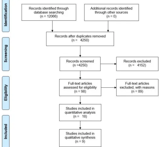
retrieved from Ellis et al. (2005) ... 10 Figure 3. Flow chart of the methodology for the search results ... 34
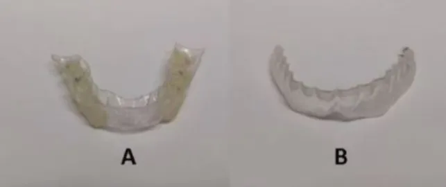
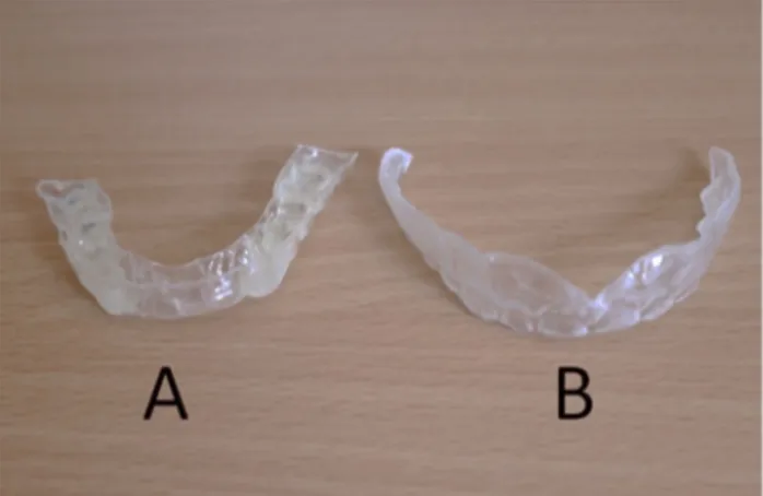
Figure 4. Oral devices tested. A - Occlusal splint; B - Placebo splint ... 51
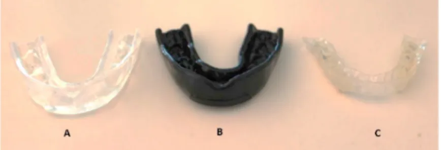
Figure 5. The occlusal devices used. A – Mandible controlled mouthguard; B –
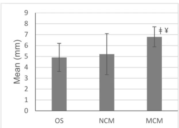
Non-controlled Mouthguard; C - Occlusal splint ... 71 Figure 6. VDO results. Significant at p ≤ 0.05: ¥ higher than OS; ǂ higher than NCM 73
Figure 7. Oral devices tested. A - Occlusal splint; B - Placebo splint ... 85
Figure 8. Occlusal devices used. A – Occlusal splints; B – Placebo splint ... 98
Figure 9. Position of the shooter on the force platform and placement of the
accelerometer on the gun barrel ... 99 Figure 10. Representative data of accelerometer and EMG analysis during a shooting
task. The onset of the shot was determined by the abrupt change in the accelerometer trace. ... 100
xv LIST OF TABLES
Table 1. Main aspects of the methodology used for each experimental study... 23
Table 2. PEDro scale results of the included studies ... 33
Table 3. Summary of the reviewed studies ... 37
Table 4. Results of the shoulder abduction / adduction tests ... 54
Table 5. Results of the shoulder external / internal rotation tests ... 55
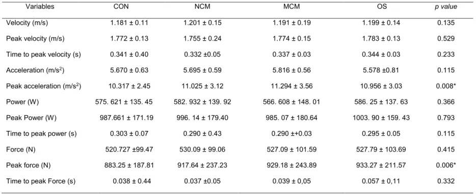
Table 6. Values of ballistic bench throws variables in the four conditions: No mouthguard (CON), Non-controlled Mouthguard (NCM); Mandible controlled mouthguard (MCM) and Occlusal splints (OS) ... 74
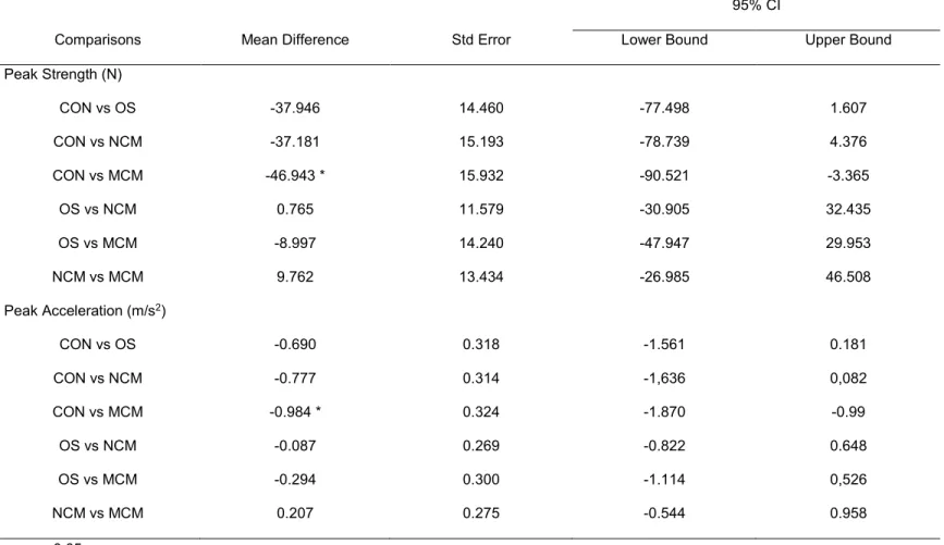
Table 7. Multiple comparisons between conditions: No mouthguard (CON), Non-controlled Mouthguard (NCM), Mandible Non-controlled mouthguard (MCM) and Occlusal splint (OS). ... 75
Table 8. Results of the three experimented conditions in gait and running. OS - Occlusal splint; PS - placebo splint; CON - no splint: control condition ... 87
Table 9. Results of COP parameters and EMG on the three experimental conditions. AP – anterior posterior; ML – medium lateral; OS – Occlusal splint; N – Normal condition; PS – Placebo splint ... 103
xvii LIST OF ABBREVIATIONS
BBT – Ballistic bench throw
CAP – Concurrent activation potentiation CMJ – Counter-movement jump
CMRR – common-mode rejection ratio CNS – Central nervous system
CON – Control condition COP – Center of pressure CR – Centric relation EMG – Electromyography LPT – Linear position transducer
MCM – Mandible controlled mouthguard N – Normal condition
NCM – non-controlled mouthguard OS – Occlusal splint
PT – Peak torque PS – Placebo splint
RVC – Remote voluntary contraction SS – Stomatognathic system
TMJ – Temporomandibular joint VD – Vertical dimension
1
CHAPTER I – Introduction
Human performance has always been a subject of research. The use of occlusal splints to increase such performance has been a discussion topic for the last years. Occlusal splints (OS) are removable appliances that cover some or all of the occlusal surfaces of the teeth (Yunus, 2009). Occlusal splint therapy may be defined as the art and science of establishing harmony in the neuromuscular system through the correction of temporomandibular joint (TMJ) and the change of functional length of related muscles (Dylina, 2001). Most splints alter the vertical dimension of occlusion. This instantaneous change that occurs in muscle behaviour can be summarized as an increase of postural position of minimal muscles activity or a reduction of activity of postural muscles (Boero, 1989). When a subject wears an OS, it causes an immediate and pronounced relaxation in the masticatory muscles, which will eventually result in the mandible repositioning and the distribution forces across the masticatory system (Yunus, 2009).
From the existing literature, two hypotheses have been put forward as possible explanations for the use of OS and changes in the human body. The first one is based on changes in neuromuscular patterns caused by changes in peripheral proprioceptive input signals in the orofacial region, when using OS (Milani, De Perière, Lapeyre, & Pourreyron, 2000). The second one is based on fascial chains, that appear to possess an ability to contract when stimulated and can distribute tension throughout the body (Schleip, Klingler, & Lehmann-Horn, 2005).
Electromyography (EMG) analysis of muscle activity has been helpful to establish a connection for the relationship between the stomatognathic system muscles and muscles that control posture, as well as muscles involved in the movements of distal segments. The relation between TMJ and human body activity appears to exist (Gangloff & Perrin, 2002; Kibana, Ishijima, & Hirai, 2002) with specific functional correlations (Valentino, Melito, Aldi, Valentino, 2002), but there is still no scientific evidence to support this relationship. However, the above-mentioned studies before, demonstrate that T stomatognathic system muscles and muscles of other body segments are linked. The effects of OS on muscle strength of upper and lower limbs (Alexander, 1999; Chakfa et al., 2002; Forgione, Mehta, McQquade, Westcott, 1992; Golem, 2012; Jung, Chae, & Lee, 2013; Lee et al., 2014) and trunk (Yates, Koen, Semenick, Kuftinec, 1984) muscles has been one of the great focus of studies in this area. Still, results reveal different effects on muscle strength, which range from significant increases to no differences at all. Posture also appears to be affected by OS in static condition (Bracco, Deregibus, & Piscetta, 2004a) and during gait. When walking, the mandible moves vertically in relation
2
to the maxilla (Flavel, Nordstrom, & Miles, 2003). If the muscles involved in TMJ are active and contracting during gait, maybe they could have an effect on body stability. Some studies point to this conclusion (Cuccia, 2011).
However, there are gaps in terms of quality and methodology of most studies that address the use of oral appliances on human performance (Hanke, Motschall, & Türp, 2007; Perinetti & Contardo, 2009), whether it is in a sport-related context or not. So, there is a need for further studies that quantify, and characterize neuromuscular patterns associated with motor performance tasks while using OS.
Objectives and Hypothesis
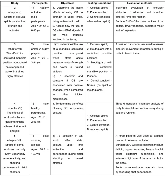
The main purpose of this thesis was to investigate the immediate changes and neuromuscular adaptations caused by the use of OS in healthy participants. In order to reach this objective, five studies were carried out, each one with its specific goal, but following a logical sequence.
The study 1 was conducted with the objectives of:
1) Evaluate the body of evidence available on the efficiency of OS in producing changes in strength in healthy participants;
2) Determine the most used protocols and assessed parameters.
To achieve these objectives, we performed a systematic review, with quality studies analysis, using keywords from the dental medical field as well as from the sports science field. We searched the literature for studies that examined the acute effects of OS on strength, regardless of the assessment protocol or the body part tested. This review was also used to determine some of the parameters assessed in studies 2 and 3.
The study 2 was performed with the following objectives:
1) Determine the acute effects of using OS on shoulder strength, through isokinetic evaluation;
2) Access how the use of OS affects neuromuscular activation patterns of muscles involved in the evaluated movements.
This second study was conducted with healthy participants. Considering the lack of unanimity and also the methodological limitations found in the experimental designs of studies that evaluated how OS affected strength in upper body, we measured isokinetic peak torque (PT) in two different arm movements. The definition of movements and conditions analysed in this study was based on some of the findings of study 1. Electromyography (EMG) of upper body muscles was also evaluated in this study, to
3
address how and if the use of OS affects neuromuscular activity of shoulder muscles responsible for these movements.
The following hypotheses were tested in this study:
a) The use of OS that repositions TMJ would affect arm strength;
b) The amplitude of EMG signals of main muscles involved in these movements increases using OS.
The study 3 aimed to address the following objectives:
1) To determine if the use of a mandible controlled position mouthguard would affect acute measurements of strength and power in rugby trained athletes;
2) To ascertain and compare if OS is associated with positive changes when compared to other mouthpieces.
This third study was performed with rugby players as participants, since they are acquainted with strength and power training and also because they usually use mouthguards during training sessions and games. Therefore, they qualify as an ideal pool of subjects for testing the acute effects of OS on trained athletes. For this study, a dynamic and ballistic movement was chosen for assessing a range of variables. We also manufactured and evaluated a custom made mouthguard that promoted the similar changes in TMJ as the OS.
Two hypotheses were formulated for this study:
a) The use of OS would affect strength in a dynamic power movement;
b) The use of custom made mouthguards that promoted the same changes in TMJ as the OS has a positive effect on strength.
The study 4 was directed to the following objective:
1) Determine the immediate effect of using OS on dynamic posture.
This fourth study examined how the use of OS could affect body dynamic posture during gait and running on a treadmill. With the aid of 3D kinematic analysis, body horizontal and vertical sway was determined and evaluated to see how it would variate with the use of OS. Therefore, we tested two hypotheses:
a) The use of OS would reduce body sway during gait; b) The use of OS would reduce body sway during running.
Finally, study 5 was performed in order to:
1) Establish if OS would affect static posture, upper limb activation and performance during pistol shooting in trained athletes.
4
Given that current literature shows that the use of OS affects static posture, this study intended to analyse if OS would affect posture and shooting performance. For this purpose, a group of national level pistol shooters was recruited for the study. The experimental protocol used in this study gaged kinetic variables, as well as EMG activity of the arm and shoulders muscles of the arm holding the pistol. Sports performance (target accuracy) was also recorded to compare between test conditions. Three hypotheses were formulated for this study, as follows:
a) The use of OS would create a positive effect on body sway, by reducing the dispersion area;
b) Muscle activity amplitude of arm and shoulder muscles would be affected when using OS;
c) Scoring accuracy would have an increment, with the use of OS
Structure of the thesis
This thesis follows a study compilation organization, and will be presented as follows:
Chapter I – Includes an introduction where the problem of the research is identified, and sets the structure of the thesis, describing the sequence of studies, their objectives and related hypothesis.
Chapter II – Displays a review of the literature, where the main concepts are defined and explained. The topics of occlusion, occlusal splints and support theories for the problem are explored and detailed.
Chapter III – Presents the methodology used in experimental design studies in a general way.
Chapter IV to VIII – Presents, in each chapter, the five studies previously mentioned. These chapters share a similar organization: Introduction, Methods, Results, Discussion, Conclusions and References.
Chapter IX – The General Discussion of the results of different studies are included in this chapter. A summary of the main findings of the studies is presented, followed by a rationale of how these results can be useful for sports performance. The limitations of the thesis are also discussed in this chapter, as well as recommendations for future research.
Chapter X – The general Conclusions of the thesis are presented here. A summary of the main findings of the studies is presented, as well as clues for future studies on this topic.
5
Chapter XI – Presents the list of references used in chapters I, II, III and IX. This chapter does not include the references used in the studies, given that each one of them has its own reference list.
7
CHAPTER II – Review of the literature
1. Temporomandibular joint anatomy
The temporomandibular joint (TMJ) is one of the last joints to appear in the uterus, emerging only at the 8th week of gestation (Bag, Gaddikeri, Singhal, Hardin, Tran,
Medina, Curé, 2014). It is considered underdeveloped at birth when compared to other body joints and continues its development in the early childhood years as the jaw is employed for sucking and chewing motions (Bag, Gaddikeri, Singhal, Hardin, Tran, Medina, Curé, 2014). When fully developed, the TMJ is one of the most complex joints in the human body. The joint provides hinging movements in one plane, (backward and forward) and consequently can be considered a ginglymoid joint. However, simultaneously provides gliding movements, which classifies it as an arthrodial joint. Thus, due to the combination of movements and classifications, it has been considered a ginglymoarthrodial joint (Alomar et al., 2007; Okeson, 2008).
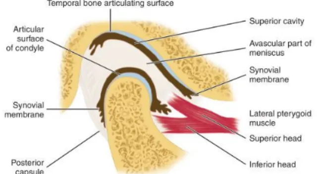
The osseous portions of the joint are composed by the concavity of the temporal mandibular bone (mandibular fossa) where the mandibular condyle fits. This concavity is designated the glenoid fossa (Walker & MacLeod, 2017). Separating these two bones from direct contact is the articular disk, which as a round or oval shape and it is biconcave. This disk has a thick anterior and posterior areas, with the intermediate zone being considerable thinner (Nelson, 2013; Walker & MacLeod, 2017). Functionally, the disk separates the joint cavity in two compartments (superior and inferior) (Tanaka & Koolstra, 2008) and serves as a nonossified bone that permits the complex movements of the joint (Okeson, 2008). The joint movement provides largely passive movable articular surface accommodating the translational movement made by the condyle (Tanaka & Koolstra, 2008). Due to this function, the TMJ is considered a compound joint (Okeson, 2008).
Figure 1. Schematic representation of the temporomandibular joint. Image retrieved from Nelson et al. (2012).
8
The capsule that encircles the TMJ contains a synovial membrane, responsible for the production of the synovial fluid that fills and nourishes the joint space. The joint has three functional types of ligaments: the collateral ligaments, the capsular ligaments and the temporomandibular ligament (Walker & MacLeod, 2017). The collateral ligaments spans from the mandible to the temporal bone and are responsible for allowing the disk to move passively with the condyle as it glides anteriorly and posteriorly. Thus, these ligaments are responsible for the hinging movement of the TMJ. The capsular ligaments are located inside the capsule and connect the disk to the poles of the condyle. These ligaments surround the TMJ and act to resist any lateral, medial or inferior forces that tend to separate or dislocate the articular surfaces. Additionally, these ligaments have a significant function of containing the synovial fluid, since they encompass the joint. Proprioceptive feedback regarding position and movement of the joint is also another function of these ligaments, since they are well innervated (Okeson, 2008). The temporomandibular ligament is composed of strong and tight fibers, overlaying the capsule, from the lateral aspect of the temporal bone to the neck of the condyle. This ligament is composed of two components: a horizontal component that resists to posterior displacement and an oblique component that limits rotational movement. This ligament is considered to play an important role in joint stabilization(Okeson, 2008; Walker & MacLeod, 2017). Additionally, two noncapsular accessory ligaments that have limited functional impact on the joint also exist: the sphenomandibular and the stylomandibular ligaments. The sphenomandibular ligament extends downward from the spine of the sphenoid bone to a small bony prominence on the medial surface of the mandible and it does not have any restrictive effects on the mandibular movement. The stylomandibular ligament arises in the styloid process and spreads downwards to the mandible. Its function is to limit excessive protrusive movements of the mandible (Okeson, 2008).
The TMJ exhibits several common features to other synovial joints, but most importantly, possesses exclusive characteristics that differentiates this joint, making it unique in several aspects. Both TMJ’s must function together, with a range of motion that has a fixed endpoint in the dentition. Contrasting to other synovial joints, that are covered by hyaline cartilage, the articular surfaces of the TMJ are covered by a layer of fibrous cartilage (Tanaka & Koolstra, 2008).
The muscular function of the TMJ is maintained by four pairs of muscles: the masseter, temporalis, medial and lateral pterygoid. All the four muscles are innervated by the anterior branch of the mandibular division of the trigeminal nerve (Walker & MacLeod, 2017).
9
The masseter originates in the zygomatic arch and has a downward profile to the lateral aspect of the lower border of the mandible, with a rectangular shape. When this muscle contracts, it elevates the mandible in such a way that teeth make contact, or in other words, its main function is the elevation of the mandible. When the mandible is protruded it can also assist in the stabilization of the condyle.
The temporalis is a muscle with a broad origin along the lateral temporal bone. It has a fan-shaped and extends downward forming a tendon between the zygomatic arch and the lateral surface of the skull that inserts on the coronoid process and anterior board of the ascending ramus. The temporalis can be divided into three different portions, according to fiber direction and function. The anterior portion is composed of fibers that are almost vertically in direction and when they contract, raises the mandible vertically. The middle portion contains fibers that are obliquely and has a function of elevation and retrusion of the mandible. The posterior portion has fibers that are aligned almost horizontally and when It contracts elevates and slightly retrudes the mandible. The temporalis is the principal positioner of the mandible during elevation, able of coordinating closing movements, due to the different angulation of its muscle fibers. The medial pterygoid muscle originates in the pterygoid fossa and extends downward, backward and outward to insert along the medial surface of the mandibular angle. Along with the masseter, it forms a muscular sling that supports the mandible at the mandible angle. When active, this muscle elevates the mandible, but also helps in the protruding of the mandible. Unilateral contraction of this muscle will create a mediotrusive movement of the mandible.
The lateral pterygoid muscle is composed of two distinct portions. The superior portion originates from the base of the greater sphenoid wing and inserts into the articular disk. The inferior head originates from the lateral surface of the lateral pterygoid plate and inserts in the pterygoid fovea, on the front of the condylar neck. Their action together serves to pull the condyle and the disk simultaneously down the articular eminence, as well as helping in the protraction, depression and contralateral abduction. It also may be active during other movements for joint stabilization (Nelson, 2013; Okeson, 2008; Walker & MacLeod, 2017).
The TMJ is innervated by the same nerve that provides motor and sensory innervation to the muscles that control it. This sensory and motor innervation is provided by the trigeminal nerve, particularly by the mandibular division, which gives rises to four terminal branches (Walker & MacLeod, 2017). Most innervation is provided by the auriculotemporal nerve, which originates the mandibular nerve behind the joint and ascends laterally and superiorly to encompass the posterior region of the joint (Okeson, 2008). This nerve provides sensory innervation and proprioception from the TMJ.
10
Additional sensory innervation is also available anteriorly from the masseteric nerve and posteriorly form the posterior deep temporal nerve. Both nerves are branches of the anterior aspect of the mandibular division of the trigeminal nerve and provide sensory information to the TMJ, before innervating the muscles from which their names are derived (Walker & MacLeod, 2017).
2. Temporomandibular joint biomechanics
The fact that the two TMJs are connected to the same bone (mandible) makes this an extremely complex system. Each joint can concurrently act separately but is always dependent on the influence of the other. Adding to its intrinsic complexity, movement caused by the TMJ involves a high degree of interactions and coordination between bilateral condyles, disks, muscles and ligaments. Most frequently, an analogy is used to classify the movement of the mandible. The mandible is classified as a class III lever (Fig. 2), where the TMJ (via the condyle) acts as a fulcrum, the bite pressure as resistance and the musculature as applied forces, ultimately transmitting variable loads to the TMJ (Choi, Conway, Taraschi, & Ben-Nissan, 2015; Ellis & Throckmorton, 2005; Walker & MacLeod, 2017).
Figure 2. Illustration suggesting that the mandible functions as a class III lever. Image retrieved from Ellis et al. (2005)
In order to achieve mechanical equilibrium, the assumption behind the class III lever is that the bite force is less than the applied force, so that the entire system can achieve a mechanical advantage equal to less of one (Choi et al., 2015). This classification is not yet fully acknowledged and the interactions within the TMJ are to this day not fully understood (Bag et al., 2014). Some controversy still stands, since some authors argue that the condyle and articular fossa are not configured to withstand the large resultant forces required by the model. Also, according to the same authors, the model does not take in consideration the centre of mandibular rotation (Roberts & Tattersall, 1974).
11
Mathematical models of the forces acting on the TMJ have been made, but also with non-complying results, since a complete model of all the forces acting in the TMJ, with all vectors determined in a three dimension model is complex (Choi et al., 2015). For this model to be completed it is required to calculate the magnitude and direction of bite force, the magnitude and direction of each muscle force as well as the length of their moment arms.
The TMJ is a compound joint, and its structure and function can be divided into two distinct systems. The first system is composed by the tissues that surround the condyle and articular disk. Because this system is tightly bound by the lateral and medial discal ligaments, the only physiologic movement that can occur between these surfaces is the rotation of the disk on the articular surface of the condyle. Therefore, the condyle–disk complex is the joint system responsible for rotational movement in the TMJ (Choi et al., 2015; Okeson, 2008). The second system consists of the condyle-disk complex mentioned before functioning against the surfaces of the mandibular fossa. Since the disk is not tightly attached to the articular fossa, free sliding movement can happen between these surfaces in the superior cavity. This movement occurs when the mandible is positioned in a forward position, referred to as translation. Therefore, translation occurs in in the superior joint cavity between the superior surface of the articular disk and the mandibular fossa (Choi et al., 2015; Okeson, 2008). The articular disk plays a role in both joint systems, since it acts as a nonossified bone, and because of this function, it justifies the TMJ classification as a true compound joint (Okeson, 2008)
3. Occlusion and occlusal splints
The Glossary of Prosthodontic Terms defines dental occlusion as “the static relationship
between the incising or masticating surfaces of the maxillary or mandibular teeth or tooth analogues” (Prosthodontics, 2005). This definition was the norm for many years and is
in consonance with a view in which assessment and description of the occlusion is in static jaw positions, such as in intercuspal, lateral or protrusive jaw positions. But, more recently an evolution of this definition was conveyed, that assesses the teeth contacts from a more functional perspective. Therefore, a more fitting definition of dental occlusion is “the dynamic biological relationship of the components of the masticatory system that
determine tooth relationships” (Klineberg & Jagger, 2004).
Occlusal splints (OS) are removable appliances that cover some or all of the occlusal surfaces of the teeth in either the maxillary or mandibular arches. The basic function of the OS is to prevent the existing occlusion from controlling the maxillo-mandibular relationship at maximum intercuspation (Yunus, 2009).
12
The use of OS may be defined as the process of establishing harmony in the neuromuscular system, when parafunctional forces are present. These parafunctional forces are harmful to the stomatognathic system and OS create a mechanical disadvantage for these forces, with removable appliances (Dylina, 2001), re-establishing a good relation of forces.
Most splints alter the vertical dimension of occlusion instantaneously. These changes can be summarized as (Boero, 1989):
a) Elevator muscles have greater length with an increase in vertical dimension of occlusion and can contract more efficiently;
b) The position of minimal muscle activity is obtained with a larger vertical dimension of occlusion;
c) An increase in the occlusal vertical dimension causes an immediate adaptation to a new freeway space;
d) The EMG activity of the postural muscles (anterior temporalis) is reduced with an increased vertical dimension of occlusion.
When a person uses an occlusal splint in his mouth, it causes an immediate and pronounced relaxation in the masticatory muscles, which will eventually result in the mandible repositioning and closing in a more retruded position without teeth interference. The OS also provides a platform for the teeth, which will allow for equal distribution of tooth contacts, immediate posterior tooth disclusion in all movements and reduced stress on the joint. An optimal function and comfort is achieved with this neuromuscular harmony, by the distribution of the force across the masticatory system (Yunus, 2009).
4. Stomatognathic system
The TMJ is part of a large system comprised of several structures. This system is designated by stomatognathic system (SS) and is a functional unit that embraces skeletal components (maxilla and mandible), dental arches, soft tissues (salivary glands, nervous and vascular supplies), and the temporomandibular joint and masticatory muscles (Cuccia & Caradonna, 2009) which are controlled by the central nervous system (Kandel, 2013). These structures act in harmony to perform different functional tasks: speech, open and closing , mastication, and deglutition as well as parafunctional actions (Prosthodontics, 2005).
13
4.1. Stomatognathic system and body posture
Posture refers to the position of the human body and its orientation in space. Posture involves muscle activation that, controlled by the central nervous system (CNS), leads to postural adjustments. These adjustments are the result of a complex system of mechanisms that are controlled by multisensory inputs (visual, vestibular, and somatosensory) integrated in the CNS. Through mechanisms of back and feed-forward, postural adjustments play a critical role in orthostatic and dynamic postural control, influencing the ability to perform daily living activities. As with reflexes, postural adjustments improve through exercise and learning (Kandel, 2013).
The SS also plays an important role in postural control, as TMJ makes muscular and ligament connections to the cervical region, forming a functional complex called the “cranio-cervico-mandibular system” (Cuccia & Caradonna, 2009).
A study that measured the electromyography (EMG) activities of the jaw closing muscles and the sternocleidomastoid muscle showed a positive correlation between those two groups of muscles. Since the sternocleidomastoid muscle is related to head posture, the authors claimed that the act of teeth clenching affects the head posture (Kibana et al., 2002).
A functional coupling between trigeminal nerve and cervical systems was demonstrated in a study (Browne, Clark, Yang, Nakano, 1993) that inhibited the sternocleidomastoid muscle with a trigeminal stimulation. EMG was used to see if an electrical stimulation of the masseter muscles would have some influence on sternocleidomastoid muscle. The results showed that sternocleidomastoid could be triggered by activating masseter muscles, showing that a link exists between the two muscles.
The existence of connections between the SS and body posture was measured in different occlusal mandibular positions, using cotton rolls, of adults who underwent anterior cruciate ligament (ACL) surgery (Tecco et al., 2006). EMG measurements were made in the masseter, temporal, sternocleidomastoid and cervical muscles, to represent neck muscles and upper and lower trapezius as the trunk muscles with different occlusal mandibular positions using cotton rolls. The same measurements were made on a control group. Body sway was measured using a force platform and EMG measurements were recorded in different muscle groups: masseter, temporal, sternocleidomastoid, cervical muscles, neck muscles, upper and lower trapezius and trunk muscles. The results showed that, compared with the healthy subjects, ACL patients, the centre of pressure (COP) significantly moved into the right and forward directions in all tests situation, and EMG activity of the masseter, lower trapezius and sternocleidomastoid muscles increased significantly. According to the authors these results may reflect the
14
extension of the muscular tensions, that is produced by the postural pathology of the knee, which appears to affect the muscle activity of neck and trunk muscles.
Subjects that had a TMJ disorder (TMD) were asked to fill a questionnaire on pain in the neck and jaw disability. This study was done to determine the relationship between pain and corresponding ineptitude for performing functional activities of daily living in patients with TMD. The results of this study demonstrated a strong correlation between jaw disability and neck disability (Olivo et al., 2010). This means that people who suffered from jaw pain, as a result, had a high level of jaw disability and also had a high disability in the neck region. A similar result was obtained when the prevalence of craniomandibular pain and cervical spinal pain was studied (Visscher, Lobbezoo, de Boer, van der Zaag, & Naeije, 2001). The authors concluded that chronic craniomandibular pain patients more often suffer from cervical spinal pain than person without craniomandibular pains.
An OS was used to access the changes in head position and postural alterations in subjects with TMD (Strini et al., 2009). The study evaluated the stomatognathic alterations before and after the installation of the occlusal splint. When analysing the results, after one week of continuous use, the patients tend to bring their head to an ideal position. After one month of use, significant differences were found in head position. Subjects also reported a major decrease in the level of pain. The authors concluded that results suggest an important interrelation between occlusion and head position, meaning that the postural position of the individual can suffer biomechanical alterations originated from stomatognathic modifications in dysfunctional individuals, causing clinically visible changes and affecting the performance of the involved structures.
The studies mentioned before appears to demonstrate that the muscular and ligament connections that exist between the TMJ and the cervical region may have an influence on head posture. Consequently, the change in head position may cause a variation in COP parameters for both static and dynamic balance. This topic will be discussed in the next point.
4.1.1. Occlusion and orthostatic position
Several studies have addressed the effect of occlusion on the body sway. Cotton rolls were used to change mandibular position and the COP was measured with a force platform with eyes opened and closed (Baldini, Nota, Tripodi, Longoni, & Cozza, 2013). The authors reported that the mandibular position had a significant influence in the sway area parameter, but did not influence sway velocity.
15
A force platform was also used to measure the effects of three different mandibular positions (centric occlusion, rest position and myocentric position) on postural stability (Bracco, Deregibus, & Piscetta, 2004b). The results support the observation that different jaws relations imply differences in body posture. In fact, the study showed a strong relationship between mandibular position and body posture: 95.8% of the subjects showed variations in load distribution when closing mouth either in centric occlusion or in myocentric position. Furthermore, 97.9% of the subjects showed changes also in the distance between theoretical and real barycenter on x axis, and all of them showed changes on y axis. The authors stated that a good balance of masticatory and head and neck muscles seems to be an important factor for postural stability.
With the use of EMG Hosoda and colleagues (Hosoda et al., 2007) were able to determine muscle activation on the masseter muscles during the maximum voluntary contractions (MVC). With this information, subjects were tested on a force platform while clenching (20% to 50% of MVC - occlusion) and without clenching (teeth not touching). To provoke body sway, an external disturbance was used. The results demonstrated only a little difference in latency with a small disturbance, between teeth clenching conditions. However, when greater disturbances were introduced, results grew apart. Latency became smaller in the presence of occlusion and greater with no occlusion. According to the authors, the time required for the beginning of the recovery in response to external disturbance in the standing position is shortened with jaw occlusion compared to without jaw occlusion, which suggests a possible role of occlusion in the improvement of balancing ability.
Other authors also scrutinized posture in the orthostatic position and its possible relation to the SS and found no differences in the results. A sample of women was divided into three sub-groups: healthy, malocclusion and TMD. Several occlusion positions were tested with the use of a force platform, in order to measure COP variability of each foot. When comparing the results of different sub-groups, there were no significant differences reported, despite a large intrasample variability. From a statistical point of view, the fact that sub-groups were small in numbers, combined with the variability of results, could have hidden between-group differences, according to the authors. They also state that it is possible that postural alterations can occur, via a complex neuromuscular mechanism, but maybe they are not detectable at the foot level (Virgilio F Ferrario, Chiarella, Taroni, & Schmitz, 1996).
Posturography was also used as a mean to access the influence of dental occlusion in COP movement, sway area, sway length and sway velocity in healthy subjects. Several occlusal conditions were tested, with eyes open and closed on a vertical force platform. A lack of significance on tests results revealed that posture was not affected by changes
16
in dental occlusion in the age range of subjects included. According to the authors, the results of the study don´t necessarily deny the existence of an influence of dental occlusion in posture, but maybe support that such a connection is smaller in importance (Giuseppe Perinetti, 2006). In conclusion, the results on the effects of TMJ manipulation and its influence in orthostatic position are still debatable. Some studies point to a connection between them, while others dismiss that link, or state that it is small in significance. More studies are required, so that a consensus can be reached on this topic.
4.1.2. Occlusion and gait
Some evidences support that dental occlusion affects posture not only in the orthostatic condition, but also when the human body is in motion.
Six different mandibular positions were tested, with the use of occlusal splints, to investigate their effect on body equilibrium and gait stability (Fujimoto, Hayakawa, Hirano, & Watanabe, 2001). Subjects underwent tests in three different walking speeds and significant differences were reported at all gait speeds, for the coefficient of variation at fast speed and ordinary speed and for the gait velocity at fast speed. According to the authors, the results of this study suggested that a change in mandibular position conditioned gait stability.
The plantar surface is important for postural control since it has receptors that convey sensory information about the ground that a person is stepping. With this idea in mind, a study was conducted to compare subjects with and without TMD in three dental occlusion conditions, while walking in a force platform (Cuccia, 2011). When comparing both groups, the authors found differences between them in postural conditions and in the plantar arch. In an intra-group analysis, differences were also found when the subjects were clenching their teeth, which instigated a load reduction and an increase in surface contact on both feet. This study was pointed by the authors as a possible explanation for the interrelationship between stomatognathic inputs and locomotion.
An experimental induced imbalance of occlusion was created with the help of cotton rolls, to investigate possible correlations between postural loading on feet during walking (Simona Tecco, Polimeni, Saccucci, & Festa, 2010). The imbalance of dental occlusion was achieved with a cotton roll on one side of the dental arch. Three conditions were tested: with a cotton roll on each side of the dental arch and normal occlusion. The results demonstrated that when using a cotton roll on one side of the dental arch, the loading on the foot of the same side was significantly lower than in habitual occlusion or with cotton
17
roll in the opposite side. These results suggest that in healthy adults, the loading on the foot may be changed with the manipulation of dental occlusion.
During gait, the mandible moves vertically relative to the maxilla during each step in all forms of locomotion in a manner that depends on the type and speed of gait. The movements occur within a small range and teeth normally don’t make contact, possibly because there are passive elastic forces that limit these movements (Flavel, Nordstrom, & Miles, 2003). It is possible that, if the muscles involved in TMJ are active and contracting during gait, they could influence body stability. Despite several studies regarding TMJ manipulation and body posture (orthostatic position and gait), some controversy exists regarding the effects of dental occlusion on human body posture. Although most results point to some kind of relationship, the strength of that relationship is unclear, as well as how that relationship happens. There is still no agreement on the subject. Taking into account the conclusions of several studies reviewed so far, future experiments should have a strong factual base, with a solid statistical design.
5. Stomatognathic system and neuromuscular mechanisms
5.1. Feedback / feedforward theory
Previously it was demonstrated that a relationship appears to exist between the SS and posture, but the possible neuromuscular mechanisms that make that connection possible have not been addressed. EMG analysis of muscle activity has been helpful to determine onset and latency of neuromuscular activations and established a connection and explication for the relationship between TMJ muscles and muscles that control posture, as well as other muscles of the distal segments.
Yokoyama (Yokoyama, 1998) studied the EMG activity in different mandibular positions while performing exercise. The results showed that the onset of EMG activity on TMJ muscles was earlier than the agonist muscles, as well as EMG amplitude was lower when subjects had a voluntary mouth opening while performing the exercise. Also, numerous subjects presented involuntary teeth clenching during the exercise. These results suggested that SS and physical exercise had an interdepend relationship and that the involuntary teeth clenching can be explained by a feedforward mechanism. TMJ muscles, as well as the sternocleidomastoid muscle, were analysed using EMG during a voluntary maximal clenching on an occlusal splint. The results demonstrated that the EMG onset of the sternocleidomastoid muscle started later, when compared to the jaw closing muscles. Since sternocleidomastoid muscle is responsible for head
18
posture, it appears that the later activation of this muscle, in relation to the jaw clenching muscles is a response/adaptation to a change that occur, via a feedback mechanism (Kibana et al., 2002).
A connection between neuromuscular mechanisms and TMJ was also established in a study by Gangloff (Gangloff & Perrin, 2002). In this study, COP displacement was monitored with eyes opened and closed in two conditions: with and without unilateral truncular anaesthesia of the trigeminal nerve. Results showed that postural control is impaired when trigeminal proprioception inputs were blocked by anaesthesia, with the head posture being modified because of masseter and SCM muscle inhibition. It appears that a connection exists between trigeminal nerve, which innerves TMJ muscles and balance control.
Another study that used anaesthesia in the right feet plantar sole, measured EMG response of the soleus and tibialis anterior muscles during a balance perturbation. The results showed that proprioceptive inputs play a role in postural control and support the feedback and feedforward hypothesis (Thoumie & Do, 1996).
Previous studies suggest that there is a connection between the SS muscles, in particular TMJ muscles and muscle activities of other body regions, including those responsible for body posture, due to a reciprocal innervation between the trigeminal and the cervical systems that produces mutual inhibition and activation. So, it seems that there is a dynamic relationship among dental occlusion, ‘space condition’ and head posture (Daly, Preston, & Evans, 1982). It was hypothesized that changes in the peripheral proprioceptive input signals in the orofacial region are transmitted to the CNS via trigeminal nerve, and then the altered output signal is transferred via spinal and autonomic nerves to the whole body system (Milani et al., 2000).
It appears that a feedback / feedforward mechanism is a viable explication for the relationship between TMJ muscles and body muscles from distal segments. Several authors have found an EMG relationship between TMJ and lower limbs (Ishijima, Hirai, Koshino, Konishi, & Yokoyama, 1998), trunk muscles (Tecco, Caputi, & Festa, 2007), and upper limbs (Ferrario, Sforza, Serrao, Fragnito, & Grassi, 2001) that support this theory, but more research is required in order to create a consensus on the topic, as well as reliable evidence.
5.2. The anatomical hypothesis – fascial contraction
A more anatomical connection appears also to exist between TMJ muscles and muscles in other parts of the body. With the aid of ultrasound, it was possible to measure muscle thickness of arm flexors and leg extensors and relate them with masseter and temporal
19
muscles thickness (Raadsheer, Van Eijden, Van Ginkel, & Prahl-Andersen, 2004). Results showed that the size of the jaw muscles was significantly related to the size of the limb muscles, suggesting that they were both influenced by the same metabolic and hormonal interactions as those reported for other skeletal muscles. In an effort to find an explanation for the effects of SS in the postural body, a review of studies was made (Cuccia & Caradonna, 2009), in order to find some common answers and also to promote an interdisciplinary approach. The authors of this review proposed the existence of muscular fascial chains as a base element of the correlation between SS and human body. Fascia is dense, fibrous connective tissue that interpenetrate and surround the human body to protect, nourish and hold organs in place (Schleip et al., 2005). The fascial system is important not only because it can passively distribute tension in the body muscles when mechanically stimulated, but also because it contains mechanoreceptors and possesses an autonomous contractile ability that influences the tension of the fasciae. According to the authors, the existence of these muscular-fascial chains could explain why TMD could influence distal musculature.
Myers (Myers, 2009) referred to fascia as myofascia and characterized them as structures that maintain their integrity due primarily to a balance of woven tensile forces continual through the structure as opposed to leaning on the continuous compressive force. According to the author, myofasciae provide a continuous network of restricting but adjustable tension around the individual bones and cartilage. This network, which the author called “tensegrity” (tension + integrity) is a structure that combines tension and compression members. Because the structure distributes strain along the lines of tension, the tensegrity structure may fail at one given point in the body, which leads to changes in other points of the body. In other words, the analysis of this model shows that an injury at any given part of the body affects other body parts, which apparently have no direct connection. This idea is identical to the one presented by another author (Cuccia & Caradonna, 2009) and can explain why an ACL injury affects EMG in cervical and trunk muscles (Tecco et al., 2006) or why EMG muscle amplitude is altered in upper and lower limbs with different jaw positions (Yokoyama, 1998).
A structural explication can be found in the fascia properties. Up until now, their role was seen as a passive contributor to biomechanical behaviour but the tensegrity model offers the possibility of active contractions by these structures, via the presence of a class of cells – fibroblasts – that can exert significant contractile force in a specific set of circumstances. Therefore, fascia may be able to adjust its degree of contraction spontaneously, in a time period that ranges from minutes to hours and therefore contribute to musculoskeletal dynamics (Schleip et al., 2005). A genetic study reinforces this supposition, since it revealed that fibroblasts, as well as chondroblasts and
20
osteoblasts are connective tissue cells with muscle (Spector, 2001).. Adding to the concept, in broad fascial sheets, mechanoreceptors are found, that are equal to the ones contained in ligaments, which provide feedback for muscle coordination. Therefore it is reasonable to assume that they have a similar function in fascias (Yahia, Rhalmi, Newman, & Isler, 1992). This contractile behaviour of fascia was called Fascial Plasticity (Robert Schleip, 2003) as some study supports this hypothesis (Staubesand & Ly, 1996; Yahia, Pigeon, & DesRosiers, 1993)
With the aid of electron photomicroscopy, the fascia cruris in humans was analysed for several years. The authors (Staubesand & Ly, 1996) reported the discovery of smooth muscles cells in the collagen fibers. They concluded that these fascial muscle cells enable the autonomous nervous system to regulate a fascial pre-tension, independently of muscle tonus. They also add that this discovery of fascia as an organ with the capability of adaptation gives it a much higher functional importance.
A contractile behaviour was also described while studying human fascial sheets in in vitro conditions. This viscoelastic property, named by the authors as “ligament contraction” was an unexpected result of an isometric stretching of human lumbar fascia (Yahia, Pigeon, & DesRosiers, 1993). When stretched for a period of time and held at a constant length repeatedly, the tissues started to slowly increase their resistance, in other words, they were becoming stiffer. The most plausible explanation, according to the authors, was the presence of smooth muscles like cells in the fascia.
While alone, the results that were presented here don’t add a definite conclusion. However, when we add them together, we can carefully state that there are smooth muscle cells embedded in the fascias that are present in the human body, and that maybe they are involved in the regulation of a muscle-fascial chain or network of fascias. The capability of regulation by this fascias can have substantial biomechanical influences (Schleip et al., 2005) and perhaps can also play an important role in how TMJ can affect distal musculature in the human body. Even though these assumptions are factual based, they are not unanimous, since some studies did not find any evidence of connection supporting the fascial contraction theory (Ambrosina Michelotti et al., 2006).
21
CHAPTER III – Methodology
1. Testing conditions
Studies on this thesis have shared similar features in their methodology (Table 1). In order to compare the ergogenic effects of OS, analogous conditions were tested in the experimental designs (chapter V to VIII): a) occlusal splint; b) placebo splint and c) control condition (no splint). In the systematic review (chapter IV) the focus of the research strategy was also in studies that assessed similar OS used in the other studies. The only exception for those testing conditions happened in chapter VI, were the placebo splint was not tested. In his place, two different mouthguards were added, one of which served as a placebo.
2. Manufacturing of the oral appliances
For the manufacture of the oral appliances used in the experimental designs, full maxillar and mandibular arch impressions were taken using irreversible hydrocoloid (Zhermack OrthoPrint, Rovigo, Italy) and poured in Type III dental stone (Blue Stone,Proal, Toledo, Spain). Facebow records were obtained using an arbitrary facebow (Artex FaceBow, AmmanGirrbach, Koblach, Austria). Maxillo-mandibular relation (Centric relation -CR) was determined after subject patient deprogramming (cotton rolls interposed between teeth arches for 4 to 5 minutes) with a Leaf Gauge (Great Lakes Orthodontics, Tonawanda, USA). Then, subjects were asked to close and slide forward/backward two or three times and then holding the most posterior comfortable, non-restrained position, without operator guidance: Subjects were asked to open and close a few times within a 10 to 15mm opening limit. The records for CR were obtained with polyvynilsiloxane bite registration material (VPS- Hydro Bite, Henry Schein,Melville, USA). After material setting, leaf gauge was removed and confirmation of record position was obtained by repeated non- guided, non-restrained repeated closure. Maxillary stone casts were related to an Artex CP semi adjustable articulator (AmmanGirrbach, Koblach, Austria) with the use of a Artex Transfer Jig (AmmanGirrbach, Koblach, Austria) and secured in place with the use of mounting plaster (Quick Rock -Protechno, Vilamalla, Spain). Mandibular stones casts were related to the maxillary casts interposing the trimmed CR records, and secured in place with the use of mounting plaster (Quick Rock -Protechno, Vilamalla, Spain).