“Deciphering the role of
α
B-crystallin in
Huntington’s Disease”
Ana Cristina Osório Marinho Oliveira
Orientador: Prof. Doutor Tiago Fleming
de OliveiraOuteiro
Co-orientadores: Prof. Doutor Steven Michael Finkbeiner
Prof. Doutor Paul Joseph Muchoswki
Doutoramento em Ciências Biomédicas
Neurociências
Universidade de Lisboa
Faculdade de Medicina de Lisboa
“Deciphering the role of
α
B-crystallin in Huntington’s Disease”
Ana Cristina Osório Marinho Oliveira
Orientador: Prof. Doutor Tiago Fleming de Oliveira Outeiro Co-orientadores: Prof. Doutor Steven Michael Finkbeiner
Prof. Doutor Paul Joseph Muchoswki Júri:
Presidente:
- Doutor José Luís Bliebernicht Ducla Soares, Professor Catedrático em regime de tenure e Vice-Presidente do Conselho Científico da Faculdade de Medicina da Universidade de Lisboa.
Vogais:
- Doutora Ana Cristina Rego, Professora Auxiliar com Agregação da Faculdade de Medicina da Universidade de Coimbra ;
- Doutora Patrícia Espinheira Sá Maciel, Professora Associada da Escola de Medicina da Universidade do Minho;
- Doutora Luísa Maria Vaqueiro Lopes, Especialista de Reconhecido Mérito e Competência, Investigadora, Group Leader do Instituto de Medicina Molecular da Faculdade de Medicina da Universidade de Lisboa;
- Doutor Joaquim José Coutinho Ferreira, Professor Associado Convidado da Faculdade de Medicina da Universidade de Lisboa;
- Doutor Tiago Fleming de Oliveira Outeiro, Professor Associado Convidado da Faculdade de Medicina da Universidade de Lisboa, (Orientador);
- Doutor Mário Miguel Coelho da Silva Rosa, Especialista de Reconhecido Mérito e Competência da Faculdade de Medicina da Universidade de Lisboa
Todas as afirmações efectuadas no presente documento são da exclusiva responsabilidade do seu autor, não cabendo qualquer responsabilidade à Faculdade de Medicina de Lisboa pelos conteúdos nele apresentados.
Este projecto foi parcialmente financiado pela Bolsa de Doutoramento SFRH/BD/65942/2009 atribuída pela Fundação de Ciência e Tecnologia (FCT).
A impressão desta tese foi aprovada pelo Conselho Científico da Faculdade
de Medicina de Lisboa na reunião de Outubro de 2016, e aprovada com
Distinção e Louvor na Defesa de Provas de Doutoramento a dia 1 de
Fevereiro de 2017.
Table of contents
Acknowledgements ……….……….………….………… vii
Abstract ………...………..…….………….. viii
Resumo ……….……….... x
List of abbreviations ………..……..……….……… xii
Chapter 1. General Introduction 1. Huntington’s disease (HD) ……… 2
1.1 Historical Background ………. 2
1.2 Epidemiology ……….. 3
1.3 The disease: Clinical Presentation …...……….……..…….… 3
1.4 Genetic Factors and neuropathology ………...………..…….…….… 4
1.5 HD models………..…………..………..… 11
1.5.1 Transgenic models ………...… 12
1.5.1.1 N-terminal huntingtin transgenic models ……...……..…..… 12
1.5.1.2 Full-lenght huntingtin transgenic models ……..………....… 13
1.5.2 Knock-in models ……….………...… 14
1.5.2.1 HdhQ111 ……….… 14
1.5.2.2 CAG140 ……….……….… 15
1.5.2.2 Hdh (CAG)150 ……….………….… 15
1.6 Diagnosis, Screening and Prevention of HD …...………….………..…..…...…. 16
1.7 Novel Strategies for Therapeutic Intervention in HD ……..…...……..…….…... 18
1.7.1 Protein Homeostasis …..………..………... 18
1.7.2 Protein Quality Control Systems ………..………..……… 19
1.7.2.1 Heat Shock Proteins (Hsps) ………..…….……….…...….… 20
1.7.2.2 Hsps families ………... 20
1.7.2.3 sHsps (small Heat Shock Proteins) family…..……..……..…. 21
1.7.2.4 αB-crystallin/HSPB5 ………..………….………..…….. 23
1.7.2.5 αB-crystallin in Neurodegenerative Diseases and other CNS pathologies ………..…….... 26
1.8 References ………... 28
Chapter 2. Aims of the project ……….…………..……... 39
Chapter 3. αB-Crystallin overexpression in astrocytes modulates the phenotype of the BACHD mouse model of Huntington’s disease 3.1 Introduction ………..………… 45
3.2 Materials and Methods …………..……….…..… 47
3.3 Results ………..… 53
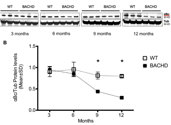
3.3.1 αB-crystallin levels decrease over time in BACHD mice ………..….... 53
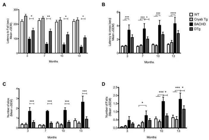
3.3.2 Over-expression of αBc in astrocytes improves motor performance in BACHD mice ………..………..……. 54
3.3.3 Expression of αBc in astrocytes improves strategy shifting in BACHD mice ………..… 59
3.3.4 αB-crystallin overexpression modulates levels of mHtt and inclusion body formation BACHD mice ……….………...…. 62
3.3.5 αB-crystallin overexpression prevents cortical and striatal neurodegeneration caused by the expression of mHtt in BACHD mice ………..….… 64
3.4 Discussion ………... 67
3.5 References ………... 70
3.6 Supplemental Data ………...………... 74
Chapter 4. Cell non-autonomous αB-crystallin potential antioxidant effects in HD 4.1 Introduction ………...………... 78
4.2 Materials and Methods ………...………... 79
4.3 Results ……….………..…... 82
4.3.1 BACHD mice do not induce up-regulation of Caspase-3 and BACHD astrocytes over-expressing αBc are more resistant to H2O2 – induced toxicity……….... 82
4.3.2 BACHD astrocytes affect neuronal carbonyl content by a non-cell autonomous mechanism ………..………… 86
4.3.3 Expression of many HD pathology related markers is not altered in the brains of
BACHD mice ………...…... 88
4.4 Discussion ……….... 89
4.5 References ………..………….………. 92
Chapter 5. Genetic deficiency of αB-crystallin does not aggravate the disease progression on the BACHD mouse model of Huntington’s disease 5.1 Introduction ……….………...……..… 96
5.2 Materials and Methods ………...….. 98
5.3 Results ……….………... 100
5.3.1 Abnormal genotype distribution in BACHD combined with αBc/HspB2 deficient mice ………...……… 100
5.3.2 αBc/HspB2 deficiency does not impair behavioral deficits in BACHD mice ..101
5.3.3 αBc/HspB2 deficiency does not alter HD related genes ………... 104
5.4 Discussion ……….……. 105
5.5 References ……….……….…….106
Chapter 6. General discussion and conclusions 6.1 Non–cell autonomous pathology in HD ……….………...…. 110
6.2 Prion-like spreading of mHtt pathology in HD ………..……….…... 113
6.3 Final Remarks ……….... 114
6.4 References ………..…..…….. 115
Chapter 7. Appendix ……….……….………. 119
7.1 αB-crystallin overexpression does not increase life span in the R6/2 mouse model of Huntington’s disease ………. 120
Acknowledgements
In many ways I consider this section to be the most important part of my thesis, as so many people directly and indirectly have helped and supported me through my five years of graduate school. Much of this time, regardless of the specifics of one’s project, is about learning to think like a scientist. While I have no doubt that I have grown tremendously as a scientist over the years, I strongly believe that I’ve grown even more as a human being. I have had the opportunity to work with incredible role models, to learn self-reliance and perseverance in the face of seemingly- constant failure, and to refine my ability to navigate a complex and often frustrating world without sacrificing my values or losing the pleasure I derive from simply being curious.
I must begin by thanking my supervisors, Tiago Outeiro, Paul Muchowski and Steven Finkbeiner for their support, guidance, confidence and for the opportunity to pursue a research project in your laboratory.
An enormous thank you to everyone I have met during this journey, and every single friend and labmate from my three distinct PhD laboratories as well as incredible people from neighboring labs I got the chance to meet and work with. You are all so special!
To my Science “outsider’s” friends goes a special “Thank you” for making me realize how lucky I am to have you around and to put all my experiments, which includes many failures, and some interesting findings, into perspective. My “University Girls”, my “Trivia team” and “Zumba team” were extreme- and uniquely important in this process.
I have had a great and special “partner in crime” for most part of this PhD, and I will be always grateful for the little surprises during crazy work weekends and long night shifts in the lab - a very special thank you goes to my favorite neighbor and my boyfriend Geno Moscetti. Thank you for your love, support and encouragment throughout my PhD.
I could not have done this without my amazing family, especially my parents and my brother, but also my great aunts and cousins. Your love and support throughout this process has been incredible. Thank you for your patience, holding your “saudades” during this time and more importantly, thank you for instilling the kind of values in me that led to this amazing opportunity! I dedicate this thesis to my beloved and unforgettable grandmothers, for being such a special inspiration during my whole life!
Abstract
Huntington's disease (HD) is caused by an expanded polyglutamine (polyQ) tract in the huntingtin (Htt) protein. The polyQ expansion increases the propensity of Htt to aggregate and accumulate, and manipulations that mitigate protein misfolding or facilitate the clearance of misfolded proteins are predicted to slow disease progression based on studies in HD models. αB-crystallin (αBc) or HspB5 is a well-characterized member of the small heat shock protein (sHsp) family that reduces mutant Htt (mHtt) aggregation and toxicity in vitro and in
Drosophila models of HD. Here, we determined if overexpressing or diminishing αBc levels in vivo modulates aggregation and the onset and progression of disease in a full-length mouse
model of HD known as the BACHD mice.Expression of sHsps in neurodegenerative disease predominantly occurs in non-neuronal cells, and in the brain, αBc is mainly found in astrocytes and oligodendrocytes. In Chapter 3, we show that directed αBc overexpression in astrocytes improves motor performance in rotarod and balance beam tests, and improves cognitive function in the BACHD mice. Improvement in behavioral deficits correlated with mitigation of neuropathological features commonly observed in HD. Interestingly, astrocytic αBc overexpression was neuroprotective against neuronal cell loss in BACHD brains, suggesting αBc might act in a non-cell-autonomous manner. At the protein level, αBc decreased the levels of soluble mHtt and decreased the size of mHtt inclusions in BACHD brain. These results support a model in which elevating astrocytic αBc confers neuroprotection through a putative non-cell-autonomous pathway that modulates mHtt aggregation and protein levels.
In Chapter 4, we attempted to elucidate the non-autonomous mechanism by which overexpressing astrocytic αBc confers neuroprotection in the progression of the disease of the BACHD mice. Several essential cellular pathways are impaired in neurological disorders such as HD. Among others, activation of caspase-3 pathways as well as formation of carbonyl species are oxidative stress hallmarks of HD pathology, and αBc has been reported to play a role in these mechanisms. Our studies, looking at mRNA and protein levels from BACHD mice as well as from primary cells (astrocytic and neuronal), found no significant differences in caspase-3 activation, and in expression of several HD markers, e.g., Glucose transporter 1 (Glut1), dopamine receptor 1 (DR1) and dopamine receptor 2 (DR2), brain-derived
neurotrophic factor (BDNF), in this disease context. Despite not finding a significant difference, when looking at the formation of protein carbonyls (derived from oxidative stress), we found a small trend towards a decrease of the levels of these species when αBc was over-expressed in astrocytes, both in brains and primary cultures of BACHD. These data may support the potential antioxidant effect of αBc in the context of HD pathology.
In Chapter 5, we hypothesized that αBc deficiency would accelerate HD disease severity and progression, as previously observed in a mouse model of Alzheimer’s Disease (AD). We tested this hypothesis by crossing mice null for αBc (and HspB2) to BACHD mice and monitoring behavioral and neuropathological symptoms of HD during disease progression. Our study showed that genetic deficiency of αBc (and HspB2) did not aggravate disease progression.
Taken together, the results presented in this thesis provide novel insight into the role of αBc and its function on essential cellular pathways in the context of HD. This work also supports the emerging significance of non-neuronal cells in chronic diseases such as HD suggesting adaptive and differential responses that might contribute to and/or provide a route to therapy of distinct aspects of neurodegeneration. Ultimately, this knowledge aims to promote and highlight the impact and role of sHsps, specifically αBc, in HD and related disorders in order to help the development of promising therapeutics for these devastating diseases.
Resumo
A doença de Huntington (HD) é causada por uma expansão de poliglutamina (polyQ) na proteína huntingtina (Htt). A expansão de polyQ aumenta a propensão da Htt para se agregar e acumular, e manipulações que mitiguem a mal-formação proteica ou que facilitem a degradação destas proteínas mal-conformadas são capazes de abrandar a progressão da doença em modelos de HD. αB-cristalina (αBc) ou HspB5 é uma proteína bem caracterizada membro da família das “small heat shock protein” (sHsp) que reduz a agregação e toxicidade da huntingtina mutante (mHtt) em estudos in vitro e em modelos Drosophila de HD. Aqui, determinámos se a sobre-expressão ou diminuição dos níveis da αBc in vivo foi capaz de
modelar a agregação e atrasar o aparecimento e a progressão da doença num modelo de longo comprimento genético de HD, os ratinhos BACHD. A expressão de sHsps em doenças neurodegenerativas ocorre predominantemente em células não-neuronais, e no cérebro, a αBc é principalmente encontrada em astrócitos e oligodendrócitos. No capítulo 3, demonstrámos que a sobre-expressão da αBc dirigida em astrócitos melhorou a performance motora nos testes de Rotarod e Balancim “Balance Beam” e melhorou a função cognitiva dos ratinhos BACHD. A melhora dos déficits de comportamento esteve relacionado com a mitigação de características neuropatológicas muito comuns observadas em HD. Interessantemente, a sobre-expressão astrocítica da αBc foi neuroprotectora contra a perda neuronal nos cérebros dos BACHD, sugerindo que αBc talvez esteja a actuar de uma forma celular não autónoma. Ao nível proteico, a αBc diminuíu os níveis de mHtt solúvel e diminuíu o tamanho das inclusoes de mHtt nos cérebros dos BACHD. Estes resultados apoiam um modelo no qual elevar a αBc astrocítica confere neuroprotecção através de um potencial mecanismo celular não-autónomo que modela os agregados e os níveis proteicos solúveis da mHtt.
No capítulo 4, tentámos elucidar o mecanismo celular não autónomo pelo qual a sobre-exressão da αBc confere neuroprotecção na progressão da doença dos ratinhos BACHD. Vários mecanismos celulares essenciais estão debilitados em doenças neurológicas como a HD. Entre muitos, a activação das vias da caspase-3, transporte e sinalização de glutamato e stress oxidativo são marcas moleculares comuns na patologia da HD, e a αBc tem um papel nestes mecanismos. Os nossos estudos baseados no mRNA e níveis proteicos dos BACHD e também em células primárias (astrócitos e neurónios), não mostraram uma
diferença significativa na activação da caspase-3, e na expressão do transportador-1 do glutamato neste contexto. No entanto, quando investigámos a formação de grupos proteicos carbonilo (derivados do stresse oxidativo), encontrámos uma pequena tendência para a diminuicão destas espécies de carbonil na presença da sobre-expressão astrocítica da αBc, tanto nos cérebros como nas culturas primárias dos BACHD. Estes dados apoiam o potencial efeito antioxidante da αBc no contexto da patologia de HD.
No capítulo 5, hipotetizámos que a deficiência em αBc iria acelerar a severidade e progressão da doença de HD, como tinha sido previamente observado num modelo de Alzheimer. Nós testámos esta hipótese, cruzando animais nulos para αBc (e para HspB2) com os ratinhos BACHD, e monitorizando sintomas comportamentais e neuropatológicos de HD durante a progressão da doença. O nosso estudo mostrou que a eliminação genética de αBc não agravou a progressão da doença de HD. Em suma, os resultados apresentados nesta tese fornecem uma nova perspectiva no papel da αBc e da sua função em mecanismos celulares essenciais no contexto de HD. Este trabalho também apoia o crescente papel e significado de células neuronais em doenças neurológicas como a HD, sugerindo respostas adaptativas e diferenciadas que podem contribuír e/ou providenciar um caminho terapêutico de aspectos distintos de neurodegeneração. Por último, este conhecimento tem como objectivos, promover e realçar o impacto das sHsps, especialmente a αBc em HD e outras doenças relacionadas para ajudar a desenvolver terapêuticas promissoras para estas doenças devastadoras.
List of abbreviations
αBc – alpha B-crystallin AD – Alzheimer’s disease
ALS - Amyotrophic Lateral Sclerosis
BDNF - brain-derived neurotrophic factor CNS – Central Nervous System
Cryab Tg – Cryab Transgene
DARPP-32 - dopamine and cyclic AMP-regulated protein DR1 – dopamine receptor 1
DR2 – dopamine receptor 2 DTg – double transgenic
EAE - Experimental autoimmune encephalomyelitis GFAP - glial fibrillary acidic protein
Glut1 – Glutamate receptor 1 GOF – gain of function
HD - Huntington's disease
Hsps - heat shock proteins HSR – Heat Shock Response Htt – huntingtin
HTT – huntingtin gene Htt – huntingtin protein
IB – inclusion bodies
iNOS – inducible nitric oxide synthase
LOF – loss of function
mHtt – mutant huntingtin
NeuN – Neuronal marker Anti-NeuN antibody
PD – Parkinson’s disease polyQ – polyglutamine
PTMs - Post-translational modifications
ROS – reactive oxygen species
RPE - retinal pigment epithelial sHsps - small heat shock proteins TNF-α- Tumor Necrosis factor alpha
Chapter 1.
_____________________________________
General Introduction
Chapter 1. General Introduction
1.
Huntington’s Disease
1.1. Historical BackgroundHuntington’s disease (HD), originally called simply 'chorea' or ‘hereditary chorea’, has been recognized as a disorder since the Middle Age. Nevertheless, it was just in 1872 that George Huntington, a young physician from Connecticut, published the first accurate description of the disease (1).
This neurological disease was described as dominantly inherited, affecting several family generations, and characterized by an excessive dance–like motor movements (the major motor abnormality occurring in HD), neuropsychological deficits, including cognitive and psychiatric impairment, and gradual deterioration that would ultimately lead to death (2). In 1983, a study conducted by a consortium of researchers – The US-Venezuela Huntington's Disease Collaborative Research Project – took place in the Lake Maracaibo region of Venezuela. This region was known for its high density of HD cases and significant consanguinity, linked a polymorphic DNA marker genetically to HD (3). More than a century passed since the first description of HD until the underlying genetic mutation was identified (4). Shortly, the polymorphic DNA marker consists of an abnormal expansion of a CAG trinucleotide repeat sequence in the coding region of the HD IT15 (Interesting Transcript 15) or HTT gene, on the short arm of chromosome 4 (4p16.3). The CAG repeat encodes a polymorphic stretch of glutamines (Q) within the N-terminal region of a high molecular weight protein, with approximately 3144 amino acids (~348 kDa), known as Huntingtin (Htt) (5). Since identification of the disease-causing gene, several approaches and a number of genetically modified cellular and animal models were developed to enable the study of the molecular underpinnings of HD, to possibly bring us closer to identify possible therapeutic strategies for this devastating disease. The first HD mouse model, called R6/2, was created in 1996 (6) and a year later, accumulation of mutant Htt (mHtt) in intranuclear inclusions was discovered in the brain of this HD mouse model and related to the pathogenic mechanism in
HD (7). Similar neuropathological features were also found in the brains of HD patients (8). Despite the intense effort on HD research since its first discovery, no clinical interventions tested in cellular and animal models to date have delayed the disease progression in humans. However, these models provide an invaluable tool for both investigating the underlying pathogenic processes and developing new effective therapies.
The details of the genetic aspects of HD and Htt - inclusion bodies formation will be described throughout this Chapter as well as some of the most promising therapeutic approaches for HD.
1.2. Epidemiology
The prevalence of HD, incorporating both genetic and clinical diagnostic studies, is approximately 10-13 in 100,000 people worldwide (9, 10), but it can vary to a great extent and can affect 700 per 100,000 individuals in some secluded regions of Venezuela (11). The age of onset is typically between 35 and 50 years old, however 15% of the cases can occur before 20 years old, called juvenile HD cases. The life expectancy of this devastating and progressive disease is typically 15-20 years from onset of symptoms till death (12).
1.3. The disease: Clinical presentation
HD is typically characterized by movement impairment (Chorea), cognitive dysfunction and decline and psychiatric disorder (13, 14). Chorea is the most common and aggressive manifestation of HD. It consists of involuntary, abnormal and irregular movements (hyperkinetic movements) that can affect most of the body, in particular the limbs, trunk and face (15). As the disease progresses, chorea often stabilizes and bradykinesia and rigidity start taking place, which is a very common feature in other neurodegenerative disorders such as Parkinson’s disease (PD) (16). Individuals in the last stages of HD suffer from gait disturbances which compromises their mobility, leading to falls (17). In the very last stage of the disease patients are not able to swallow and ultimately they are incapable of eating and also breathing (15, 18). Regarding cognitive dysfunction in HD, dementia is a common feature in the majority of the cases, varying from the designated subcortical dementia in
which individuals are not able to learn new motor skills and their visuospatial memory is also affected to a very broad of cognitive decline (13, 19). Lastly, the psychiatric disturbances in HD are extremely diverse, but the most typical in patients are irritability, apathy, anxiety, depression and impulsivity (13). HD patients also commonly suffer from progressive weight loss, alterations in sexual behavior, and disturbances in the wake-sleep cycle that occur very early in the course of the disease and may partly be explained by hypothalamic dysfunction (20). Suicide is also prevalent among HD patients (21, 22).
1.4. Genetic Factors and neuropathology of HD
As previously mentioned, HD is a devastating autosomal-dominant neurodegenerative disease caused by an abnormal expansion of the CAG triplet in the exon1 of the IT15 gene, which encodes a polyglutamine (polyQ) segment in the Htt protein (5, 14).
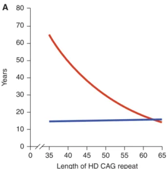
Unaffected alleles have CAG repeat size ranging from 10 to 35. With a median size of 18, the most common alleles in all populations contain repeats of 15-20 CAGs (15). HD-causing alleles are classified as: 1) Reduced- penetrance HD-HD-causing alleles with 36-39 CAG repeats. An individual with an allele in this range is at risk for HD but may not develop symptoms. 2) Full-penetrance HD-causing alleles with 40 or more CAG repeats. Alleles of this size are associated with development of HD with great certainty (3). Individuals with adult-onset HD usually have a CAG expansion from 40 to 55. In that range, ~50-70% of age of symptom onset appears to be explained by the length of the polyQ stretch, whereas the remaining variance is plausible to occur by other modifying genes and environmental factors (23–25). In longer polyQ stretches, the length of the polyQ stretch explains a greater proportion of age of symptom onset. Like other polyQ disorders, there is an inverse correlation between the length of CAG repeats and the age of onset. Thus, the longer the CAG stretch, the earlier symptoms typically appear (Figure 1). HD juvenile onset cases have CAG expansions greater than 60 that are often inherited from the father (paternal transmission) (3, 26).
Figure 1. The role of the CAG expansion in IT15 on HD pathogenesis. (A) Correlation of HD
CAG-repeat length with age at onset. Best-fit curves for age at neurological onset (red) and duration of disease from onset to death (blue), plotted against CAG- repeat length for the expanded mutant allele from HD patients. Age at onset is strongly correlated with the CAG-repeat length (r2 ¼ 0.54; p < 0.001), and duration of disease shows no correlation with the CAG-repeat length, suggesting that, after onset of HD, factors independent of the original trigger of pathogenesis determine the rate of progression. Adapted with permission from (5).
Although HD pathology has been observed in peripheral tissues (27) like other polyQ disorders, HD is predominantly a central nervous system (CNS) disorder, characterized by cell loss and atrophy (28), classified in five grades (0–4) designated in ascending order of severity (7, 29).
Within the brain, atrophy of the striatum (the largest component of the basal ganglia system) is the most prominent (17) (Figure 2), which primarily include a selective degeneration of medium spiny neurons (MSNs) (which represent about 96% of striatal neurons, and receive synaptic input primarily from the cortex) in the caudate nucleus and putamen, while aspiny interneurons (which represent around 2% of the striatal neurons) of the striatum are generally spared (30). Counts of neurons in the caudate nucleus revealed that 50% are lost in grade 1 and that 95% are lost in grade 4 (29).
Nevertheless, gradual MSN’s degeneration in the caudate nucleus and putamen appears to be distinctive. At early stages of HD, striato-globus pallidus (GP) internal segments (GPi) medium spiny projection neurons (which express substance P/gamma-aminobutyric acid (GABA) and projects to and inhibit GPi resulting in thalamus disinhibition and subsequent excitation of upper motor neurons in the motor areas of the cortex, which increase movement - direct pathway) are relatively spared. In contrast, striato-GP external segments (GPe) (which express enkephalins and projects to and inhibit GPe resulting in
subthalamic nucleus inhibition and consequent less inhibition of substantia nigra (SN) pars reticulata (SNr) and GPi which inhibits thalamus resulting in the inhibition of upper motor neurons; thus decreasing movement - indirect pathway) and striato-SNr (which express substance P/GABA and projects to and inhibit SNr - direct pathway) medium spiny projection neurons degenerate (31); loss of striato-GPe neurons was also demonstrated in presymptomatic HD (32). The early and relatively selective loss of GPe and striato-SNr neurons was suggested to be a plausible explanation for the chorea and oculomotor abnormalities that are prominent clinical features of early HD (33). However, in later stages of adult HD, both populations of striatal projection neurons are affected, with concomitant loss of markers of the direct pathway (substance P/GABA-containing neurons), including dopamine D1 receptors and substance P (31, 34). In addition, in juvenile cases of HD, degeneration of both direct and indirect pathway striatal neurons was observed (34). Thus, the degeneration of both the direct and indirect pathways was suggested to be functionally correlated with bradykinetic rigid phenotype observed in late stage and juvenile HD patients (34). Also, within the spared aspiny interneurons in HD there appears to be a differential level of vulnerability (35). In addition, non-striatal brain regions can also be affected, particularly in latter stages of the disease, and can include the SN, hippocampus, and mostly various regions of the cortex (36). Pyramidal neurons of deeper cortical layers appear to be more affected in HD, which seem to be correlated with dementia and personality changes in HD patients (17).
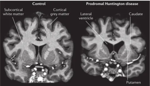
As previously discussed, brain atrophy and degeneration with marked neuronal cell loss in the regions mentioned above, putamen and caudate nucleus as well as cortex can occur before specific or severe motor symptoms appear in HD - asymptomatic patients – this period is defined by prodromal stage (Figure 2). At this point, affected patients can still perform usual functions although distress and discomfort may be felt (17).
Figure 2. Atrophy in prodromal HD shown using 7T MRI. Bilateral atrophy of the caudate and
putamen, and a concomitant increase in size of the fluid-filled lateral ventricle, is observed in the gene carrier compared with the control. This prodromal participant has only subtle signs and symptoms that are insufficient for diagnosing manifest HD. There are also subtle changes in the cortical grey matter and overall atrophy of subcortical white matter (26).
The reason why there is selective brain degeneration in HD remains unclear. However, it may be due to the role and importance of the Htt protein might have in the brain. The Htt polyQ tract begins at the 18 amino acid and is followed by a proline rich sequence (polyP) composed by 38 amino acids, which is considered to be important in Htt solubility (37). The first 17 amino acids of Htt are important for nuclear shuttling since they interact with TPR (translocated promoter region), a protein of the nuclear pore that actively exports proteins from the nucleus. Htt nuclear accumulation was observed when the first 17 amino acids were eliminated (38). Htt also contains multiple regions of so-called HEAT (Huntingtin, Elongation factor 3, a subunit of protein phosphatase 2ª nd the lipid kinase TOR) repeats, a sequence of ~40 amino acids named after the first four proteins in which it was discovered (39, 40) (Figure 3). Although the exact function of HEAT repeats is still unclear, studies have suggested that these domains play a role in a variety of interactions between proteins, including transportation in the cytoplasm and nucleus, microtubule dynamics and chromosome segregation (39).
Figure 3. Structure of the Htt full-length protein (A) and of the N-terminal exon 1 of the gene (B). Panel (A)
indicates the position of the three clusters of HEAT repeats, the caspase (black) and calpain (red) cleavage sites and some of the post-translational
modification sites along Htt protein (C214 palmitoylation, S421,
S434 phosphorylation, K444 acetylation). Panel (B) shows the N-terminal product of the exon 1 of the Htt gene, with the 17aa N-terminal tail (green) and the post-translational modifications reported to occur on the indicated residues (T3, S13, S16 phosphorylation; K6, K9, K15 ubiquitination and/or SUMOylation); Q(n) indicates the polyglutamine tract (red) and the upper insert shows the correlation between polyQ size and HD severity; the polyproline region downstream polyQ stretch is indicated in purple (41).
Htt mRNA and protein are ubiquitously expressed throughout development and in adulthood, in a variety of cells and peripheral tissues, and homogenously in the brain where it is higher expressed (8, 42), with a predominance of neuronal over glial expression (43–45). In the brain, Htt mRNA was detected in both grey and white matters (lowest expression levels), and the highest expression levels of Htt were found in the cortex (with differential expression between cortical layers), hippocampus, SN and cerebellum, followed by the striatum (43, 45). There are no differences in the distribution and levels of Htt mRNA between symptomatic HD patients and control individuals, except in the striatum where the intensity of labeling seems significantly reduced (42). However, presymptomatic HD brains revealed a striatal expression similar to controls and surviving striatal neurons in more advanced HD had an expression of Htt mRNA within normal limits. Therefore, HD brain selective degeneration does not seem to result from altered Htt mRNA expression (43). Wild-type (WT) Htt protein within neurons can be found mainly in the cytoplasm, but also in neurites and synapses. WT Htt can be associated with various organelles and structures, such as microtubules, plasma membrane, endosomal and endoplasmic compartments, clathrin-coated vesicles and mitochondria (46–48). Although mostly located in the cytoplasm, Htt is also detected in the nucleus (47). Due to its subcellular localization Htt interacts with numerous proteins involved
in gene expression, intracellular transport, signaling and metabolism, axonal trafficking of vesicles, transcriptional regulation and it also plays important functions in apoptosis and embryonic development (14, 49, 50). WT Htt is involved in embryonic development, since homozygous HTT locus knockout mice are lethally affected at early embryonic development stages (51–53). In contrast, the presence of one fully functional allele (at least 50% Htt expression) is compatible with life in humans and HD is not caused by a simple loss of function of the HTT gene (54). In fact, patients with Wolf-Hirschhorn syndrome, that have a partial deletion of chromosome 4 that comprises the CAG triplet repeats region and therefore contain only one copy of the HTT gene, are born and do not develop HD (3). WT Htt has an anti-apoptotic function in a gene and protein-dependent manner (55). In addition, WT Htt acts downstream mitochondrial cytochrome c release (a protein at the mitochondrial inter-membrane space that when released from mitochondria can bind to caspases to activate the cell death process), preventing the activation of both caspase-3 (56) and caspase-9 (57). Also, WT Htt physically interacts with active caspase-3 and inhibits its activity (58). Furthermore, Htt has been suggested to have a role in transcriptional regulation. For example, Htt was shown to regulate the genetic transcription of brain-derived neurotrophic factor protein (BDNF), a signaling neurotrophin essential for striatal neuronal survival and for the activity of cortico-striatal synapses, by binding and trapping in the cytoplasm (inhibit) the BDNF transcriptional repressor REST/NRSF (RE-1 silencing transcription factor/neuron- restrictive silencer factor) (14, 59–61). Apparently, WT Htt facilitates a de-repression of BDNF transcription but polyQ-expanded Htt does not (60). Thus, several studies have implicated that a reduction of BDNF at the transcriptional level is an important contributor to HD pathogenesis (60, 62).
In the disease context, the Htt protein with an expanded and abnormal polyQ, now denominated mutant Htt (mHtt) becomes aberrant and toxic that leads to neurodegeneration (6, 26). At the cellular level, HD neuropathology is characterized by the presence of neuritic, cytoplasmic and nuclear inclusions of mHtt (7, 8). mHtt accumulation and toxicity are associated with neuropathological damage characterized by brain atrophy and neuronal cell death, even at prodromal stages (Figure 2). How this polyQ-expanded mHtt causes selective and progressive dysfunction and neurodegeneration remains unclear. HD is one of at least nine genetic diseases caused by an abnormal expansion of a polyQ stretch, highlighting the
toxicity of polyQ-mediated protein misfolding in disease pathogenesis (54). However, increasing evidence points to the fundamental role that host protein context plays in pathogenesis. Expansion of its polyQ stretch beyond 36 repeats causes the protein to misfold and confers a toxic gain of function (GOF) (63). However, there is also evidence to support a contribution to disease pathogenesis from a loss of function of the WT allele (54). While a GOF is consistent with the autosomal dominant inheritance, there is also evidence to support a contribution to pathogenesis from loss of function (LOF) of the WT allele (14, 54). mHtt appears to have at least certain properties of WT Htt. In fact, mHtt can rescue the embryonic lethal phenotype seen in Htt-null knockout mice (64, 65). However, it is still not clear if neuronal cytotoxicity in HD is due to a LOF or GOF of Htt or actually both. Additionally, WT Htt has been implicated in a multitude of other cellular processes, including intracellular signaling, metabolism, gene expression, and intracellular transport, but its precise native function is still unknown (20, 21). Post-translational modifications (PTMs), in particular, greatly influence the toxicity of several different disease-associated polyQ proteins, including Htt (66). For instance, 4 several PTMs localized to the N-terminal 17 amino acids of Htt can modulate its toxicity (37, 67, 68). These residues, immediately preceding the polyQ stretch, are well positioned to alter the expansion’s propensity to cause protein misfolding via both direct and indirect means. However, other Htt PTMs farther away from the polyQ repeat region by primary sequence also have been reported to modify mHtt toxicity (49, 69).
The proteolytic cleavage of mHtt into N-terminal fragments containing the polyQ stretch and their subsequent translocation to the nucleus and formation of intranuclear aggregates (8, 70) is an hallmark of the disease detected in post-mortem HD human brains. The aggregates can be found before the onset of the first symptoms (7), and the rate of aggregate formation was shown to correlate with the length of the polyQ repeat (71, 72). However, the toxicity of mHtt nuclear inclusions remains controversial, since their formation is correlated with disease progression, but it is not directly linked with neuronal degeneration as initially thought. In fact, several cellular studies, including many from our laboratory, have demonstrated that large mHtt inclusions can be protective against mHtt-induced toxicity and increase cell survival (8, 73, 74). Interestingly, exposure of mHtt-transfected striatal neurons to conditions in which nuclear localization of mHtt was blocked and consequently, suppressing its ability to form intranuclear inclusions, resulted in increased cell death,
suggesting that the formation of intranuclear inclusions might reflect a cellular mechanism to protect against mHtt-induced cell death (74). On the other hand, the decrease of proteolytic cleavage of mHtt reduced its toxicity and slowed disease progression (75, 76). In addition, expression of smaller N-terminal mHtt fragments resulted in increased toxicity in cultured cells (77) and transgenic animals (8, 78, 79), when compared to the full-length mHtt expression with the same polyQ expansion. These findings suggested that the susceptibility to neuronal death is greater with decreased protein length and increased polyQ size. Therefore, several HD models have been generated to mimic the neuropathological features that occur in humans. These models express only a fragment or a full-length (fl) mHtt to study the cellular and molecular mechanisms of mHtt-induce neurodegeneration.
1.5. HD models
A wide variety of species, including the invertebrate worm (Caenorhabditis elegans) and fly (Drosophila melanogaster) models, non-mammalian species as zebrafish (Danio rerio) and mammals, such as mouse (Mus musculus) and rat (Rattus norvegicus), have also been genetically engineered to express the HD mutation (80–83). A large number of mouse models of HD that show different degrees of similarity to the human condition have been generated. Models that express either truncated, full-length human or mouse mHtt display significant phenotypic differences that may be caused by the influence of the protein context, mouse strain, or regulatory sequences between the mouse and human HTT genes (80). To overcome these problems, a group of researchers also considered the generation of large HD genetic models such as sheep (84), mini-pig (85), and the non-human primate (86, 87). With their size, organ capacity, and physiology resembling in several aspects that of humans, these models may be well suited for preclinical trials and long-term safety studies. However, in some cases, ethical concerns have been raised. In this section, we describe especially the most common HD mouse models that have particularly enlightened in the search for targets and for compounds capable of interfering with mHtt toxicity.
1.5.1. Transgenic models
In transgenic mouse models, full-length or a fragment of the mutant HTT gene is inserted randomly into the mouse genome, leading to the expression of a mutant protein in addition to the endogenous Htt. Thus, transgenic mouse models of HD express N- terminal mHtt fragments or full-length human HTT gene with an expanded polyQ tract (28).
1.5.1.1. N-terminal huntingtin transgenic models
The first transgenic mice model of HD, named R6/2 mice, include the insertion of a fragment of the human HTT gene. R6/2 mice contain a mutant N-terminal segment of the exon 1 of the human HTT gene with ∼144-150 CAG repeats (6). These mice display choreiform-like movements and mHtt inclusion formation as early as at 4-5 weeks. At 6-8 weeks their physical appearance deteriorates and motor symptoms become very severe, followed by an early death around 12-14 weeks. Despite their severe motor impairment and brain atrophy, these mice display fewer neuronal death when compared to human HD patients (6, 79, 81). Due to their high number of CAG repeats, R6/2 mice seem to recapitulate the disease features of juvenile cases of HD (88). In parallel, another transgenic model was developed - the R6/1 model of HD. This mouse model expresses a truncated HTT gene with around 115 CAG repeats, and exhibits a progressive pathology and lower expression of the mutant transgene. A marked decline in rotarod performance and other locomotor activities is observed at 13-20 weeks, correlating with the numbers of striatal neurons exhibiting intranuclear inclusions of mHtt, and with death occurring within 4-5 months of age (8, 28). The R6/1 model is not used as often as the R6/2 model and is therefore less well understood.
The N171-82Q mouse model of HD contains a longer N-terminal fragment of mHtt (exon 1 and exon 2) with 82 CAG repeats. Its lifespan is ∼17-20 weeks with HD-like symptoms beginning at 10-12 weeks of age. In these mice, neuropathological features are more similar to human HD, therefore brain atrophy and neuronal loss is more prominent and seems more selective for the striatum (78, 81). HTT is driven from a prion promoter, restricting expression solely to neurons and not glia. But in human HD, Htt is expressed in both neurons and glia (80).
1.5.1.2 Full-lenght huntingtin transgenic models
Transgenic mice expressing full-length htt have in some cases been more successful than N-terminal fragment models regarding neuronal deterioration and loss and the capability to recapitulate more closely the sequence of events leading to HD. Two genomic transgenic models expressing full-length mHtt from the human genomic locus on a yeast artificial chromosome (YAC) (64, 89, 90) or on a bacterial artificial chromosome (BAC) (91) were generated. A series of YAC transgenic models of HD expressing full-length human mHtt with 18, 46, 72, and 128 CAG repeats (i.e., YAC18, YAC46, YAC72, and YAC128) were created after microinjection of YAC DNA construct into the friend virus B strain (FVB/N) pronuclei and maintained on the inbred mouse FVB/N background strain; in contrast to the C57BL/6 background strain common to most HD mouse models, the FVB/N strain shows higher neuronal loss when exposed to excitotoxic stress after injection of kainic- or quinolinic acid (QA) (64, 89, 90). YAC128 mice are the latest of the series and exhibit by far the most robust phenotypes among all the YAC models; therefore, YAC128 is used as a preclinical model in HD (89). At the protein level, the YAC128 line expresses human mHtt at about 75% of the level of the endogenous murine Hdh (80). YAC128 mice exhibit hyperactivity at 2 months of age and hypoactivity at 8–12 months of age. They also exhibit rotarod deficits at 4 months of age, which become more prominent at 6 months of age (92–94). mHtt nuclear localization is detected in the striatum at 1-2 months and in the cortex and hippocampus at 3 months of age. Nuclear inclusions are detected only at 18 months in the striatum. As in humans, selective atrophy in the striatum and cortex, but not in cerebellum is detected (93).
BACHD, the model we have used in this study, is a more recent generated transgenic mouse model of HD and also maintained in the FVB/N background strain. Under the control of endogenous HTT regulatory machinery on the BAC, the 97 polyQ repeat within full length mHtt in BACHD mice is encoded by a mixed CAA-CAG repeat, which is stable in both the germline and somatic tissues including the cortex and striatum at the onset of neuropathology (91). The BACHD mice exhibit progressive motor deficits and selective late-onset neuropathology without somatic repeat instability in the aged brain. BACHD mice exhibit mild rotarod deficits at 2 months, with progressive deficits and hypoactivity at 6 months of age. At 12 months of age, cortical and striatal atrophy is detected. However, unlike previous
full-length mHtt mouse models, BACHD mice do not show early and diffuse mHtt inclusions (91, 95). By 12 months of age, BACHD brains have only a few small aggregates predominantly in the neuropil in the cortex and small aggregates in the striatum (82), suggesting that diffuse nuclear accumulation of aggregated mHtt in striatum and cortex is not necessarily associated with the slowly progressive and selective pathological process in the BACHD mice. Importantly, a relatively steady-state level of predominantly full-length mHtt and a small amount of mHtt N-terminal fragments, present in both the nucleus and cytoplasm, may be responsible for the onset of neuropathology (91, 96). Thus, BACHD mice represent a robust in vivo paradigm to study HD pathogenesis and treatment.
1.5.2. Knock-in models
Knock-in HD models were generated by targeting an expanded polyQ repeat and/or adjacent human mHtt exon 1 sequences (including the polyP region) to replace the corresponding sequences in the endogenous murine HD homologue gene (Hdh) (97). Therefore, differently from the transgenic model, the mHtt in the Knock-in models is expressed from the endogenous Hdh locus in a similar manner to the expression in HD patients, making this model considered one of the most precise genetic HD mouse models (98). When compared to N- terminal mHtt fragments expressing models, Hdh knock-in mice display slow progression and moderately mild phenotypes, and their lifespan is usually normal. This mouse model is important to evaluate early pathological processes induced by mHtt in humans (95). The first knock-in models generated were disappointing to the community because their extended stretch of 50 or 80 CAG repeats into the endogenous mouse Hdh gene (HdhQ50; HdhQ80) showed no behavioural phenotypes or abnormalities, or even mHtt aggregates (99, 100). For these reasons, other knock-in models were generated to better represent the human pathology.
1.5.2.1. HdhQ111
The HdhQ111 model is a knock-in mouse model of HD with an insertion of a chimeric murine Hdh/human mHtt exon 1 into the endogenous Hdh locus; the human mHtt portion
includes 111 CAG repeats and a polyP region (88). The behavioral phenotypes of HdhQ111 are very mild and slowly progressive. Rotarod, clasping or open field abnormalities were not detected in heterozygous or homozygous HdhQ111 mice until 17 months of age, and gait abnormalities were detected only at 24 months of age (101). HdhQ111 homozygous mice show selective and progressive accumulation of nuclear mHtt at 2.5 months of age, nuclear inclusion formation at 10 months of age, and reactive gliosis in the striatum at 24 months of age (101).
1.5.2.2. CAG140
The CAG140 HD knock-in mice model was generated by replacing the endogenous murine Hdh exon 1 with a chimeric mouse and human exon 1 with 140 CAG repeats (102). CAG140 HD mice display early hyperactivity at 1 month of age, followed by hypoactivity at 4 months of age, gait abnormality at 12 months of age, with nuclear mHtt micro-aggregates in the striatum and cortex, and nuclear inclusions in the striatum at 4 months of age and in the cortex at 6 months (103). However, nuclear micro-aggregates were also observed at 6 months of age in the cerebellum, a relatively spared region in human HD brain, but presented no cell loss or brain atrophy (95, 103).
1.5.2.3. Hdh(CAG)150
Hdh(CAG)150 HD knock-in mice was developed by a replacement of the short CAG repeat in the murine Hdh exon 1 with a stretch of 150 CAG repeats (104). Homozygous Hdh(CAG) 150 mice revealed several slowly progressive motor abnormalities (105, 106). Homozygous HdhCAG(150) mice display progressive rotarod deficits at 18 months of age and mHtt aggregates in the striatum and hippocampus at 6 months and widespread in the brain at 10 months of age (107). Striatal neuronal loss is observed at 100 week-old (105).
1.6. Diagnosis, Screening and Prevention of HD
HD is easily diagnosed based on the appearance of physical symptoms specific to the disease. As previously described, the motor symptoms are the first clinical features to be diagnosed, specifically abrupt, excessive and random timing of involuntary movement (26). Characterized by its dominant inheritance, the available genetic test for HD consists of a simple blood examination to confirm the number of CAG repeats in the HTT gene. However, a positive result is not considered a differential diagnosis since it can be obtained before the onset of symptoms. This test is very important to rule out the disease and also because its result may dramatically influence personal and family planning decisions. Longitudinal analyses show a consistent increase in the prevalence of HD over the past two decades, coinciding with wider availability of the genetic test. Since HD is an autosomal dominantly transmitted disorder, there is a 50 % chance of children from an affected individual inheriting the disease (15, 26). Prenatal testing can also be performed using non-invasive techniques to determine the carrier status of the fetus. The progression of the disease can be measured based on the unified HD rating scale (UHDRS) that characterizes the relevant clinical features of HD (108). Medical functional imaging, such as fMRI and PET, are very important in the early detection of HD since they can show changes in the brain activity before symptoms onset in addition to monitoring the disease progression (15, 26).
HD is devastating to patients and their families and unfortunately, at the present, there is no effective treatment to stop or slow the disease progression.
The current available therapies act mainly on motor symptoms in order to improve patients’ quality of life. These therapies are mainly focused to treat movement disorders such as chorea and dystonia, as well as gait, speech, swallowing and fine motor tasks (16). The only drug currently approved and specifically licensed by the US Food and Drug Administration (FDA) in 2008 to treat chorea in HD patients is tetrabenazine (109). As a synaptic vesicular amine transporter inhibitor, tetrabenazine acts by reducing the quantity of dopamine released from vesicles in the brain by reversibly inhibiting the monoamine transporter type 2 (VMAT2) and therefore depleting central monoamines (110). Tetrabenazine is also responsible for reducing dopamine by blocking dopamine receptors. Unfortunately, this drug is also associated with some adverse side effects, namely, sedation
and somnolence, agitation, depression, akathisia, anxiety and hyperkinesia (108, 110). Several studies are underway to investigate other potential treatments for chorea in patients with HD, including pallidal deep-brain stimulation (111, 112) and a deuterated tetrabenazine molecule (111, 113, 114). In fact, many reviews have emphasized the weak evidence supporting any other pharmacological intervention in the treatment of HD (115, 116).
In an effort to diminish therapeutic nihilism in the absence of established and accurately proven treatments, a series of algorithms for the treatment of chorea, irritability and obsessive–compulsive behaviors were reported in 2011 by an international group based on surveys of HD experts (117). Until better treatments arise, the clinicians must adopt an attitude that therapeutics providing benefit to patients without HD, who have neuropsychiatric symptoms, may be also expected to help HD patients that share the same symptoms. As an example, some pharmacological approaches to treat or ameliorate the neuropsychiatric symptoms in HD include: olanzapine for weight loss, selective serotonin reuptake inhibitors and mirtazapine for depression, antipsychotic drugs for psychiatric and behavioral aspects of this pathology. Although these pharmacological compounds are not specific for HD (116), clinicians should proceed thoughtfully to optimize the patients’ quality of life with available medications and supportive therapies.
1.7. Novel Strategies for Therapeutic Intervention in HD
In this section I briefly describe new possible and promising therapeutic targets as well as potential disease-modifying therapies underlying the pathobiology of mHtt, that are currently being studied. Figure 4 highlights some of the current and potential disease-modifying strategies for HD. Special emphasis will be given to chaperone-mediated folding and aggregation, and also pathways involving protein homeostasis, since it is the central topic for this thesis work.
Figure 4. Current priority preclinical therapeutic targets under investigation for Huntington disease. Several targets have been identified for potential exploitation in therapy, with strategies that
include Htt lowering and immunomodulation (118) . Ac, acetyl group; ASO, antisense oligonucleotide; BDNF, brain-derived neurotrophic factor; CB2, cannabinoid receptor 2; EAAT2, excitatory amino acid transporter 2; GM1, monosialotetrahexosylganglioside; HDAC4, histone deacetylase 4; JNK, c-Jun N-terminal kinase (MAPK8, MAPK9 and MAPK10); KMO, kynurenine 3-monooxygenase; MAPK, mitogen-activated protein kinase; NMDA, N-methyl-d-aspartate; P, phosphate group; p38, mitogen-activated protein kinase (MAPK11, MAPK12, MAPK13 and MAPK14); PDE, phosphodiesterase; PPAR-γ, peroxisome proliferator- activated receptor-γ; RNAi, RNA interference; Su, sumoyl group; TrkB, tyrosine receptor kinase B. Figure adapted from (26).
1.7.1. Protein Homeostasis
Protein homeostasis is essential for cell function and survival, because proteins are involved in all aspects of cellular function varying from cell metabolism and cell division to the cellular response to environmental stressors. Protein homeostasis is tightly regulated by
the synthesis, folding, trafficking and clearance of proteins, all of which act in an orchestrated manner to ensure proteome stability (119). The protein quality control system is composed of stress response pathways, which take action whenever the proteome is challenged by environmental or physiological stress. Aging, however, damages the proteome, and such damage is thought to be associated with age-related diseases. In the next sections I will discuss the different cellular processes that define the protein quality control system and focus on their role in protein conformational diseases.
1.7.2. Protein Quality Control Systems
Dysregulation of protein quality control is a key event in HD pathogenesis. The accumulation of misfolded proteins is a hallmark of many human diseases, including several incurable neurological disorders, such as HD. In HD, expansion of the polyQ stretch within the first exon of the Htt protein leads to Htt misfolding, aberrant protein aggregation, and disease onset (50). The accumulation of aberrant mHtt and other polyQ disease proteins form insoluble aggregates and, as mentioned above, the role that aggregation has in the pathogenesis of polyQ diseases remains contentious (50, 73, 122). Neurons, as post-mitotic cells with a high metabolic activity, are extremely sensitive to protein accumulation and the presence of aberrant proteins lead to neuronal cell death. Thus, the post-mortem confirmation of the presence of protein aggregates in the brain of patients with neurodegenerative diseases such as HD emphasizes the importance of protein turnover in neuronal homeostasis. To prevent the deleterious process of misfolded proteins accumulation, cells have specific quality control mechanisms that include the molecular chaperones, including the heat shock proteins (Hsps), the ubiquitin-proteasome system (UPS) activation, autophagy-mediated protein degradation. While chaperones help other proteins in the folding process, refolding misfolded proteins to their native conformation, the UPS and autophagy are responsible for the targeting and degradation of unfolded or mutant proteins (123). This degradation mechanism is critical to clear the cytosolic space from these toxic proteins that might disturb the essential cellular processes, and also to recycle amino acids (119, 123). Impairment of the cell quality control mechanisms is commonly associated with neurodegenerative diseases such as HD, and therefore the focus of intensive research in the field (119).
1.7.2.1 Heat Shock Proteins (Hsps)
Hsps, are able to bind specifically and non-covalently to interactive proteins surfaces that are transiently exposed during cellular processes (124). The controlled binding and release of substrate proteins facilitates the correct fate of the protein in the cell, whether this be correct folding assembly into oligomers, transport to a specific subcellular compartment, membrane translocation, or even degradation (125). Hsps protect the cells against a variety of stresses by preventing abnormal protein aggregation and by keeping proteins in a state competent for either refolding or degradation (121). Some chaperones interact with a variety of polypetide chains, whereas other act on specific targets via substrate specific binding domains. For example the 70 kDa Hsps recognize hydrophobic protein surfaces or amino acid residues that are generally exposed by non-native proteins and newly synthesized polypeptide chains (126). The ClpX/Hsp100 family can illustrate an example of specificity of some molecular chaperones. Proteins of this family form ring-like subunit architectures and are able to disassemble multimeric or aggregated forms of certain proteins in an ATP dependent fashion (127). Two members of this family (Clp X and ClpaA) recognize specific C-terminal sequences on target proteins. This property has been extensively characterized for the interaction between E.coli Clp X and the transposase of phage Mu (MuA protein) (128). Molecular chaperones were originally termed Hsps due to their initial discovery in Drosophila melanogaster cells exposed to elevated temperatures. Despite the name, the Hsps are also known to be induced by a wide variety of environmental or metabolic stresses including anoxia, ischemia and viral agents (121, 129). Hsps can be divided into ATP dependent and ATP independent families as well as being subdivided into families according to their molecular weight. The ATP-dependent Hsps require ATP hydrolysis to facilitate the folding process (129).
1.7.2.1 Hsp families
The name of the members of the Hsp family is derived from the molecular weight of the main representative protein (Table 1) (121, 130). Additionally, the nomenclature is based on systematic gene symbols that have been assigned by the HUGO Gene Nomenclature Committee (HGNC) (131).
Table 1. Major families of Hsps
Different Hsps families recognize structural features that are specific to immature and unstable proteins at different of folding and unfolding. The Hsp70 chaperones, rin-shaped chaperonins and Hsp90 are involved successively more advanced stages of protein folding and function, whereas the small Hsps and Hsp100 act predominantly during stress (132).
1.7.2.3. Small heat shock proteins (sHsps) family
Small heat shock proteins (sHsps) have molecular weights between 12 and 43 kDa, distinguishing them in size from larger Hsps (133, 134). There are ten human sHsps: HSPB1– HSPB10 (134, 135). They share common structural characteristics, including a highly conserved 90 amino acid long Hsp20 domain, usually referred to as the alpha-crystallin (α-crystallin) domain, and the capacity to form large dynamic oligomers (136). Thus, sHsps can be found as homo- or heteromeric complexes, consisting of 2-40 subunits (137). The α-crystallin domain consists of two layers of three and five anti-parallel strands, respectively, connected by a short inter-domain loop, forming a β-sheet (138). The α-crystallin domains of two monomers interact tightly to form dimeric building block of the sHsp oligomers. The terminal region contains α-helical components and is variable in structure (139). The N-terminal of many of the sHps contains a small prolinephenylalanine-rich region with one or two WD/EDF motifs. This region seems to be important in oligomeric complex formation and also for chaperone activity (140). The C-terminal tail is highly motile and flexible, and is also involved in stabilizing the oligomeric structure through contacts between a conserved motif in the C-terminal region and a hydrophobic patch in the α-crystallin domain of a neighboring subunit (139).
As chaperone proteins, sHsps bind misfolded proteins and prevent them from aggregating. However, they are unable to actively refold the protein themselves because in contrast to other Hsps families, the sHsps do not require ATPase activity for their chaperone function (141). Instead, sHsps sequester the misfolded proteins within the cell, forming large dynamic oligomeric structures that are able to bind several non-native proteins per oligomeric complex, to prevent aggregation until a large Hsp, as Hsp70 or Hsp40, can assist in refolding (134, 137, 142) (Figure 5). Figure 5. Mechanism of sHsp chaperone interaction with amorphously aggregating target proteins sHsps selectively interact with and stabilize slowly aggregating,
intermediately folded target proteins on their amorphous off-folding
pathway. The
intermediates (I2 ) are relatively disordered in structurewith only some elements of secondary structure in place.The dynamic nature of the
equilibrium between the monomeric and aggregated I2 species facilitates interaction with the similarly dynamic sHsp. There is some evidence that the dissociated, dimeric form of the sHsp is the chaperone-active species which interacts with I2 and is subsequently sequestered into a high-mass complex containing both proteins. It is possible to recover natively folded target protein (N) via the action of another chaperone, for example by Hsp70 action coupled to ATP hydrolysis (124, 137).
Historically, sHsps have been extensively studied in the context of being intracellular molecular chaperones. However, recent studies looking at the role of sHsps in neurological diseases have demonstrated that despite being found in areas of damage, sHsps show therapeutic effects by transgenic overexpression and exogenous administration in various experimental disease paradigms (143). In addition to acting as molecular chaperones and preventing protein aggregation, sHsps have been suggested to be strongly involved in
apoptosis inhibition, anti-inflammation, organization of the cytoskeleton (134, 137). The most intensively studied sHsps are the Hsp27 or HSPB1 and αB-crystallin (αBc) or HSPB5 (144).
1.7.2.4. αB-crystallin/HSPB5
αB-crystallin (αBc) or HSPB5 is a ubiquitously expressed protein and is a major constituent of the eye lens (145), and in the brain is mainly expressed in astrocytes and oligodendrocytes (146).
αBc acts as a molecular chaperone by preventing the aggregation of unfolded proteins and is know to be a multi-tasking chaperone by playing a role in several cellular functions: protein turnover, redox homeostasis, apoptosis and inflammation inhibition, cytoskeletal assembly and architecture (147). It is associated with cytoskeleton proteins, for example, αBc modulates the assembly of the intermediate filament protein vimentin, of the glial fibrillary acidic protein (GFAP) and stabilizes actin filaments in a phosphorylation-dependent manner (148). In addition, αBc also associates with tubulin, and microtubule-associated protein (MAP) and this is mediated through the α-crystallin domain (149, 150). αBc might play a role on proteosome degradation, UPS pathway, since it has been found to interact with FBX4, and F-box-containing protein that is a component of the ubiquitin-protein isopeptide ligase SCF (SKP1/CUL1/F-box). This interaction is dependent on phosphorylation of αBc (151). The chaperone activity or cytoprotective activity of αBc can be regulated by phosphorylation. Interestingly, in the brain, some of the αBc isolated from the proteinaceous aggregates of patients with amyloid plaques and Lewy bodies is phosphorylated (152). However, it remains unclear whether there is a preference for the phosphorylated forms of αBc to interact with the proteins associated with these deposits (153).
αBc has three phosphorylation sites, serines 59, 45, and 19 (153). The triple phosphomimic of αBc (i.e. S19D, S45D, S59D αBc or 3D αBc) is a more effective chaperone in preventing fibril formation by the natively unstructured target proteins k-casein and α-synuclein compared to the WT protein, but is less effective against the ccb-Trp peptide which adopts a well-structured coiled-coil configuration in its native state prior to fibril formation (154).
At the least two pathways are implicated in αBc phosphorylation: the MAPKAPK2 kinases phosphorylate serine 59 whereas serine 45 may be regulated by p42/p44 MAPK. The kinase involved is the phosphorylation of serine 19 is still unknown (140) (Figure 6).
Figure 6. Domain structure and biochemical properties of αB-crystallin (αBc).
A) Light box - conserved region in N-terminus; black box - α-cystallin domain; grey box – WDPF domain; ^^^ - flexible domain; P – phosphorylated serine residues. Arrows indicate positions of point mutations that are responsible for pathologies. B) Biochemical properties of sHsps. Large oligomeric structures are favored under conditions of stress and associate with unfolded proteins. Phosphorylation decreases the size of the oligomers. Large non-phosphorylated oligomers of αBc (and Hsp27) (>300 KDa) can protect cells through their chaperone activity, and small oligomers may act at the level of F-actin polymerization/depolymerization (133).
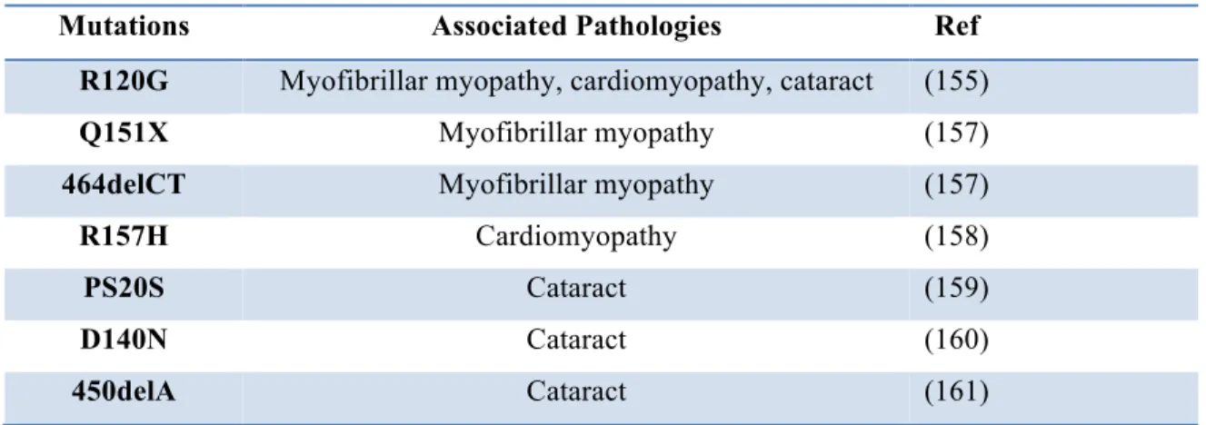
Mutations in the sHsps, namely in αBc, have been reported in a number of human diseases. Some of these mutations have been found as genes responsible for human degenerative myopathy, cardiomyopathy and congenital cataracts (Table 2). Regarding the myopathies, recent studies have revealed the importance of αBc towards desmin network (148, 155). The majority of mutations found in αBc (and other sHsps) are contained in the conserved α-crystallin domain, suggesting that oligomerization and chaperone function of αBc is impaired (156).