Universidade de Lisboa
Faculdade de Farmácia
Distinct strategies to overcome severe forms of PKU:
The p.G46S as a model to identify small molecules modulators of protein
aggregation and evaluation of an enzyme replacement approach using a
nanoparticulate system
Ana Carolina Ramos Costa
Dissertação orientada pela Professora Doutora Ana Paula Costa dos Santos
Peralta Leandro e, coorientada pelo Doutor João Paulo Travassos Leandro
Mestrado em Ciências Biofarmacêuticas
The studies presented in this thesis were performed at Research Institute for Medicines and Pharmaceutical Sciences (iMed.ULisboa), Faculty of Pharmacy, University of Lisbon under the scientific supervision of Professor Ana Paula Costa dos Santos Peralta Leandro and Professor João Paulo Travassos Leandro
TABLE OF CONTENTS
Acknowledgements... IX
Abstract... XI
Resumo... XIII
Abbreviations... XV
Chapter I • INTRODUCTION... 1
1. New approaches to the treatment of Inborn Errors of Metabolism ... 3
1.1. Mutation Specific Therapy ... 4
1.1.1. Premature Termination Codon Read-through Therapy ... 4
1.2. Non-Mutation Specific Therapy ... 10
2. Phenylketonuria ... 13
2.1. Metabolic Pathway and Clinical Phenotype ... 13
2.2. From Gene to Protein ... 15
2.2.2. PAH Protein ... 16
2.3. PKU Treatment ... 22
Chapter II • AIMS
... 27
Chapter III • MATERIAL AND METHODS
... 33
1. Materials ... 35 1.1. Reagents ... 35 1.2. Expression vectors ... 35 1.2.1. pTrcHis ... 35 1.2.2. pMAL-c2X ... 35 1.2.3. pEXP51 ... 36
1.3. Bacterial Strains and Eukaryotic Cell Lines ... 36
1.4. Molecular chaperones ... 36
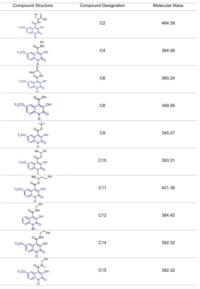
1.5. Tested compounds ... 37
2. Methods ... 40
2.1. Expression and Purification of hPAH-G46S ... 40
2.2. Expression and Purification of hPAHwt ... 41
2.3. SDS-PAGE Protein analysis ... 42
2.4. Protein Quantification (Bradford/BCA Methods) ... 42
2.5. FXa Inhibition Assay ... 43
2.6. hPAH-G46S Aggregation Assay ... 43
2.7. Nanoparticles Preparation ... 44
2.8. Nanoparticles Characterization ... 44
2.9. Eukaryotic Cell Culture ... 45
2.10. Co-immunoprecipitation Assays ... 46
2.11. Immunocytochemistry Assays ... 47
2.12. Western Blot Analysis ... 47
Chapter IV • RESULTS AND DISCUSSION
... 51
1. Effect of 3HQs on p.G46S aggregation ... 53
1.1. In vitro studies ... 53
1.2. In cellulo studies ... 60
2. Studies of hPAH-G46S and molecular chaperone interactions ... 64
2.1. Optimization assays ... 64
2.2. Co-Immunoprecipitation Assays ... 66
3. In cellulo evaluation of hPAHwt loaded Nanoparticles ... 67
3.1. Expression and Purification of hPAHwt ... 67
3.2. Properties of Nano Encapsulated hPAHwt ... 69
3.3. Evaluation of hPAHwt content and function in different cellular fractions ... 70
3.4. Stability of hPAH activity ... 73
Chapter V • CONCLUSION AND FUTURE PERSPECTIVES
... 77
REFERENCES
... 83
Acknowledgements
Em primeiro lugar gostaria de agradecer à Professora Doutora Ana Paula Costa dos Santos Peralta Leandro por ter aceite orientar a minha tese de mestrado, por me ter recebido de braços abertos no seu grupo de investigação e por me proporcionar todas as condições necessárias à realização deste trabalho. Obrigado por toda a partilha de conhecimento científico ao longo deste ano, em especial, pela oportunidade de participar no 13º Simpósio Internacional da Sociedade Portuguesa de Doenças Metabólicas. A sua genuína paixão pela ciência aliada ao seu carácter humano permitiram que ao longo deste ano de trabalho pudesse contar sempre com a sua amizade, disponibilidade e preocupação, pelo que me sinto eternamente agradecida.
Ao Doutor João Paulo Travassos Leandro, co-orientador desta tese, por toda a sua dedicação e trabalho desenvolvido ao longo destes anos em torno da PKU, que serviu de ponto de partida para a realização deste trabalho. Embora longe, esteve sempre presente e disponível. À Professora Doutora Lídia Maria Diogo Gonçalves, um especial agradecimento pela sua amizade, profissionalismo e partilha de rigor e conhecimento científico. Por me ter recebido no laboratório Nano2B, pela sua contribuição e acompanhamento no desenrolar de grande parte do trabalho prático e pela sua incansável disponibilidade em me receber sempre com um sorriso.
À Raquel por todo o apoio, companheirismo e todos os ensinamentos nos primeiros dias de laboratório e à Hana por ser sempre tão prestável. À Ana e à Dona Lurdes pela boa disposição e pelo apoio e preocupação em disponibilizar todo o material necessário à execução do trabalho.
A toda a minha família, em especial aos meus Pais, a quem devo tudo o que sou e a quem agradeço o apoio incondicional desde sempre. À minha irmã e ao meu irmão por estarem sempre ao meu lado em todos os momentos. Obrigada por tudo!
Ao Diogo, agradeço por me desafiar diariamente a ser mais e melhor a todos os níveis e por tudo o que compartilhamos ao longo destes anos, para os quais não há palavras suficientes.
Abstract
This work explores distinct strategies to overcome phenylketonuria (PKU; OMIM 261600), the most common autosomal recessive disorder of amino acid metabolism caused by a deficiency of the hepatic human phenylalanine hydroxylase enzyme (hPAH; EC 1.14.16.1) and for which the available therapies rely mainly in a dietetic restriction. PKU is considered a conformational disease, with loss-of-function, where the normal balance between folding and degradation machineries (proteostasis) is displaced towards the accelerated degradation of the misfolded hPAH variants due to their decreased stability and high tendency to aggregate. Therefore, small molecules modulating either the conformation (pharmacological chaperones, PC) or the interaction of misfolded proteins with the cellular pathways involved in protein homeostasis (proteostasis regulators; PR) might enhance the concentration and/or location of the target proteins thus contributing to alleviate disease pathogenesis. From the more than 600 different disease-causing mutations identified to date, the hPAH p.G46S is an excellent model of aggregation-prone variants as it promotes self-association and fibril formation in vitro. Alternatively, enzyme reposition therapy (ERT) would be an universal therapeutic approach as it could rescue the full spectrum of PKU phenotypes. As such, this project aimed to: (i) hint a new class of PC, from an in-house compound library, inhibiting hPAH aggregation and using p.G46S as the study model; (ii) optimize the experimental conditions for further studies to validate PR involved in p.G46S degradation and; (iii) evaluate a potential drug delivery system, previously developed by the research group aiming to develop a ERT for PKU.
By performing in vitro aggregation studies on recombinantly produced p.G46S and immunocytochemistry assays on transfected eukaryotic cells, two 3-hydroxyquinolin-2(1H)-one derivatives (C6 and C18) were identified as aggregation inhibitors. These are promising compounds to be used as scaffolds for further structure refinement. Additionally, co-transfection and co-immunoprecipitation experimental conditions were established in order to study the interactions of molecular chaperones and p.G46S, a fundamental step to further identify PR. Our assays, regarding the protective effect of chitosan-nanoparticles showed that in fact the nanoencapsulated hPAHwt presented a higher activity (after 4 h incubation in culture medium, at 37°C) than the naked protein (in the same experimental conditions). Overall, the obtained results will allow to continue to pursuit novel therapeutic strategies to PKU, which in case of the adaptation of cellular proteostasis approach, might be transversal to others conformational disorders.
Key words: Inborn errors of metabolism, Phenylketonuria, Pharmacological Chaperones,
Resumo
Este trabalho explora diferentes estratégias para tratamento da fenilcetonúria (PKU; OMIM 261600), o erro hereditário do metabolismo dos aminoácidos mais comum, o qual é causado pela deficiência da enzima fenilalanina hidroxilase (hPAH; EC 1.14.16.1) e onde a restrição dietética continua a ser a principal abordagem terapêutica. A PKU é considerada uma doença conformacional, por loss-of-function da hPAH, em que o balanço normal entre o folding e a maquinaria de degradação (proteostase) se encontra deslocado no sentido de acelerar a degradação de proteínas misfolded devido à sua instabilidade e tendência para agregação. Assim, pequenas moléculas que modulem a conformação (chaperones farmacologócios, PC) ou a interação de proteínas misfolded com as vias celulares envolvidas na homeostase (reguladores da proteostase; PR) poderão modular a quantidade e/ou localização de proteínas alvo e desta forma, contribuírem para melhorar a patogénese da doença. Das mais de 600 mutações diferentes, identificadas até à data como causadoras de PKU, a hPAH p.G46S é um excelente modelo de variantes associadas a agregação proteica uma vez que,
in vitro, promove auto-associação e formação de fibrilas. Alternativamente, a terapia
enzimática de reposição (ERT) seria uma abordagem terapêutica universal, pois poderia atuar sobre qualquer fenótipo PKU. Deste modo, este projeto teve como objetivo: (i) desenvolver uma nova classe de PC, a partir de uma biblioteca de compostos sintetizados
in-house, com capacidade para inibir a agregação da hPAH, usando a variante p.G46S como
modelo de estudo; (ii) otimizar as condições experimentais para identificar as vias de proteostase envolvidos na degradação da p.G46S de modo a identificar PR e; (iii) avaliar um potencial sistema de entrega de fármacos, previamente desenvolvido pelo grupo de investigação, que permita alcançar uma abordagem terapêutica para a PKU através de ERT.
A p.G46S foi produzida em E. coli e os ensaios de agregação permitiram identificar dois derivados 3-hydroxyquinolin-2(1H)-one (C6 e C18) inibidores de agregação. Estes resultados foram corroborados em ensaios in cellulo, onde os mesmos compostos demonstraram ser promissores para serem utilizados como scaffold para refinamento da estrutura molecular. Além disso, foram otimizadas as condições experimentais de transfeção e co-imunoprecipitação que permitirão estudar as interações entre chaperones moleculares e a p.G46S, um passo fundamental para a futura identificação de PR. Relativamente ao efeito protetor das nanoparticulas de quitosano, a proteína hPAHwt nanoencapsulada apresentou maior atividade (após 4 h de incubação em meio de cultura, a 37 ° C) do que a proteína livre (nas mesmas condições experimentais).
Em suma, os resultados obtidos com este trabalho permitirão continuar em busca de estratégias terapêuticas para a PKU. De realçar que os estudos que envolvam a proteostase celular, poderão ser transversais a outras doenças conformacionais.
Palavras chave: Erros hereditários do metabolismo, Fenilcetonúria, Chaperones
Abbreviations
ACT – Aspartase kinase, Chorismate mutase and TyrA AAAHs – Aromatic Amino Acid Hydroxilases
ASO – Antisense Oligonucleotide therapy BCA – Bicinchoninic acid
BH4 – 6(R)-L-erythro-5,6,7,8-tetrahydrobiopterin BSA – bovine serum albumin
CS – Chitosan
DDS – Drug Delivery System DLS – dynamic light scattering DMSO – dimethyl sulfoxide DTT – dithiothreitol
ERT – Enzyme Replacement Therapy
EURODIS – European Organization for Rare Diseases FXa – factor Xa
GMP - Glycomacropeptide HPA – Hyperphenylalalinemia
hPAH – human phenylalanine hydroxilase
hPAHwt - human phenylalanine hydroxilase wild type
hPAH G46S – human phenylalanine hydroxilase mutant G46S HPLC – high performance liquid chromatography
Hsp – Heat shock protein
IEM – Inborn Error of Metabolism
IMAC – Immobilized Metal Affinity Chromatography IMD – Inherited Metabolic Disorders
IPTG – isopropyl-β-D-thiogalactoside LNAs – Locked Nuclei Acids
LNAAs – Large Neutral Amino Acids L-Phe – L-phenylalanine
L-Trp – L-tryptophan L-Tyr – L-tyrosine
MBP – maltose binding protein Min – Minute(s)
MM – molecular mass NP – Nanoparticle ON – Overnight
PAL – Phenylalanine Ammonia Lyase PC – pharmacological chaperone
PEG – Polyethylene glycol PEI - Polyethylenimine PKU – Phenylketonuria PN – Proteostasis Network PR – proteostasis regulator
PTC – Premature Termination Codon
SDS-PAGE – sulphate polyacrilamide gel electrophoresis SEC – size exclusion chromatography
tlag – lag time
TPP - Tripolyphosphate
UPR – Unfolded Protein Response λ – wavelenght
1. NEW APPROACHES TO THE TREATMENT OF INBORN ERRORS OF
METABOLISM
The term “inborn error of metabolism” (IEM) was firstly presented by Sir Archibald E. Garrod, in 1908 during his studies on alkaptonuria (1) . Since then, the number of diseases identified as IEM has increased, due to the development of new diagnostic techniques for the various biochemical phenotypes, and, since then, more than five hundred different IEM have been described (2).
Inborn errors of metabolism, or inherited metabolic disorders (IMD), are monogenic disorders and classically they result from the lack or impaired activity of a specific enzyme or a transport protein. The consequences of the protein deficiency include: (i) accumulation of the enzyme substrate, usually present in small amounts; (ii) the deficiency of critical intermediary products; (iii) the deficiency of specific final products; (iv) the toxic excess of products of alternative metabolic pathways (3). The differences in the phenotypic manifestations observed depend on the severity of the underlying gene mutation, the type and function of the affected protein, post translation mechanisms, cellular processes, genes in other loci that may also be affected and environmental factors. For all these factors, patients with IMD present a complex assembly of symptoms and some IEM are thus considered multifactorial (2).
IMDs are classified as rare disorders and according to the European Organization for Rare Disease (EURODIS), a disease is considered rare when it affects less than 1 in every 2,000 inhabitants. About 6,000 to 8,000 rare disorders have been described and although the individual frequencies are low, collectively IMD affect around 6 to 8% of the EU population (4). As stated above, IMDs are inherited in a monogenic or Mendelian form, since only one gene plays a predominant role in the determination of disease. The majority of IMDs (67%) are of autosomal recessive inheritance, 21% are of autosomal dominant inheritance, while 6% are X-linked and another 6% are related with defects in mitochondrial genome. In general, a high heterogeneity is observed and the kind and intensity of clinical manifestations are related to the type of mutations detected (5).
As monogenic disorders, IMDs gene mutations can be classified into four major groups: missense, nonsense, splicing and frameshift. Missense mutations are typically single nucleotide changes that either alter the amino acid in the translated protein or do not alter the amino acid (silent mutation). Nonsense mutations are point mutations in a sequence that create a premature stop codon (UAA, UAG or UGA) in the coding region of the mRNA, resulting in premature translation termination and, usually, a non-functional or rapidly degraded protein is expressed. Splicing mutations result in disruption of critical sequences for splicing and abolishment of the usual splice sites, or creation of aberrant or cryptic splice sites, which in turn resulting in aberrant proteins. Finally, frameshift mutations are commonly caused by deletion or insertion of a number of nucleotides that alter the reading frame for any
subsequent downstream codons (6).
According to the pathophysiology, IMDs can be divided into three main categories; (i) disorders that give rise to intoxication, (ii) disorders involving energy metabolism and (iii) disorders involving complex molecules (7). The first group comprises inborn errors of intermediary metabolism since usually they are characterized by an acute or progressive intoxication due to a metabolic block and accumulation of toxic compounds. This group also includes inborn errors of amino acid catabolism, such as phenylketonuria (7).
In terms of therapy, the main goal in a IMD is to re-establish the metabolic balance and for that, many strategies can be applied either isolated or in combination. The first approach is to either decrease the accumulating substrate with diet restriction or to inhibit the activity of enzymes involved in preceding steps of the metabolic pathway. Other common strategies are the fast elimination of the toxic products from the body, an increase of the residual enzymatic activity (e.g. by cofactor administration) and the supplementation with the reaction end product in shortage (8).
Considering IMD medical care, the treatment represents a significant challenge to the public system, even with the scientific achievements of the last years. Until now the majority of the available therapies is not definitive and only ameliorates the patient’s symptoms. For these reasons, several novel pharmacological treatments are arising and being largely investigated. They can be divided into two main groups: mutation specific therapies and non-mutation specific therapies.
1.1. MUTATION SPECIFIC THERAPY
The existence of a common type of mutations prompted the hypothesis of common type-specific molecular pathogenesis, in other words, nonsense, splicing and frameshift mutations usually lead to loss of or unstable protein whereas missense and in-frame insertion or deletion mutations produce nonfunctional or partially functional proteins. The comprehension of the underlying molecular mechanism associated with each mutation type led to the development of approaches based in the mutations nature, so that a therapeutic strategy developed against a certain type of mutation will be effective against similar mutations, regardless the gene (6,9).
1.1.1. PREMATURE TERMINATION CODON READ-THROUGH THERAPY
Approximately 1800 inherited human diseases are caused by nonsense mutations thus leading to the presence of a premature termination codon (PTC) in the transcript (10). Suppressing, by reading through the resulting PTC with compounds, allowing translation to continue to the true end of the transcript, is a promising approach for correcting this type of mutations (Figure I.1). The read-through compounds reduce ribosome termination at the PTC,resulting in the insertion of a random amino acid and the translation of the remainder of the correct full length protein (11). PTCs that occur more than 50 nucleotides upstream of the final exon-intron junction generally induce transcript degradation through the nonsense-mediated mRNA decay surveillance pathway (12), which make any nonsense mutation that does not trigger significant nonsense-mediated mRNA decay a good candidate for this approach.
FIGURE I.1. Representation of the termination and readthrough processes. Termination is a
process that occurs when a ribosome encounters a PTC and the release factors eRF1 and eRF3 enter the ribosomal A site to promote translation termination and ribosomal subunits dissociation. Readthrough is an alternative process where a near-cognate tRNA forms a less-than-optimal interaction with a codon decoding the PTC, leading the ribosome to carry on translation until reaching the next stop codon. Adapted from (13).
One of the PTC read-through compounds are aminoglycoside antibiotics. They bind to the decoding site of the 16S ribosomal RNA, inducing a local conformational change that allows translation through what would otherwise be read as a PTC (14). Paromomycin, geneticin (G418) and gentamicin are aminoglycosides that have demonstrated to partially restore the full-length protein of nonsense mutations in mammalian cells (paromomycin and G418) (14) and in clinical trials for the treatment of cystic fibrosis, Duchenne muscular dystrophy and hemophilia (gentamicin) (15,16). However, for an effective read-through a high concentration of aminoglycosides is needed which is often toxic to cells and consequently restricts their uses in humans. Furthermore, most antibiotics do not cross the blood-brain barrier efficiently and would be of limited use for treating central nervous system diseases. To reduce aminoglycosides’ cell toxicity while retaining the read-through activity, some attempts have been made in redesign their structure (17).
The non-aminoglycoside antibiotic PTC124 is a nonsense-suppression read-through compound recently identified, that was systemic delivered into mouse models for Duchenne muscular dystrophy and cystic fibrosis showing restoration of protein and function in vivo without any obvious toxicity (18). PTC124 has demonstrated specific read-through activity to the PTC rather than to the true termination codons, promoting selective and specific read-through of disease-causing premature stop codons (18). Du L et al., have identified more than
50 non-aminoglycoside chemicals with read-through activity which may contribute to a rapid expansion of the discovery aspect of read-through chemicals (19).
1.1.2. ANTISENSE OLIGONUCLEOTIDE THERAPY
Antisense oligonucleotide therapy is based on an antisense oligonucleotide (ASO), which are single-stranded deoxyribonucleotides (typically 20 bp in length) that are complementary to the target mRNA (20). Hybridization of ASO to the target mRNA via Watson-Crick base pairing can result in specific inhibition of gene expression by various mechanisms, depending on the chemical make-up of the ASO and location of hybridization, resulting in reduced levels of translation of the target transcript. The ASO is not only a useful tool for studies of loss-of-gene function and target validation, but also highly valuable as a strategy to treat genetic diseases in which decreasing the levels of a mutant protein would favourably alter the phenotype (9). ASO induced protein knockdown is usually achieved by induction of RNase H endonuclease activity that cleaves the RNA phosphodiester bonds of the RNA–DNA heteroduplex, leading to the degradation of target mRNA while leaving the ASO intact (Figure I.2) (21). Another ASO mechanism include translational arrest by steric hindrance of ribosomal activity, interference with mRNA maturation by inhibiting splicing and destabilization of pre-mRNA in the nucleus (22).
FIGURE I.2. Antisense Oligonucleotides Mechanism. (A) Steric hindrance for inhibiting gene
expression through an antisense oligomer that binds mRNA and blocks translation. (B) Binding of a DNA-RNA hybrid oligonucleotide that is recognized by RNase H, which leads to the mRNA cleavage. Adapted from (23).
Since ASO first use, many chemical modifications have been made to improve resistance to nucleases and consequently, to improve their function and to limit their toxicity. One of the most successful modifications was the substitution of ribose moieties by a morpholine ring. Morpholinos, also known as phosphorodiamidate morpholino oligos (PMOs), normally consist of a 25 nucleotides chain with high specificity, water solubility and resistance to a wide range of nucleases (24,25). Morpholinos do not freely cross the cells’ membrane, their entry in the cell is mediated by endocytosis and need to be aided by a delivery mechanism, either electroporation, conjugation with a weakly-basic polyamine (ethoxylated polyethylenimine (EPEI)) or the commercially available agent Endo-Porter® (26). Due to this fact, Morpholinos cannot be used in in vivo experiments. In order to circumvent this problem, Morpholino molecule have been attached to a transporter, resulting in a dendritic structure called vivo-morpholino that allows an effective cell membrane penetration (27,28).
Locked nucleic acids (LNAs) are another type of oligonucleotides widely used. In LNAs a modified ribonucleotide is used, where the ribose moiety presents an extra bond between the 2’-oxygen and the 4’-carbon. As such, LNAs present a rigid structure with resemblance with RNA, high affinity with complementary RNA as well as ssDNA, high specificity toward the target, high stability in vivo, lack of toxicity and good resistance to degradation by 3’-exonucleases. In addition, it is possible to synthetize oligomers with lower sizes (6 to 20 nucleotides) and with varied ratios of LNA, allowing different mixtures like LNA/LNA, LNA/DNA and LNA/RNA. LNAs act mainly by recruiting RNase H to degrade the aberrant mRNA, although non-RNase H based mechanisms have also been reported (29).
Currently, the ASOs Fomivirsen (Vitravene® from ISIS Pharmaceuticals) have been approved for the treatment of cytomegalovirus infection and homozygous familial hypercholesterolemia, respectively. Other ASOs are already in phase 2 and 3 of clinical trials (30). These advances in antisense therapy are encouraging and contributing to the use of ASO in metabolic disorders.
1.1.3. PHARMACOLOGICAL CHAPERONES AND PROTEOSTASIS
REGULATORS
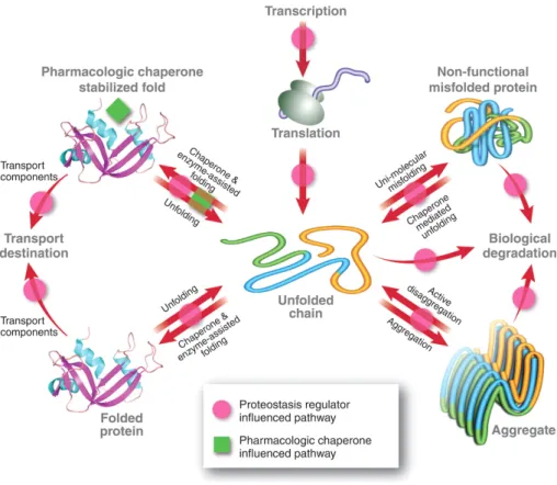
Pharmacological Chaperones (PC) and Proteostasis Regulators (PR) are two classes of distinct small molecules that act directly or indirectly to promote the stabilization of nonfuncional or partial functional unstable proteins (Figure I.3), originated by specific mutations.
FIGURE I.3. Influence of pharmacological chaperones (green squares) and proteostasis
regulators (magenta dots) in protein homeostasis pathways (red arrows). Proteostasis regulators can restore protein homeostasis and ameliorate conformational diseases through manipulation of proteostasis network, while pharmacological chaperones assist and enhance protein folding through a distinct mechanism from the innate biological pathways. Adapated from (31).
Pharmacologic chaperones are small molecules which bind to and stabilize intermediate folded states of a specific misfolding-prone protein, thereby increasing the concentration of native-like states of the mutant protein resulting in increased function or proceed to its destination environment to resume its function (32). It is considered a heterogeneous group of small compounds as it comprises molecules that bind weakly to a specific target protein, competitive inhibitors, ligands, agonists/antagonists and protein cofactors including metal ions. Interestingly, interactions with cofactors, either covalent or non-covalent bonds, are among those that contribute significantly to the maintenance of the tertiary structure of a protein (33). The downside of this approach is the small molecules specificity as they have to be tailored for each non-homologous protein linked to a loss-of-function misfolding disease (32). Currently, PC have been developed for several inherited metabolic disorders such as Gaucher disease (34), infantile Batten disease (35) and Fabry disease (36), which means that they have been adopted as a therapeutic strategy to ameliorate protein misfolding diseases. When it comes to small molecules to rescue folding defects in proteins, the term chemical
chaperone often arises. The slight difference between chemical and pharmacological chaperones comes down to the unspecific action for the former and the more specific direct action, over a particular target protein, observed in the latter (33). Chemical chaperones are mainly osmolytes and include polyols (e.g. glycerol, sorbitol), sugars (e.g. trehalose), methylamines (e.g. trimethylamine-N-oxide), free amino acids (glycine, taurin) or its derivatives (e.g. ectoine and gama-aminobutyric acid) and also other low molecular weight compounds with a chaperone-like function, such as dimethyl sulfoxide (DMSO) (33). As mentioned before, chemical chaperones have an unspecific mode of action and do not bind directly to proteins, their stabilization action results from the hydration effect which results from the ability of water molecules to establish favourable interactions with polar groups from the protein backbone, thus increasing protein compactness (37,38). Chemical chaperones had shown being effective in rescuing misfolding proteins in diseases such as cystic fibrosis, characterized by the impairment of a transmembranar protein (cystic fibrosis membrane conductance regulator (39), and also in several mild forms of phenylketonuria (40,41).
Proteostasis Regulatores act on the machinery responsible for maintaining the cellular protein homeostasis (proteostasis) including molecular chaperones among other components. Molecular chaperones are defined as any protein that interacts with and aids in the folding or assembly of another protein without being part of its final structure (42). They are central elements of the proteostasis network (PN), which is considered a fully integrated, layered system, unique to each cell type/compartment (43), comprising nearly 1000 molecular chaperones. Molecular chaperones regulate protein synthesis, folding, trafficking, disaggregation and degradation (32). The PN is used by cells to respond to proteome insults through stress sensors and inducible pathways which include the heat shock response (HSR), the unfolded protein response (UPR), oxidative stress pathways and growth factor and diet sensitive pathways (44–46). For this reason, molecular chaperones are designated as stress proteins or heat shock proteins (Hsp) and were initially named according to their molecular weight monomers (Hsp40, Hsp60, Hsp70, Hsp90, Hsp110 and small Hsp), although most of them exist as oligomers. Based on the sort of interaction with client proteins, molecular chaperones are also classified in holdases, foldases, and disaggregases (47). Foldases and disaggregases are molecular chaperones that function broadly in de novo folding and refolding (i.e. the chaperonins, Hsp70s and Hsp90s). They are ATP regulated and recognize segments of exposed hydrophobic amino acid residues, which are later buried in the interior of the natively folded protein. Binding to hydrophobic segments enables these chaperones to recognize the non-native states of many different proteins. Folding is then promoted during ATP- and cochaperone-regulated cycles of binding and release of non-native protein. In this mechanism of kinetic partitioning, (re)binding to chaperones blocks aggregation and reduces the concentration of free folding intermediates, whereas transient release of bound
hydrophobic regions is necessary for folding to proceed. Holdades are ATP-independent chaperones (such as the small Hsps) that buffer aggregation. They can recognize and stabilize partially folded proteins, preventing their aggregation and presenting client proteins to foldases (47–49).
Because protein molecules are highly dynamic, constant chaperone surveillance is required to ensure proteostasis. Recent advances suggest that unbalanced cell proteostasis are in the origin of aging and also of pathological states related to a wide range of diseases such as oncological diseases, Alzheimer’s, Parkinson’s and metabolic disorders. Presently, the Hsp90 inhibitor 17-allylaminogeldanamycin (17-AAG) is the first modulator to enter clinical trials (50) due to evidences of recent work that identified the molecular chaperone Hsp90 as being involved in the stabilization and conformal maturation of many signaling proteins that are deregulated in cancers, showing that its inhibition results in the proteosomal degradation of these client proteins and consequently leads a possible antitumor activity (51). In addition, celastrol and MG-132, heat shock transcription factor 1 (HSF-1) enhancers, have been described to increase cytosol’s proteostasis capacity and might induce one or more of the three pathways of the unfolded protein response (UPR) that remodels the proteostasis network and were stated to be candidates to treat two loss of function diseases (52).
Hence, while a decline of the PN is detrimental to cell and organismal health, a controlled perturbation of this network may offer new therapeutic avenues against human diseases (53,54).
1.2. NON-MUTATION SPECIFIC THERAPY
The development of strategies where mutation nature is not considered has also showed very interesting outcomes in a wide range of genetic diseases, including metabolic disorders, neurodegenerative, haematological, immunological and ocular. This type of therapeutic approach is considered transversal and effective against different type of genetic diseases, for which the gene causing the underlying condition has been identified, regardless how well the underlying pathophysiology is understood. Hence, non-mutation specific therapies emerge as a promise of providing lasting therapies and even cures for diseases that were previously untreatable.
1.1.1. GENE THERAPY
Gene therapy is the delivery of genetic material into an individual’s cells and tissues to treat inherited or acquired diseases. It is seen as a promising molecular approach that directly delivers into the host cell nucleus a gene aimed to repair the genetic defect, in order to cure or ameliorate the clinical phenotype (55,56). Although, gene therapy can provide treatment for
complex genetic diseases and acquired genetic diseases, its ideal target are monogenic diseases (56,57). According to literature, gene therapy has been shown to be effective in treating metabolic diseases (58), immune-deficiencies (59,60), eye (61) and coagulation disorders (62). These type of disorders are characterized by the dysfunction of a single specific gene, where the complete absence or the reduction in activity of the gene product causes the pathological phenotype. Thus, the introduction of one or more copies of the healthy gene is able to restore the genetic defect (63).
A number of methods have been stablished to accomplish gene delivery, taking into account not only the characteristics of the different genetic diseases, such as the size of therapeutic gene to be transferred and the tissue affected, but also several characteristics of what is an ideal vector: efficient and specific transduction of the target cell regardless of cell cycle, a therapeutic level and proper duration of gene expression, no associated genotoxicity, absent pre-existing immunity against the vector and transgene and a non-invasive delivery route (58). Effective strategies for clinical gene therapy are based in either in vivo or ex vivo gene delivery methods. The in vivo method involves direct introduction of the vector, carrying the therapeutic gene, into the patient, either into or near the target organ. Although this procedure might lead to inadvertent gene transfer into tissues and cell types that are not proper targets and may elicit immune responses towards the transgene and the vector or even damage to healthy genes, it may be ideal for metabolic diseases for which liver transplantation is a treatment (64). The ex vivo gene therapy implies the isolation of target cells, from donors or patients, to be genetically modified in vitro. Cells are harvested and transduced with a vector to express the therapeutic gene at normal or even supra-normal levels. Subsequently, to restore the healthy phenotype, the gene-corrected cells are infused into the patient where they can proliferate (57). This approach is appropriate for tissue specific diseases for which cell expansion or transplantation have been stablished as a treatment and for this reason has largely been focused on genetic diseases that involved hematopoetic derived cells (65,66). Different types of gene delivery systems have been exploited as useful vehicles for gene delivery: viral and non-viral vectors. The most common viral vectors are derived from pathogenic viruses: retroviruses, lentiviruses, adenoviruses and adeno-associated viruses (58). Actually, viral vectors arise from its innate ability of deliver genetic material into infected cells. The principles for the generation of viral vectors are the elimination of the viral toxic and infective functions, without altering the capacity to efficiently infect cells and deliver new transgenes (67).
Non-viral vectors, when compared with viral vectors, tend to be less immunogenic, to have larger packing capacity and easier to be produced. However, the cellular uptake is not very efficient. Several strategies, such as the use of polymeric systems and compounds or physical delivery methods have been used in order to overcome non-viral vectors limitations. Those
efforts may, in the future, result in a shift from the use of viral delivery to the use of non-viral delivery, but, in the present, viral delivery is the preferred approach in gene therapy(58). In fact, new vectors are constantly under development with the aim of targeted integration and DNA editing. At the beginning, early clinical trials for gene therapy were initiated with the hope that a new era for the treatment of inherited diseases would begin, but, only more recently, gene therapy clinical trials have demonstrated safety and efficacy in treating genetic diseases. Gene therapy is certain to represent a new and exciting therapeutic option, when one considers that there are no or inadequate treatment for a majority of patients with genetic diseases (58).
1.1.2. ENZYME REPLACEMENT THERAPY
Enzyme Replacement Therapy (ERT) is a medical treatment to reintroduce an enzyme into a patient who has deficiency of a specific enzyme, and is usually associated with inherited metabolic diseases. The concept of systemic delivery of an enzyme, to rescue cellular function, first started with Lysosomal Storage Diseases (LSD) and it derives from early cell-culture experiments by Neufeld and her group (63,68). ERT is usually performed by infusions of an enzyme that is purified from human or animal tissue or blood or produced by novel recombinant techniques. Typically, the enzyme is modified to allow for a longer half-life, more potent activity, resistance to degradation or targeting to a specific organ, tissue or cell type (69). One of the first successful ERT was for alpha-1-antitrypsin (A1AT) deficiency using derived purified human A1AT (70) and the second one was developed for type I Gaucher disease, an inherited deficiency of the lysosomal enzyme β-glucocerebrosidase that leads to accumulation of the substrate in lysosomes. Initial ERT for Gaucher disease Type I used highly purified placenta-derived glucocerebrosidase and later on recombinant technology, constituting now the standard therapy for this disease (71).
Currently, ERT is also available to treat other enzyme deficiency syndromes such as Fabry disease, Pompe disease, Hurler and Hunter syndrome, lysosomal acid lipase deficiency and several of the rarer forms of mucopolysaccharidoses (69).
Despite the advances in ERT, several technological constraints limit the use of ERT as protein formulation due to the inherent instability of these macromolecules. Besides that, no enzyme given intravenously crosses the blood-brain barrier, which makes ERT inappropriate for metabolic diseases that affects the brain (72).
Emerging strategies to mitigate ERT limitations include the use of immune tolerance regimens (73), covalent PEG attachment strategies (74), modified targeting procedures or complementary therapeutic methods, such as those involving pharmacologic chaperones or substrate reducing agents (75,76).
2. PHENYLKETONURIA
Phenylketonuria (PKU; OMIM 211600), the most common inborn error of amino acid metabolism, is an autosomal recessive disorder caused by a deficiency of the hepatic human phenylalanine hydroxylase enzyme (hPAH; EC 1.14.16.1) (77). PKU was first described by Asbjørn Følling, one of the first Norwegian physicians to apply chemical methods to the study of medicine (78). Nowadays it is known as a worldwide genetic disease with marked regional and ethnic variation in its incidence, with a reported prevalence of 1 in 10,000 live births in Caucasians (77).
The deficiency in hPAH causes high levels of L-phenylalanine (L-Phe) concentration in plasma and, if left untreated, this condition is accompanied by progressive mental retardation, neurological and behavioral problems due to the neurotoxic effect of hyperphenylalaninemia (HPA) (77). To date, more than 600 disease causing mutations have been identified in the
PAH gene (see PAH Mutation Analysis Consortium database; http://www.pahdb.mcgill.ca/)
and most of them are associated with PKU (79).
The discovery of PKU by Dr Asbjørn Følling was an important milestone in medicine as the PKU model was, since then, used to link neurological effects to metabolic abnormalities and has also been used as a template to shed light on over 200 other IEM (80).
2.1. METABOLIC PATHWAY AND CLINICAL PHENOTYPE
The amino acid Phe exists as D and L enantiomers, being L-Phe one of the essential amino acid required for protein synthesis in humans, once is not synthesized endogenously (81). Dietary intake of L-Phe along with endogenous recycling of amino acid stores are the main sources of Phe, whereas, in the cell, the consumption of L-Phe occurs via integration into proteins, oxidation to L-Tyrosine (L-Tyr) or conversion into other metabolites (Figure I.4) (80). The initial and rate-limiting step in the complete catabolism of L-Phe to CO2 and water is its
hydroxylation to L-Tyr that occurs mainly in the liver. This reaction is catalysed by the complex Phe hydroxylating system, consisting of hPAH, the pterin cofactor (6R)-L-erythro-5,6,7,8-tetrahydrobiopterin (BH4), and several enzymes that serve to regenerate BH4 (dihydropteridine
reductase and 4α-carbinolamine dehydratase) (80). This catabolic pathway accounts for approximately 75% or more of the disposal of dietary phenylalanine, which explains the major role of hPAH in maintaining L-Phe homeostasis (80).
FIGURE I.4. L-Phe metabolism pathways in humans. L-Phe pool results from diet intake and
from recycling trough endogenous amino acid pools from protein degradation. In the main pathway, L-Phe is hydroxylated by phenylalanine hydroxylase in the presence of its cofactor BH4 and molecular O2 producing L-Tyr. The alternative metabolism produces various
metabolites, by decarboxilation or transamination, which are excreted in urine. Adapted from (80).
The benzene ring of L-Phe cannot be ruptured without first being hydroxylated in the para position. However, the alanine side-chain of the amino acid can be metabolized in the absence of the ring hydroxylation step. This alternative pathway is initiated by transamination or descarboxilation of L-Phe to phenylpyruvate or phenylethylamine, respectively, followed by conversion of the latter compounds to metabolites such as phenyllactate, phenylacetate and o-hydroxyphenylacetate, which are posteriorly excreted in the urine. The alternative pathway is relevant only when the cell is not able to hydroxylate the L-Phe ring in the para position (82). The compromised activity of hPAH results in the failure to convert the L-Phe into L-Tyr, both indispensable aromatic amino acids with a requirement set at 25 mg/kg per day. The hPAH deficiency results in high levels of L-Phe and low levels of L-Tyr (83). Consequently, in untreated scenery, high levels of circulating L-Phe (HPA) give rise to a spectrum of disorders (84).
Any degree of HPA could be referred as a “phenylketonuric” phenotype and would be a risk factor to be managed appropriately. According to the HPA degree, various classification schemes have emerged. For example, one proposed by Kayaalp and collaborators (85) uses a simple nomenclature where PKU is related to (i) most severe type (classical PKU) which, in an untreated state, is associated with plasma L-Phe concentrations higher than 1000 µmol/L
and dietary L-Phe tolerance of <500 mg/day; (ii) non-PKU HPA which is associated with plasma L-Phe concentrations consistently above normal (>120 µmol/L) but lower than 1000 µmol/L when an individual is on a normal diet; and (iii) variant mild group where individuals do not fit the description for either PKU or non-PKU HPA. Another classification scheme proposed by Guldberg and co-workers (86) subdivides PAH deficiency into four categories based on L-Phe tolerance before the age of 5 years. However, despite the widespread utilization of these schemes, the classification is not always straightforward because L-Phe concentrations and L-Phe tolerance are not easily and accurately measured in newborn babies. Indeed, some authors claim that there may be little value in trying to classify this disorder based on current knowledge, as there is no clear clinical application (87).
Clinically, untreated PKU children with the classical PKU phenotype, show impaired brain development. Signs and symptoms include microcephaly, epilepsy, severe intellectual disability and behaviour problems. The excretion of excessive L-Phe and its metabolites can create body odour and skin conditions such as eczema. The mechanisms by which elevated blood L-Phe concentrations disturb cerebral metabolism and cognitive function are not fully understood. However, there is a proposed mechanism that includes a potential effect of disturbed large neutral amino acid (LNAA; which includes L-Phe, L-tryptophan (L-Trp), L-Tyr, L-leucine; L- isoleucine and L-valine) transport from blood to the central nervous system (CNS) reflecting the competitive nature of the transporter for LNAA at the blood-brain barrier (BBB). The low levels of L-Trp and L-Tyr in the CNS will impact the synthesis of neurotransmitters namely serotonin and dopamine (from L-Trp) and epinephrine and norepinephrine (from L-Tyr). In addition, affected individuals have a decreased myelin formation and protein synthesis. White matter pathology will occur (88) and further problems can emerge later in life (89). In the third or fourth decade of life, individuals late- or never-treated may develop severe behavioral or psychiatric problems such as depression, anxiety and phobias (90).
Early treatment in individuals with PKU prevents severe complications as the ones described previously. However, a growing evidence suggests that individuals with strict adherence to diet may still have some sequelae and suboptimal cognitive outcome (91).
2.2. FROM GENE TO PROTEIN
2.2.1. THE PAH GENE AND PKU GENOTYPES
The PAH gene spreads over 90 kb and consists of 13 exons (representing 3% of the genomic sequence), with introns varying from 1 to 20 kb. It maps to chromosome 12, region 12q23.2. Chromosome 12 is particularly rich in disease-associated loci, with 487 loci accounting for 5.2% of known ‘‘disease genes” (77). The locus for the gene encoding the enzyme hPAH,
covers 1.5 Mbp and harbours five other genes of known and unknown function (International Human Genome Sequencing Consortium, 2001). The cDNA and full-length genomic sequences are both visible online in PAHdb (www.pahdb.mcgill.ca/).
As mentioned before, hundreds of disease-causing mutations in the PAH gene have been identified and registered in the Human PAH Mutation Knowledgebase (hPAHdb; www.pahdb.mcgill.ca/) (Scriver et al. 2003) (77). The current spectrum of PKU mutations consists of ~60% missense mutations; ~13% of deletions; ~11% of splice mutations; ~6% silent mutations; ~5% nonsense mutations and ~2% of insertions (87). Mutations have been found in all 13 exons, but the majority are localized in the 3’-half of the gene, namely between exon 5 and 12 (92). In addition, repetitive DNA sequences are quite abundant and are now seen as causes of previously unidentified large PAH gene deletions and duplications (93), as well as, the CpG dinucleotides (n51198), which are potential sites for recurrent mutation in the
PAH gene (94).
Although mutations at the human locus affecting hPAH, generally, are the cause of the disease PKU or related forms of HPA, approximately 1-2% of HPA results from mutations in genes encoding for the enzymes involved in the de novo synthesis of BH4 from guanosine
triphosphate (GTP) and in the regeneration system of BH4 (94).
Apparently, the HPA phenotype of the patient is determined by the position and nature of the mutation, which interfere with the activity of the PAH enzyme. However genotype-phenotype correlations may not be a robust predictor (95), because each individual has a personal genome and even those with similar mutant PAH genotypes may not have similar “PKU” phenotypes (87). Besides, hPAH, as a homomeric enzyme, exhibit interallelic complementation (IC), a phenomenon that occurs in heteroallelic states when particular combinations of two different mutant alleles, at a given locus, produces a less (positive IC) or a more severe (negative IC) phenotype than their homoallelic counterparts (96). Thus, IC is of particular importance in PKU, since ~75% of the patients are heteroallelic for PAH mutations, being classified as compound heterozygous, which means that they have a different mutation for each allele (94). Experimental evidences support that genotype does predict biochemical phenotype (i.e. by Phe loading tests) but does not predict clinical phenotype (i.e. occurrence of intellectual disability) (97).
2.2.2. PAH PROTEIN
The PAH protein belongs to a small group of enzymes, known as aromatic amino acid hydroxylases (AAAHs) which also include tyrosine hydroxylase (EC 1.14.16.2) and tryptophan hydroxylase (EC 1.14.16.4). These enzymes catalyse the hydroxylation of aromatic rings of the amino acids L-Phe, L-Tyr and L-Trp in the presence of the cofactor BH4 (98). In addition,
for the catalytic reaction, AAAHs require molecular dioxygen (O2) as well as a non-heme iron
in the ferrous state (Fe(II)) at the active site (98).
Human PAH is a cytoplasmic enzyme present mainly in the liver, but also expressed in some extent in the kidney and epidermis (99,100). Due to its role in the degradation of L-Phe from diet, is considered a catabolic enzyme but it also provides an endogenous source of L-Tyr to the organism, converting an essential amino acid (L-Phe) into a non-essential one (L-Tyr) (101).
In terms of structure, hPAH is a homotetrameric enzyme that in vitro is found in equilibrium with a dimeric form. However, the tetrameric form has been considered the biological active form. Each monomer presents 452 amino acids, a molecular mass of ~52 kDa (102), adopts an a/b structure and presents three functional and structural domains: (i) an N-terminal regulatory domain (residues 1-142), containing the serine residue which is thought to be involved in activation by phosphorylation (Ser16 in hPAH); (ii) the catalytic domain (residues 143-410), containing the non-heme iron atom; and (iii) C-terminal domain (residues 411-452), which consist in a dimerization and tetramerization motif (Figure I.5). The C-terminal domain assembles to form the functional dimeric and tetrameric forms of the enzyme (94).
FIGURE I.5. Structure of human phenylalanine hydroxylase full-length composite model. (A)
Structure of a monomer where the iron atom is shown in red; the N-terminus starting over the active site, as well as the rest of the regulatory domain is highlighted in blue; the catalytic domain in shown in yellow and the tetramerization domain is depicted in green; (B) Structure of the native tetrameric form of the enzyme. Adapted from Erlandsen and Stevens (1999) (92). The regulation of hPAH activity is known to occur at several levels and includes allosteric activation by the substrate L-Phe, inhibition by the cofactor BH4 and an additional activation
by phosphorylation of Ser16 (103). The tetrameric form of the enzyme displays substrate activation and positive cooperativity for L-Phe binding, involving all three functional domains and all subunits (104). BH4 acts as a negative regulator by blocking L-Phe activation when the
binding to PAH is without L-Phe forming an inactive PAH-BH4 complex. However, recent
studies demonstrate that when PAH is Phe-ativated, the BH4 binding results in positive cooperativity (105). Phosphorylation acts as a mediator of L-Phe activation by decreasing the L-Phe concentration required to activate hPAH (104).
Some of these regulatory properties are mediated by the regulatory domain. In addition, recent studies propose that L-Phe not only binds the catalytic domain but also an allosteric site localized in the hPAH regulatory domain (106,107). In fact, the hPAH regulatory domain has the ACT (Aspartate kinase, Chorismate mutase and TyrA) domain, a structural motif, common in several allosteric proteins, which is involved in the binding of small activator molecules, usually amino acids and pyrimidines. Recent studies supported an allosteric regulation of hPAH, involving the stabilization of this ACT domain upon binding of L-Phe, during hPAH activation (103). This could be an important discover to open new ways of protein activation by small molecules.
2.2.3. PKU AS CONFORMATIONAL DISEASE
The term conformational disease arises to describe disorders that are caused by the expression and/or accumulation of unfolded or misfolded proteins and to date it comprises more than 40 different conditions (108). The cell biological mechanism by which abnormal protein structures lead to conformational diseases is still unclear. However it is known that improper protein folding (misfolding) as well as accrual of unfolded proteins can lead to the formation of disorder (amorphous) or ordered (amyloid fibril) aggregates (109).
Apparently, diverse disorders can be classified as conformational diseases as their aetiology involves abnormal unfolding followed by aggregation of an underlying protein. Taking into account the pathology, proteins can be classified as: (i) naturally unfolded, proteins prone to self-aggregation due to their specific amino acid sequence; (ii) proteins that become aggregation prone only after post-translational modifications; and (3) proteins that require specific mutations to become aggregation prone (108).
A common feature of conformational diseases with gain-of-functon is that the aggregates are usually devoid of helical regions, present in the form of b-sheets and contain high percentage of post-translational modifications (109).
It is presently assumed that more than one half of all sequence alterations in genetic diseases, like PKU, are missense mutations, meaning that single amino acid substitutions can affect the structure, stability and folding of a protein turning it into a misfolded protein.
Expression and characterization of several PAH missense mutations in in vitro systems have identified at least three main groups of enzymatic phenotypes, which differ in their kinetic behaviour and/or stability. The first group include structurally stable mutations with altered
kinetic properties; the second group include mutations with normal or almost normal kinetic properties, but reduced stability both in vitro and in vivo; and the third group include mutations affecting both kinetic and stability properties of the enzyme(96). These mutations are located in different regions of the three-domain structures, described earlier. As, the main molecular pathogenic mechanism of PAH deficiency is protein misfolding causing protein instability and low intracellular levels, due to specific mutations, PKU is now considered a Conformational disease with loss-of-function (96,110).
For protein misfolding diseases therapeutic strategies under investigation, aim to rescue the native protein conformation or to induce stabilization of intermediate or misfolded protein states. The use of pharmacological chaperones (PC) and proteostasis regulators (PR) to rescue folding defects in proteins involved in conformational diseases have been recently explored (33,42). Actually, PC are small molecular weight compounds that usually resemble natural ligands of the target proteins and they act by stimulating protein renaturation or scaffolding the final folded structure. For example, in the case of PKU, the cofactor BH4 is a
natural ligand acting as a PC, especially when given as therapeutic supplementation (111). There are groups that have been working on the identification of PCs in order to treat PKU (111,112). One of the groups performed a high-throughput ligand screening and, from the over 1000 pharmacological agents testes, they identified 4 compounds that improved hPAH thermal stability and did not show relevant inhibition of hPAH activity (111). The other group, developed PCs by virtual screening using BH4 as query structure and identified 84 candidates
with potential to bind the active site of hPAH (112).
Although these studies demonstrate interesting perspectives in terms of increasing hPAH activity, recently, Patel et al. disclosed the possibility to develop a new generation of PCs that specifically target the regulatory domain. This emerging model of PAH allosteric regulation, proposes that L-Phe not only bind the hPAH catalytic domain but also a site at the hPAH N-terminal regulatory domain, to activate the enzyme via an unclear mechanism (113). The synthesis of L-Phe like molecules that could act in such manner would be a stabilization strategy to target the allosteric domain and may constitute an alternative approach for the treatment of hPAH missense mutation that are non-responsive to BH4 supplementation and
that belong to the group of mutants with normal or almost normal kinetic properties, but reduced stability both in vitro and in vivo.
2.2.4. THE p.G46S MUTANT AS A MODEL TO STUDY SEVERE FORMS
OF PKU
As mentioned, the majority of variant hPAH, when over-expressed in prokaryotic systems, have a propensity to self-associate and form soluble/insoluble aggregates but when produced in eukaryotic cells are rapidly degraded (114). There are no evidences about the exactly
cellular mechanism that leads to protein degradation or which are the cellular protein quality control pathways and the molecular chaperones involved in this homeostatic response in this type of variants.
The substitution of a glycine by a serine in residue 46 of the hPAH, leads to the p.G46S variant (hPAH-G46S). This protein belongs to the group of enzymatic phenotypes with reduced stability, both in vivo and in vitro, which results in a severe form of PKU (115). The p.G46S variant results from a G to A transition in cDNA position 136 (c.136g>a) of exon 2 and was first identified by fluorescence-based single-strand conformation polymorphism (F- SSCP) analysis on PKU haplotype 5.9 alleles (115). The G46 residue is located in the regulatory domain of hPAH that contains the ACT module. Residue Gly46 is located at the start of α-helix 1 (Ala47– Glu57), in a five residue (Leu41–Gly46) loop stabilized by a network of hydrogen bonds and a salt-bridge (Lys42– Glu44). It has been proposed that the G46S substitution will lead to the extension of α-helix 1 that then perturbs the α-β sandwich of the ACT module in the regulatory-domain (Figure I.6) (114). The prokaryotic expression of the hPAH G46S variant as a fusion protein coupled to the maltose binding protein (MBP) via a peptide sequence recognized by the factor Xa restriction protease (MBP-pep(FXa)-PAH-G46S), gave rise to a metastable tetrameric form characterized by a near normal catalytic efficiency (114). However, destabilization and self-association of the G46S tetramer (and dimer) and the formation of higher-order oligomers and large twisted non-amyloid fibrils were evident (Figure I.6), after the cleavage of the MBP fusion partner by the restriction protease factor Xa (114).
FIGURE I.6. Representation of the human phenylalanine hydroxylase monomer with the
localization of residue Gly46 (A) and the self-association of the G46S tetramers (B). In panel (A) the interactions established by the Gly46 residue are shown; the regulatory domain is shown in green while the catalytic and tetramerization domains are depicted in red. Panel (B) shows the time-course of the self-association of pep(FXa)-hPAH-G46S () and MBP-pep(FXa)-PAH-WT (p) following the cleavage by factor Xa; the graphic inset represents SDS-PAGE analysis of the cleavage reaction at time 0 (lane 2), 10 (lane 3), 20 (lane 4) and 30 minutes (lane 5); the electron micrographs of negatively stained hPAH-G46S at the maximum of light scattering (120 min; arrow) are also shown. Adapted from Leandro et al (114).
The variant p.G46S has been well characterized by Leandro and collaborators (114) in terms of the intrinsic physico-chemical properties, as well as extrinsic factors such as ionic strength, temperature, pH, protein concentration, phosphorylation state and deamination and it is considered a well-controlled model to study not only PR involved in protein degradation but also new PC that specifically target the regulatory domain as a stabilization strategy, with aim of to prevent self-association and to restore hPAH activity (79,96,114).
2.3. PKU TREATMENT
Currently, as there is no cure for PKU, the main goal in PKU treatment is to lowering L-Phe levels to prevent intellectual disability but maintaining the minimum levels required for normal growth. Therefore, the prevailing treatment is dietary restriction of L-Phe, supplemented with specifically designed medical foods (116).
The establishment of newborn screening programmes along with prompt institution of dietary treatment has prevented intellectual disability, although neurophysiological and neuropsychological impairments may still persist in treated patients (117–121). This dietetic restriction has, as major advantage, the applicability towards all hPAH mutations with an adequate outcome, but, as disadvantage, this strict long term diet can lead to social boundary, nutritional deficiencies (in vitamin B12, vitamin D, calcium, iron and unsaturated long chain fatty acids), which may exacerbate the neurological problems, as well as non-compliance due to poor palatability, despite the recent improvement of low-Phe dietetic products (122,123). Therefore, there is an urgent need for exploring new approaches and alternatives to partially or totally substitute the L-Phe diet.
2.3.1. CURRENT TREATMENTS
Besides, dietary restriction of L-Phe intake, which remains the mainstay of treatment for PKU since its introduction in 1953 (124), the cofactor BH4, LNAA and glycomacropeptide (GMP)
supplementation, are the current available approaches for PKU treatment (116).
The responsiveness to BH4 therapy is likely to be associated with mutations in the PAH gene
resulting in some residual enzyme activity, characteristic from patients presenting the mild PKU phenotype, however genotype-phenotype correlations are inconsistent (125). Sapropterin dihydrochloride (KuvanÒ, Biomarin Pharma) is a synthetic formulation of the active 6R-isomer of the naturally occurring hPAH cofactor, orally administered to patients for PKU treatment. The mechanism of action in lowering L-Phe levels, in patients with PKU, has not been fully elucidated, but appears to be related, in part, to its effect in stabilizing mutant hPAH enzymes, resulting in increased clearance of L-Phe from the body. In patients with BH4
deficiency, its mechanism of action is presumed to be secondary to replacement of endogenous tetrahydrobiopterin (126).
Kuvan has received Orphan Drug status for the treatment of PKU, since phase II and III clinical data demonstrated that it was a safe and effective therapy (127). In fact, this therapeutic approach has proven successful in significantly increasing L-Phe tolerance allowing patients to relax their diet and in some cases discontinuing the L-Phe restrictive diet (128,129). However, for 90% of patients with classical PKU, who comprise about 50-80% of patients detected by newborn screening, this therapy has no beneficial effect (130). So, alternative treatments for which efficacy is not dependent on genetic variations in the PAH gene may benefit these patients.
The LNAAs had been shown to reduce CNS L-Phe concentrations and consequently, cerebral damage, despite the observed increased plasma levels. This might be due to the fact that the administered LNAAs will compete with L-Phe to cross the BBB (89). Although LNAA treatment appears to have beneficial effect on executive functioning, this approach is only suitable for adults who are not adhering to a low Phe diet (131). In addition, regarding long term outcome, clinical data using this treatment strategy are not yet available (131), requiring additional studies to prove its safety and efficacy (132).
Glycomacropeptide (GMP) is a protein derived from cheese whey, naturally low in L-Phe, rich in valine, isoleucine and threonine, that when supplemented with the essential amino acids (L-Tyr, L-Trp, arginine, cysteine and histidine) can be a useful adjunct to the L-Phe restricted diet also improving palatability (133). It has been shown that the potential benefits of having GMP included in PKU diet are reduced ureagenesis, improved protein retention and L-Phe utilization (134), decreased levels of postprandial ghrelin concentration when compared to an amino acid diet (135) and increased metabolic activity (in PKU mice) (136). From this last study, the authors conclude that the GMP diet provides a more natural and physiological low L-Phe source of intact protein as compared with the synthetic amino acid (136). Although, the present data are promising, further studies are needed to investigate the effect of the GMP diet in humans, once the majority of the studies were performed in mice. This will allow further evaluation of the safety and efficacy of GMP consumption for long term.
2.3.2. FUTURE TREATMENT STRATEGIES
All the previous mentioned data highlight the need for alternative strategies for PKU treatment. Enzyme replacement therapy with PAH-based fusion proteins and protein delivery systems, along with genetic therapy and pharmacological chaperones are some of the therapeutic strategies that have been studied in order to answer this demand.
Enzyme substitution therapy using phenylalanine ammonia lyase (PAL; E.C.4.3.1.5) has been suggested as a possible therapeutic approach for PKU. PAL is an enzyme that catalyses the