Universidade de Lisboa
Faculdade de Farmácia
Departamento de Bioquímica e Biologia Humana
Biological membranes – biophysical properties
and aquaporin function
Ana Paula Cavaco da Silva Martins
Tese orientada pela Professora Doutora Graça Soveral e co-orientada pela Professora Doutora Catarina Prista, elaborada para a obtenção do grau de Doutor em Farmácia, especialidade em Bioquímica.
i
Biological membranes – biophysical properties
and aquaporin function
₪₪₪₪₪₪₪
Membranas biológicas – propriedades biofísicas e função
das aquaporinas
Dissertação apresentada à Faculdade de Farmácia da Universidade de Lisboa para obtenção do grau de Doutor em Farmácia (especialidade de Bioquímica)
Ana Paula Cavaco da Silva Martins
Lisboa 2014
ii
SUPERVISÃO
Supervisor: Prof. Dr. Graça Soveral Co-supervisor: Prof. Dr. Catarina Prista
INSTITUIÇÕES PARTICIPANTES
The studies presented in this thesis were performed at the:
- Research Institute for Medicines (iMed.ULisboa) and Department of Biochemistry and Human Biology, Faculty of Pharmacy, University of Lisbon, Lisbon, Portugal and Requimte, FCT-UNL, 2829-516 Caparica, Portugal, under the supervision of Prof. Dr. Graça Soveral. - Centro de Botânica Aplicada à Agricultura (CBAA); Laboratório de Bioenergética Microbiana, Instituto Superior de Agronomia, Universidade de Lisboa, Lisboa, Portugal, under the supervision of Prof. Dr. Catarina Prista.
FINANCIAMENTO
This work was supported by Fundação para a Ciência e Tecnologia, Portugal (SFRH/BD/65046/2009).
iii
CONTRIBUTO PESSOAL NOS TRABALHOS DE INVESTIGAÇÃO APRESENTADOS NESTA DISSERTAÇÃO
De acordo com o disposto no ponto 1 do artigo nº41 do Regulamento de Estudos Pós-Graduados da Universidade de Lisboa, deliberação nº93/2006, publicada em Diário da República – II série nº153 – 5 de Julho de 2003, a autora desta dissertação declara que participou na concepção e execução do trabalho experimental, interpretação dos resultados obtidos e redação dos manuscritos.
v
vii
LIST OF ORIGINAL PUBLICATIONS
This thesis is based on the following original publications:
1. Martins AP, Lopes PA, Martins SV, Madeira A, Santos NC, Prates JA, Moura TF, Soveral G. Conjugated linoleic acid reduces permeability and fluidity of adipose plasma membranes from obese Zucker rats. Biochem Biophys Res Commun. 2010 Jul 23;398(2):199-204.
doi: 10.1016/j.bbrc.2010.06.059.
2. Martins AP, Lopes PA, Costa AS, Martins SV, Santos NC, Prates JA, Moura TF, Soveral G. Differential mesenteric fat deposition in bovines fed on silage or concentrate is independent of glycerol membrane permeability. Animal. 2011 Dec;5(12):1949-56.
doi: 10.1017/S1751731111001091.
3. Martins AP, Lopes PA, Madeira MS, Martins SV, Santos NC, Moura TF, Prates JA, Soveral G. Differences in lipid deposition and adipose membrane biophysical properties from lean and obese pigs under dietary protein restriction. Biochem Biophys Res Commun. 2012 Jun 22;423(1):170-5. doi: 10.1016/j.bbrc.2012.05.108.
4. Martins AP, Marrone A, Ciancetta A, Galán Cobo A, Echevarría M, Moura TF, Re N, Casini A, Soveral G. Targeting aquaporin function: potent inhibition of aquaglyceroporin-3 by a gold-based compound. PLoS One. 2012;7(5):e37435.
doi: 10.1371/journal.pone.0037435.
5. Martins AP, Ciancetta A, de Almeida A, Marrone A, Re N, Soveral G, Casini A. Aquaporin inhibition by gold(III) compounds: new insights. ChemMedChem. 2013 Jul;8(7):1086-92.
doi: 10.1002/cmdc.201300107.
6. Sabir F, Leandro MJ, Martins AP, Loureiro-Dias MC, Moura TF, Soveral G, Prista C. Exploring three PIPs and three TIPs of grapevine for transport of water and atypical substrates through heterologous expression in aqy-null yeast. PLoS One. 2014 Aug 11;9(8):e102087.
doi: 10.1371/journal.pone.0102087.
7. Noronha H, Agasse A, Martins AP, Berny MC, Gomes D, Zarrouk O, Thiebaud P, Delrot S, Soveral G, Chaumont F, Gerós H. The grape aquaporin VvSIP1 transports water across the ER membrane. J Exp Bot. 2014 Mar;65(4):981-93.
doi: 10.1093/jxb/ert448. Other publications
Soveral G, Martins AP, Martins SV, Lopes PA, Alfaia CM, Prates JA, Moura TF. Effect of dietary conjugated linoleic acid isomers on water and glycerol permeability of kidney membranes. Biochem Biophys Res Commun. 2009 May 22;383(1):108-12.
doi: 10.1016/j.bbrc.2009.03.136.
Abreu-Rodríguez I, Sánchez Silva R, Martins AP, Soveral G, Toledo-Aral JJ, López-Barneo J, Echevarría M. Functional and transcriptional induction of aquaporin-1 gene by hypoxia; analysis of promoter and role of Hif-1α. PLoS One. 2011;6(12):e28385.
2
TABLE OF CONTENTS
ABBREVIATIONS ... 5
ABSTRACT ... 7
SUMÁRIO ... 11
GENERAL AIMS AND THESIS OUTLINE ... 15
PART I – Lipid Bilayer, biophysical properties ... 19
INTRODUCTION ... 21
1. Composition and properties of the lipid-bilayer. ... 21
2. Dietary fatty acids. ... 25
3. Reduced protein diets (RDP). ... 28
4. Adipose tissue and obesity. ... 29
5. Genetic background and animal models. ... 30
Aim of part I: ... 31
CHAPTER 1 - Influence of diet on cell membrane composition and biophysical properties ... 33
Publication 1: Conjugated linoleic acid reduces permeability and fluidity of adipose plasma membranes from obese Zucker rats. ... 37
Publication 2: Differential mesenteric fat deposition in bovines fed on silage or concentrate is independent of glycerol membrane permeability. ... 43
Publication 3: Differences in lipid deposition and adipose membrane biophysical properties from lean and obese pigs under dietary protein restriction. ... 51
PART II – Aquaporins ... 57
INTRODUCTION ... 59
1. Aquaporin classification and selectivity. ... 60
2. Aquaporin structure. ... 61
3. Aquaglyceroporin structure. ... 64
4. Aquaporin regulation. ... 65
5. Physiological relevance. ... 65
6. Plant aquaporins. ... 70
7. Aquaporin heterologous expression in yeast ... 71
8. Functional studies to assess aquaporin activity. ... 71
Aim of part II ... 73
CHAPTER 2 – Aquaporins as Drug Targets ... 75
Publication 4: Targeting aquaporin function: potent inhibition of aquaglyceroporin-3 by a gold-based compound. ... 79
3 CHAPTER 3 - Aquaporin Functional Assessment in the Yeast Cell Model ... 101
Publication 6: Exploring three PIPs and three TIPs of grapevine for transport of water and atypical substrates through heterologous expression in aqy-null yeast. ... 105 Publication 7: The grape aquaporin VvSIP1 transports water across the ER membrane. ... 119 GENERAL DISCUSSION AND CONCLUSIONS ... 133 FUTURE PRESPECTIVES ... 141 REFERENCES ... 145
5
ABBREVIATIONS
AQP Aquaporin
CLA Conjugated linoleic acid
DHA Docosahexaenoic acid (22:6n-3)
DMPC 1,2-dimiristoyl-sn-3-phosphocholine EPA Eicosapentaenoic acid (20:5n-3)
FA Fatty acid
FFA Free fatty acids
LA Linoleic acid
MUFA Monounsaturated fatty acids
OA Oleic acid
PUFA Polyunsaturated fatty acids
RBC Red blood cells
RPD Reduced protein diet
7
ABSTRACT
9
ABSTRACT
Membranes are barriers that assure the selective communication of the internal media with the external environment, a process that is crucial for life. Membrane components, lipids and proteins, are the main players responsible for the membrane selective permeability and its regulation. The work presented in this thesis encompasses different aspects of biological membranes features.
The first part of this thesis is dedicated to the study of the influence of dietary lipids in membrane biophysical properties, namely fluidity and permeability. We used animal models and designed experiments where the effect of the diet supplementation with fatty acids, PUFA and the conjugated linoleic acid (CLA) on membrane composition, fluidity and permeability, were analysed. In addition, since the animal genetic background is known to have an important contribution to the regulation of several biochemical pathways and resulting in different phenotypes, this variable was included in the study.
Within the same animal species all adipose membranes were found richer in SFA independently of their genetic background or diet. The lipid adipose membrane composition of Zucker rats is highly dependent on the lipid composition of the diet. Regarding the adipose membrane biophysical properties, we were able to correlate a decrease in membrane fluidity and the concomitant decrease on permeability to water and glycerol with the incorporation of t10,c12 CLA isomer into adipose membranes of Zucker rats. Interestingly, this CLA isomer is the one with suggested fat lowering properties in several animal studies. The animal genetic background in obese pigs was also found to play a determinant role on the fluidity of adipose membranes, in accordance with the reported for a wide variety of cell membranes from obese mice and rats. Interestingly, the same increase in membrane fluidity was observed for pigs fed a low protein diet and was correlated with the ratio oleic /linoleic acid. A similar effect was consistently reported in adipose membranes of genetically obese mice pointing to a clear compensatory mechanism to maintain membrane biophysical properties.
The second part of this thesis is dedicated to the study of membrane protein channels, aquaporins, aiming at identifying new modulators with potential pharmacological use. Furthermore, building an experimental cell model able to characterize individual aquaporin activity and selectivity as well as to validate modulators’ effect and potency was another goal of this work.
Using the human erythrocyte that expresses one orthodox aquaporin (AQP1) and one aquaglyceroporin (AQP3), we reported on the potent and selective inhibition of human AQP3 by a water-soluble gold(III) coordination compound, Auphen.
From molecular modelling studies and docking approaches, we were able to propose a mechanism of action of Auphen for human AQP3 inhibition, where Cys40 is the crucial residue for binding. Inhibitory assays with other metal complexes, namely phenantroline derivatives of Pt(II) and Cu(II), showed that the AQP3 inhibition potency decreased drastically in the order Au(III) > Cu(II) >> Pt(II). Interestingly, no inhibition effect was achieved when incubating the cells with gold(I) compounds, therefore,
10
demonstrating the necessity of gold(III)-based scaffolds to achieve protein binding and blockage of the channel. Our mechanistic hypothesis was based on the possibility for Auphen and analogues to bind to certain amino acid residues in the channel close to the selectivity filter domains, thus acting as a “cork” preventing the passage of glycerol.
Given the diversity of pathologies associated with dysfunction of the different aquaporins, these proteins are now emergent drug targets. However, cells and tissues frequently express more than one aquaporin isoform in the plasma membrane and unless a specific isoform is mutated or overexpressed, its function is not easily discriminated. To overcome this difficulty, we developed a yeast heterologous system for expression and functional assessment of aquaporin isoforms. Such a cell system expressing individual AQPs would also be very useful for analysis of function and regulation of aquaporins from the plant kingdom. Plants express numerous aquaporin isoforms, but for most their physiological relevance is still unclear.
Using this approach, grapevine aquaporins VvPIPs, VvTIPs and VvSIPs were expressed in yeast and functionally analysed. We demonstrated that only some isoforms are functional while others are not, and they may transport other small molecules of physiological importance such as ammonia, boron, CO2, hydrogen peroxide and urea. In
particular for VvSIP1, we disclosed for the first time its ability to transport water but not glycerol, urea, sorbitol, glucose, or inositol.
11
SUMÁRIO
13
SUMÁRIO
As membranas biológicas comportam-se como barreiras que asseguram a comunicação seletiva do meio interno com o ambiente externo, um processo que é crucial à vida. Os componentes membranares, lípidos e proteínas, são os principais responsáveis pela permeabilidade seletiva das membranas e sua regulação. O estudo apresentado nesta tese abrange diferentes aspetos característicos das membranas biológicas.
A primeira parte desta tese foi dedicada ao estudo da influência dos lípidos da dieta nas propriedades biofísicas das membranas, em particular fluidez e permeabilidade. Foram utilizados modelos animais e concebidos ensaios experimentais onde se analisou o efeito da suplementação da dieta em ácidos gordos, PUFA e ácido linoleico conjugado CLA, na composição lipídica, fluidez e permeabilidade das membranas do tecido adiposo. Ainda, uma vez que a herança genética tem uma contribuição importante para a regulação de vários processos metabólicos resultando em fenótipos diversos, esta variável também foi incluída neste estudo.
Na mesma espécie animal, as membranas de tecido adiposo são mais ricas em ácidos gordos saturados independentemente da sua raça ou dieta. A composição lipídica das membranas de tecido adiposo de ratos Zucker é muito dependente da composição lipídica da dieta. Relativamente às propriedades biofísicas, foi possível correlacionar um decréscimo da fluidez da membrana e concomitante decréscimo da permeabilidade à água e ao glicerol com a incorporação do isómero de CLA t10,c12 nas membranas do tecido adiposo de ratos Zucker. Curiosamente, em diversos estudos animais têm sido atribuídas propriedades de emagrecimento a este isómero CLA.
A herança genética (raça) também tem um papel importante na fluidez das membranas do tecido adiposo, em concordância com o já descrito em vários estudos em membranas celulares de ratinhos e ratos obesos. Também se verificou um aumento da fluidez membranar em porcos alimentados com restrição proteica, o que foi correlacionado com a razão dos ácidos oleico/ linoleico. Um efeito semelhante foi já reportado para membranas de tecido adiposo de ratinhos geneticamente obesos, apontando para um mecanismo compensatório que leva à manutenção das propriedades biofísicas das membranas.
A segunda parte desta tese foi dedicada ao estudo de canais proteicos membranares, as aquaporinas, com o objetivo de identificar novos moduladores com potencial utilização farmacológica. Ainda, pretendemos desenvolver um modelo experimental para a caracterização individual da atividade e seletividade de aquaporinas e para a validação do efeito e da potência de moduladores.
Utilizando eritrócitos humanos que expressam uma aquaporina ortodoxa (AQP1) e uma aquagliceroporina (AQP3), descrevemos a forte inibição seletiva da AQP3 por um composto de coordenação de ouro(III) hidrossolúvel, o Auphen.
14
Através de estudos de modelação molecular e de docking, foi possível propor um mecanismo de ação do Auphen para a inibição da AQP3 onde o resíduo de cisteína cys40 é crucial para a ligação.
Os ensaios de inibição com outros complexos metálicos, nomeadamente derivados da fenantrolina de Pt(II) e de Cu(II), revelaram que a inibição decresce drasticamente na ordem Au(III) Cu(II) >> Pt(II). Curiosamente, não se observou inibição quando as células foram incubadas com compostos de gold(I), demonstrando assim a importância dos esqueletos dos compostos de gold(III) para a ligação à proteína e bloqueio do canal. Assim, a nossa hipótese de mecanismo é baseada na possibilidade do Auphen e análogos se ligarem a certos resíduos de aminoácidos no canal, perto do filtro de seletividade, atuando deste modo “como uma rolha” que impede a passagem do glicerol.
Devido á diversidade de patologias relacionadas com a disfunção de diferentes aquaporinas, estas proteínas são atualmente consideradas alvos terapêuticos. Contudo, na membrana plasmática das células e tecidos é frequentemente expressa mais do que uma isoforma de aquaporina, pelo que a sua função não é facilmente discriminada a não ser que essa isoforma específica seja mutada ou sobre-expressa. Para ultrapassar esta dificuldade, foi desenvolvido um sistema de expressão heteróloga em leveduras para a expressão e análise funcional de aquaporinas. Tal sistema também é muito útil para a análise da função e regulação de aquaporinas do reino vegetal. As plantas expressam numerosas isoformas de aquaporinas mas na maior parte dos casos, a sua relevância fisiológica ainda não foi estabelecida.
Utilizando esta abordagem, as aquaporinas de uva VvPIPs, VvTIPs e VvSIPs foram expressas em leveduras e a sua função foi analisada. Foi demonstrado que somente algumas isoformas são funcionais, e que algumas podem estar envolvidas no transporte de outras moléculas com importância fisiológica tais como amónia, boro, CO2, peróxido
de hidrogénio e ureia. Em particular para a VvSIP1, foi pela primeira vez revelada a sua capacidade para transportar água mas não glicerol, ureia, sorbitol, glucose ou inositol.
15
GENERAL AIMS AND THESIS OUTLINE
17
GENERAL AIMS AND THESIS OUTLINE
The plasma membrane is the boundary of the cell, separating its content from the environment. This semipermeable membrane is composed of a lipid-bilayer where thousands of different lipid molecules interact dynamically, forming transient or stable structures used by many proteins as platforms for their activity and to enhance their interactions with other proteins. One fundamental cell membrane function is to quickly respond to intra and extracellular events, accomplishing transmembrane fluxes fundamental to the physiology of all living organisms.
Water is the major component of all living organisms. The controlled flow of water through biological membranes not only plays a key role in cellular homeostasis but also contributes to cell survival. Triggered by osmotic and/or pressure gradients, water crosses the membrane both through the lipid-bilayer and specialized membrane channels called aquaporins (AQP), which facilitate highly selective and efficient flow of water and, in some cases, other small solutes such as glycerol and urea.
This work is focused in the study of membrane characteristics that affect the permeability of water or small uncharged solutes such as glycerol. Within this scope, membrane biophysical properties modulated by lipid membrane composition and by the activity of aquaporin channels play a central role.
This work is divided in two parts, Part I (containing chapter 1) and Part II (containing chapter 2 and 3).
The first part is dedicated to study the influence of genetic background and diet composition on adipocyte membrane composition and its outcome on membrane biophysical properties such as fluidity and permeability to water and glycerol.
It is known that dietary fatty acids incorporate into cell membrane phospholipids. The health beneficial effects attributed to dietary compounds such as polyunsaturated fatty acids (PUFA) are vast. Examples are their protective effects on hypertension, cardiovascular diseases, and cancer, among others 1. Albeit several candidate
mechanisms have been suggested, the means by which they elicit their effects remain largely unknown. In view of the broad range of effects attributed to these compounds, it is expected that, at least in part, they act at some fundamental cellular level common to all organism.
Our previous results 2 have shown that kidney plasma membranes from Wistar
rats, fed with palm oil-based diets supplemented with conjugated linoleic acid (considered a fat lowering fatty acid ), showed altered membrane permeability, an effect that was correlated with the incorporation of this fatty acid into the plasma membrane. With this starting point, this work aims at further understanding the effects of fatty acids and food components on cell membranes and their outcome on membrane composition and biophysical properties such as permeability and fluidity.
In chapter 1 we present 3 publications where we studied the influence of dietary lipids, protein restriction conditions and genetic background on adipocyte membrane composition and its outcome on membrane biophysical properties such as fluidity and permeability to water and glycerol (publications 1, 2 and 3).
18
The second part is divided in two chapters (chapters 2 and 3) in which human and plant aquaporins were studied.
AQPs are a family of small transmembrane proteins ubiquitous in nature. These proteins form highly selective channels for water and, in the case of the sub-family aquaglyceroporins, other small solutes such as glycerol and urea.
In chapter 2, human aquaporins were investigated regarding the screening for specific inhibitors.
Thirteen different mammalian AQPs have been identified so far, present in almost all organs and tissues. It is clear that they play fundamental roles in human physiology and pathophysiology, their dysfunction or aberrant expression is correlated with several diseases such as kidney diseases, brain oedema, obesity and cancer3.
Numerous reports have highlighted the possible areas where AQP modulators could be useful in treating human diseases. Yet, only a few pharmaceutically relevant compounds have been identified to date and none of them proved to be suitable for clinical trials.
Using a screening system based on permeability assays of human red blood cells (RBC), we investigated modulators of aquaporin activity (publications 4 and 5).
In chapter 3, the activity of different isoforms of plant aquaporins natively located in plasma membrane and in intracellular membranes of grapevine, was characterized. To assess their individual activity and contribution to membrane permeability, we used a yeast heterologous expression model that was optimized for the expression and functional analysis of aquaporins and is now suitable to be used as inhibitor screening system (publications 6 and 7).
19
PART I – Lipid Bilayer, biophysical properties
21
INTRODUCTION
The plasma membrane represents the barrier of life, the structure that separates the cells from their surroundings. This semipermeable membrane is composed of a lipid-bilayer embedded with proteins accomplishing fundamental processes central to the physiology of the cell. In this context, the lipid composition of the cell membrane and the transporters within, play a central role. Part I will deal with the bilayer composition of the membrane and its biophysical properties.
The Lipid-Bilayer
This part of the work aimed at studying plasma membrane lipid composition and its outcome in biophysical properties such as fluidity and permeability to water and different solutes in response to different dietary compositions, namely, fatty acids (FA) content and protein restriction.
1. Composition and properties of the lipid-bilayer.
Biological membranes have a similar general organisation in sheet-like structures, composed of a heterogeneous and asymmetric lipid-bilayer firmly established as the universal basis for cell-membrane function. Herein, thousands of different lipid molecules interact dynamically, forming transient or stable structures used by many proteins as platforms for their activity and to enhance their interactions with other proteins4,5.
Membrane lipids are divided in three classes of amphipathic lipids: phospholipids, glycolipids, and sterols. The amount of each amphipathic lipid depends upon the type of cell, but in the majority of cases phospholipids are the most abundant, while glycolipids are found exclusively in the outer layer of the membrane. Cholesterol is also abundant in eukaryotic cell membranes - up to one molecule for every phospholipid molecule.
Phospholipids and glycolipids share a similar structure containing a hydrophilic headgroup and one or two hydrophobic tails. Phospholipids are derived from either glycerol (phosphorglycerides) or sphingosine (sphingolipid), having the following components: a glycerol or sphingosine backbone, two or one FA chains, a phosphate group and (usually) an alcohol (e.g. choline, ethanolamine, inositol) (Figure 1). The FA tails can differ in length (containing normally between 14 and 24 carbon atoms) and may be saturated or unsaturated. The number of double bonds present in their hydrocarbon chains can vary from none, saturated fatty acid (SFA), one, monounsaturated fatty acid (MUFA) to several, polyunsaturated fatty acid (PUFA).
Each double bond creates a small bend in the tail. Differences in the length and saturation degree of the FA tails influence their ability to pack against one another, thereby affecting the fluidity and other biophysical properties of the membrane6.
22
Figure 1 – Schematic drawing of common membrane lipids.
The variety of headgroups and aliphatic chains allows the existence of more than 1000 different lipid species in any eukaryotic cell7. This diversity is only beginning to be
characterized while lipidomics is becoming an important tool in cell and developmental biology, molecular medicine and nutrition8. The membrane composition differs
throughout inner and outer sheets of lipid bilayer, cell organelles and cell type7 The
assortment of lipid species allows to adapt the lipid composition of membranes to fulfil specific functions. How the organism regulates lipid composition and the exact mechanisms of how compositional complexity affects cell homeostasis and its regulation remains poorly understood8.
The effect of lipid composition on biological membranes points to an extensive list of perturbations induced on membrane lipid structure including changes in membrane fluidity, phase behaviour, permeability, membrane fusion, lateral pressure and flip-flop dynamics7,5. In this manner FAs influence the membrane function at several
levels including membrane microdomain organization, membrane proteins activity, cell signalling and ultimately human health5.
1.1. The fluidity of a lipid-bilayer depends on lipid composition.
Three main factors, besides temperature and the head group of membrane lipids, contribute to membrane fluidity: membrane lipid tail length, the degree of unsaturation of lipid tails and the amount of cholesterol.
The tail length affects fluidity as lipids with longer tails exhibit more friction when moving around in the membrane. In other words, the longer the tails, the higher the attractive van der Waals forces between them. As a result, membranes with longer lipid tails tend to be less fluid.
The average degree of unsaturation of lipid tails also affects membrane fluidity, as unsaturated tails do not pack as tight as saturated ones, therefore increasing membrane fluidity.
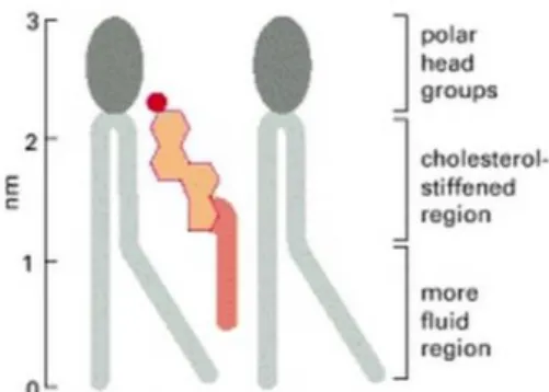
Cholesterol molecules contribute to membrane fluidity enhancing the permeability-barrier properties of the lipid bilayer. They orient themselves in the bilayer
23
with their hydroxyl group close to the polar head groups of the phospholipid molecules. In this position, their rigid, plate-like steroid rings interact with, and partly immobilize, those regions of the hydrocarbon chains closest to the polar head groups (Figure 2). By decreasing the mobility of the first few CH2 groups of the hydrocarbon chains of the
phospholipid molecules, cholesterol makes the bilayer less deformable in this region and thereby decreases its permeability. Although cholesterol tends to make lipid bilayers less fluid, at the high concentrations found in most eukaryotic plasma membranes it also prevents the hydrocarbon chains from coming together and crystallizing. In this way, it inhibits possible phase transitions6.
Figure 2 - Schematic drawing of a cholesterol molecule interacting with two phospholipid molecules.
Cholesterol and length and unsaturation degree of fatty acyl chains are not the only players regarding membrane fluidity. The head group of phospholipids also influences membrane behaviour. For example, choline is a bulky headgroup causing phosphatidylcholine (PC) to have a cylindrical molecular geometry. In contrast, ethanolamine is a smaller headgroup, with phosphatidylethanolamine (PE) assuming a conical molecular geometry, allowing a tighter packing of the phospholipids. Thus, when the PE/PC ratio increases there is a reduction in membrane fluidity7.
1.2. Effect of fatty acids on membrane permeability, membrane proteins activity and on micro-domain organization.
FAs can influence membrane permeability, an effect that has been associated with disorder in the membranes' interior and the interaction of the incorporated lipid with the polar head group of phospholipids9. It is known that at the gel-to-fluid phase
transition of 1,2-dimiristoyl-sn-3-phosphocholine (DMPC) bilayers, when the largest number of defects in the lipid matrix appears, membrane permeability reaches its maximum10. It has also been demonstrated that the dietary FA, docosahexaenoic acid
(DHA) increases permeability more effectively than its metabolic precursor, linoleic acid (LA) or oleic acid (OA)11. DHA is incorporated into lipid membranes either as a FFA or as
part of a phospholipid and can increase the permeability of phospholipid vesicles and tumour cells12.
Since integral membrane proteins are surrounded by lipids, they are affected by this adjacent environment. Effects of lipid structure on membrane protein function can
24
be described either in terms of molecular interactions or in terms of physical properties of the lipid bilayer such as lipid fluidity, membrane tension, etc13. For example, it was
proposed that FAs perturb the lipid bilayer and disturb the protein-lipid complementarity of the human erythrocyte membrane14, inducing changes in the morphology of the
membrane and its fluidity, leading to changes in the activity of the human erythrocyte membrane sodium pump15,16. It has also been shown that sphingomyelinase activity in
red blood cells is modified by membrane bending (surface tension)17, which in turn, is
intimately linked with its shape. Curvature-sensing lipids and proteins localize to curved regions where they accumulate introducing a local curvature18.
Biological membranes are heterogeneous structures organized in domains and microdomains, characterised by different lipid composition. Proteins may alter the lipid organization via protein-lipid interactions influencing the relative distribution of lipids and proteins in the membrane18. It’s likely that lipid microdomains form, at least in part, as a
consequence of the distinct affinities between lipids5. One particular type of lipid domain
is the lipid raft. Rafts are sphingolipid- and cholesterol-enriched liquid-ordered domains floating in “a sea” of liquid-disordered phospholipids19,20. Building on a wide variety of
biophysical studies, it was proposed that a dietary FA with important effects in human health, DHA, is incorporated in membrane phospholipids affecting the organization of plasma membranes, inducing changes in raft and non-raft domains21,22. DHA is the
longest, most unsaturated FA representing an extreme example of dynamical shape, rapidly interchanging between multiple configurations. The high degree of disorder introduced by DHA severely impacts lipid packing and hence membrane physical properties. Moreover, results obtained with model membranes show that DHA and cholesterol push each other away, leading to segregation of DHA containing phospholipids in highly disordered membrane domains23,24. These domains are
compositionally and organizationally opposite to lipid rafts that are ordered domains predominantly enriched in sphingolipids "glued" by cholesterol. It was hypothesized that DHA-rich domains formed in the plasma membrane are responsible, at least in part, for the diverse range of health benefits associated with DHA by altering cellular biochemical activity, including essential signalling pathways21,22.
1.3. Regulation of membrane lipid composition.
The incorporation of FFA within membranes may occur quite rapidly. They can be detected either as free entities interlaid between the membrane lipids or as part of the membrane phospholipids after undergoing esterification. For example, OA molecules can be detected embedded within the phospholipids of membrane vesicles under 3 minutes25,26. Concerning the esterification of FFA, some examples inculde, LA added to
culture media is processed through the endoplasmic reticulum and appears in the plasma membranes phospholipids of neuroblastoma cells between 2–10 min after addition27.
Another example is eicosapentaenoic acid (EPA) that may be detected as part of phospholipids and triacylglycerols in rat liver, brain and heart within 5 min after intravenous infusion28. Moreover, the synthetic FFA, 2-hydroxyoleic acid, has also been
25
this lipid, causing a marked remodelling on the cell membrane FA composition. These changes lead to an important modulation of the cell membrane microdomain structure29.
As referred above, insertion of FA into membranes and their incorporation in more complex molecules enable these acyl chains to induce changes in the structure of lipid bilayers with the concomitant change in their biophysical properties. Yet, regulation of membrane lipids composition with an appropriate balance between SFA, MUFA, and PUFA phospholipids acyl chains is critical for membrane organization and to maintain membrane biophysical properties tuned to optimize cellular function. That composition is regulated by complex mechanisms involving several transcription factors, such as sterol regulatory element binding proteins, whose activity is in turn modulated by sensitive sensory systems in response to changes in lipid levels30–32.
2. Dietary fatty acids.
The presence of FAs In human diet is essential for healthy growth and organism homeostasis inasmuch as FAs have important functions in energetic metabolism, signal transduction, molecular recognition processes and phospholipid membrane formation7.
Intake of suitable dietary raw materials is a requirement for endogenous synthesis of appropriate lipids, which in turn is critical for the formation of membrane bilayers.
After ingestion and until the incorporation of new lipids into the membranes, FAs face a complex set of processes involving digestion, absorption, transport, storage and metabolizing. During these processes FAs ingested in the diet are subjected to several alterations according to the body necessities. In the body, FAs derived from the diet and from de novo lipogenesis can be further elongated (via elongases) and unsaturated (via desaturases), generating a variety of lipid species that can then provide a specific lipid composition30. This variety of lipids is illustrated by the fact that cells use approximately
5% of their genes to synthesize all of these lipids7. Thus, dietary FAs may influence
membrane composition, although due to this complex set of processes together with membrane composition regulatory mechanisms, substantial changes in diet are often required for relatively small changes in membrane composition33.
As some FAs (essential FAs) cannot be endogenously synthesized they have to be provided in diet rendering them important dietary components.
2.1. Essential fatty acids.
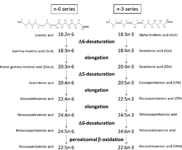
The n-3 and n-6 (or omega-3 and 6) FAs are a family of naturally occurring PUFAs (Figure 3). Since n-6 and n-3 double bonds cannot be inserted into FAs by animal enzymes allowing their endogenous de novo synthesis, the simplest members of n-6 and n-3 series, linoleic (18:2n-6) and α-linolenic (18:3n-3) FAs, are considered essential34. Symptoms of
essential FA deficiency, similar to other essential nutrient deficiencies, have been reported in mammals35.
In the cell, linoleic and α-linolenic acids are converted to longer chain FAs upon desaturation and elongation giving rise to the n-3 and n-6 series34(Figure 3).
26
Figure 3 – Metabolic pathways of essential linoleic (18:2n-6) and α-linolenic (18:3n-3) fatty acids. Different n- 3 and n-6 fatty acids are produced through elongation and desaturation of the essential linoleic and
α-linolenic acids, respectively. The addition of double bonds and the elongation of the acyl chains occur in the endoplasmic reticulum, while the final step in the synthesis of the n-3 DHA and the n-6 docosopentaenoic acid consists of a single reaction from β-oxidation in the peroxisome. Carbon backbones of linoleic and α-linolenic acids are depicted. Carbons are numbered (in blue) from the first methyl group. Names include: number of carbons : number of double bonds n-carbon number of the terminal double bond (i.e. that closest to the methyl end of the hydrocarbon chain).
The PUFAs of the n-3 and n-6 series play a significant role in health and disease. Both series are involved in the synthesis of potent modulatory molecules important for inflammatory responses, including eicosanoids (prostaglandins and leukotrienes) and cytokines (interleukins), affecting the gene expression of various bioactive molecules36,37.
In general, the n-6 eicosanoids are considered pro-inflammatory while n-3 are much less so. The n-6 eicosanoids family of inflammatory mediators is generated from 20 carbon PUFA as the arachidonic acid (20:4n-6), liberated from cell membrane phospholipids37.
Increased consumption of the long chain n-3 PUFA, such as the EPA (20:5n-3) and DHA (22:6n-3) acids, decreases the amount of arachidonic acid in cell membranes diminishing arachidonic acid-derived eicosanoids38.
Since n-6 and n-3 PUFAs cannot be synthesised de novo, their availability to the composition of cell membranes is ultimately determined by the fraction present in the
27
diet. Actually, membrane composition was found to be more responsive to n-6 and n-3 PUFA levels in the diet and most sensitive to n-3 PUFA and to the n-3/n-6 ratio33,39.
2.2. Effect of dietary fatty acids in human health.
A link between diet and disease is being increasingly recognized. Among dietary components, FAs have especially gained recognition in affecting health. The amount and type of FAs consumed are directly involved in the etiology of various diseases. While the ingestion of excessive amounts of SFAs and trans-fatty acids is considered to be a risk factor for cardiovascular diseases, insulin resistance, dyslipidemia, and obesity1, MUFAs
and PUFAs are recommended, for example, for their cardio-protective benefits40. In
particular, essential FAs from n-6 and n-3 series and their derivatives have varied biological actions and seem to be involved in several physiological and pathological processes such as obesity, hypertension, diabetes mellitus, coronary heart disease, schizophrenia, Alzheimer's disease, atherosclerosis, and cancer41. For example,
numerous studies have shown that high olive oil intake reduces blood pressure; it was proposed that this effect would be caused by its high OA content, increasing OA levels in membranes and thus regulating membrane lipid structure in such a way as to influence the localization and/or activity of signalling proteins involved in hypertension42,43. DHA
and EPA have also been associated with the prevention of cardiovascular diseases and cancer44. Gamma linolenic acid (GLA) (18:3n-6), a n-6 PUFA, has been suggested for
potential applications as an anti-inflammatory nutrient or adjuvant36.
Understanding the mechanisms by which FAs exert their biological effects is important in unravelling the pathogenesis of many disorders, ultimately providing effective preventive measures. Given the large number of processes and health benefits where FAs seem to be involved, it is reasonable to think that they may act at some fundamental level common to the whole organism. Their implications on membrane structure, may explain, at least partially, the modulation exerted by some natural FAs on cell function. In fact there is some evidence that a few metabolic pathologies are related to altered lipid composition. For instance, alterations in the FA composition of membrane phospholipids of human erythrocyte membranes have been linked to obesity and insulin resistance45. Another study in rats fed with a diet rich in SFA suggested that intake of the
beef tallow diet promotes body fat accumulation by reducing lipolytic activities resulting from lower β-receptor binding and sympathetic activity in adipose tissues. The decreases in β-receptor binding affinities were correlated to changes in membrane fluidity46.
2.3. Conjugated linoleic acid.
Another interesting group of FAs is that formed by the conjugated linoleic acid (CLA). This is a family of 28 naturally occurring isomers of linoleic acid, with 18 carbons and 2 double bonds either on trans or cis configuration. CLA is produced by bio-hydrogenation in ruminants or by bacteria from the gastro-intestinal tract of humans. Although as minor component, consumption of ruminant meat (beef and lamb) and dairy products (milk and cheese) is the main source of dietary CLAs47. Where the predominant
28
isomer formed is cis-9,trans11 (c9,t11) and unlike other trans double bond-containing FAs, it may have beneficial effects on human health48.
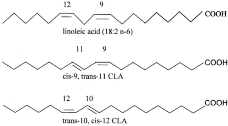
Dietary supplements of CLA have been attracting consumers’ interest because of the alleged body fat-lowering effects, together with the perception of a natural compound devoid of harmful effects. These preparations present CLA mostly as a mixture of (c9,t11) and trans-10,cis-12 (t10,c12) isomers (Figure 4).
Figure 4 - Structures of linoleic acid, and CLA isomers (c9,t11) and (t10, c12)
There are several indications that various isoforms might have different biological actions. The c9,t11-isomer was implicated as the active form responsible for the protective effects against tumorigenesis. The t10,c12-isomer seems to be the active form that affects energy metabolism and body fat deposition and composition49. Despite
numerous studies on CLA their true effect and the mechanism of action in such processes remain unclear49,50,51,52.
It was reported CLA incorporation into membrane phospholipids of pigs fed with a commercial CLA mixture, where the distribution of CLA isomers in liver phospholipids showed an increase compared to that present in the diet53.
3. Reduced protein diets (RDP).
An equilibrated diet contains 10-15% of the total daily energy provided from protein. Reducing the proportion of protein relative to energy in the diet is known to increase fat deposition. Using pigs as animals models it was shown that plasma cholesterol increased dramatically in lean pigs fed low protein diets54 and that reduced
dietary energy and protein in growing pigs significantly increased intramuscular fat55,
while having a minor effect on the amount of subcutaneous adipose tissue56,57. One
possible explanation for this effect is that the low protein content restricted muscle growth, resulting in excess energy being converted into intramuscular lipids rather than in endogenous protein synthesis56.
29
Since RDP affects FAs deposition in the body it would be interesting to investigate the effect of RDP on membrane composition and biophysical properties.
4. Adipose tissue and obesity.
Adipose tissue is a complex organ that regulates and coordinates energy homeostasis. This heterogeneous tissue is primarily composed of adipocytes surrounded by fibroblasts, fibroblastic preadipocytic cells, endothelial cells, nerves and immune cells. Although adipose tissue was originally thought to just be an energy storage site, studies in recent years have revealed that it carries out many key endocrine functions. Indeed, dysfunction of the adipose compartment is central to the pathology associated with metabolic diseases such as obesity, type 2 diabetes, cancer cachexia and lipodystrophies58.
There are two main types of adipose tissue in mammals, white adipose tissue (WAT) and brown adipose tissue (BAT). Adipocytes from WAT are optimized to store energy and are characterized by containing a large single cytoplasmic lipid droplet and a “squeezed” nucleus. BAT adipocytes dissipate energy for thermogenesis and are characterized by being polygonal cells with a roundish nucleus and several cytoplasmic lipid droplets. Thus, white and brown adipocytes are quite different in their morphology and physiology: white adipocytes store energy for the metabolic needs of the organism, whereas brown adipocytes burn energy for thermogenesis59. WAT is the main type of
adipose tissue found in adult humans and is distributed throughout the body in subcutaneous regions, surrounding visceral organs and in the face. It is an active endocrine organ that mostly regulates insulin sensitivity, lipid metabolism and satiety58.
During caloric excess periods, adipocytes readily convert glucose into FAs storing them along with those collected from the extracellular space. During periods of caloric deficit, stored triacylglycerols are hydrolysed to FFAs and glycerol60, which then leave the
cell towards other tissues.
Obesity is characterized by excess body fat, which is predominantly stored in the adipose tissue leading to adipose cells expansion. As adipose cells expand, more phospholipids have to be incorporated into cellular membranes. The specific mechanisms that may lead from obesity towards the higher risk of metabolic complications such as insulin resistance and type 2 diabetes remain elusive. One hypothesis is the adipose tissue expandability that states that adipose tissue has a limited maximal capacity to increase in mass. When the adipose tissue expansion limit is reached, it stops storing lipids appropriately. The excess lipid accumulates in organs such as muscle, liver, and pancreas, causing metabolic disease with insulin resistance61.
While the idea that lean and very obese people may develop insulin resistance through the same pathogenic paradigm of exhaustion of adipose tissue expandability is controversial61, it is actually well supported in rodent models; for example, studies in
mouse have demonstrated that even in overweight or obese mice, a genetic limit on adipose tissue expansion can exacerbate insulin resistance62,63. In humans, lipidomic
analyses of adipose tissue of lean and obese (but metabolically healthy) individuals, identified multiple changes in membrane phospholipids. Using computer modeling, it was shown that “lean” and “obese” membrane lipid compositions have the same physical
30
properties despite their different compositions64. These changes in lipid membrane
composition were suggested to occur in order to protect the physical properties of the membranes.
The expandability limit is determined on an individual basis by environmental and genetic factors61. Since dietary FAs may incorporate into the membranes and/or serve as
precursors to other FAs they may also have a role in the expansion capacity of adipocyte cell membranes.
5. Genetic background and animal models.
It is well accepted that the genotype influences the metabolism of nutrients in particular lipid metabolism. How the membrane biophysical properties are affected by the genetic background and regulated to match the specific needs of the individual is still obscure.
Considering problems in collecting tissue samples and the multifactorial etiology of obesity in human patients, suitable animal models are essential for a better understanding of the metabolic onset of obesity. Pigs display several anatomical-physiological and metabolic similarities to humans65. Additionally, taking advantage of
pigs genetic background (fat or lean, genetic or diet induced obesity), pigs are considered a valuable animal model. Zucker rats are also commonly used as a genetic model for human obesity66. The fa/fa Zucker rat is a strain of laboratory animal descendent from
the wild model rodent Rattus norvegicus67, used often to study genetic obesity problems.
This animal model develops morbid obesity, through the fa/fa mutation in the extracellular domain of the leptin receptor, which inhibits completely leptin action. This leads to an appetite increase promoting severe insulin resistance68. The rat is a highly
valuable model for the investigation of cardiovascular diseases, metabolic disorders (e.g. lipid metabolism, diabetes), cancers, renal dysfunctions, neurologic pathologies, and so many other diseases.
In addition, in these studies we also used bovines that are models for animal production studies. This approach allows investigating the possible correlation of dietary fats with membrane features and possible outcome on meat quality.
31
Aim of part I:
The work presented in part I - chapter 1 of this thesis includes publications 1, 2 and 3 that are focused on the study of the influence of genetic background and diet on adipocyte membrane composition and its outcome on membrane biophysical properties such as fluidity and permeability to water and glycerol.
33
CHAPTER 1 - Influence of diet on cell membrane composition and
biophysical properties
35
CHAPTER 1 - Influence of diet on cell membrane composition and
biophysical properties
The information contained in this chapter is included in the following original publications:
Publication1
Martins AP, Lopes PA, Martins SV, Madeira A, Santos NC, Prates JA, Moura TF, Soveral G.
Conjugated linoleic acid reduces permeability and fluidity of adipose plasma membranes from obese Zucker rats.
Biochem Biophys Res Commun. 2010 Jul 23;398(2):199-204. doi: 10.1016/j.bbrc.2010.06.059.
Publication 2
Martins AP, Lopes PA, Costa AS, Martins SV, Santos NC, Prates JA, Moura TF, Soveral G.
Differential mesenteric fat deposition in bovines fed on silage or concentrate is independent of glycerol membrane permeability.
Animal. 2011 Dec;5(12):1949-56. doi: 10.1017/S1751731111001091.
Publication 3
Martins AP, Lopes PA, Madeira MS, Martins SV, Santos NC, Moura TF, Prates JA, Soveral G. Differences in lipid deposition and adipose membrane biophysical properties from lean and obese pigs under dietary protein restriction.
Biochem Biophys Res Commun. 2012 Jun 22;423(1):170-5. doi: 10.1016/j.bbrc.2012.05.108.
37
43 Publication 2: Differential mesenteric fat deposition in bovines fed on silage or concentrate is independent of glycerol membrane permeability.
51 Publication 3: Differences in lipid deposition and adipose membrane biophysical properties from lean and obese pigs under dietary protein restriction.
57
PART II – Aquaporins
59
INTRODUCTION
One of the fundamental tasks of the plasma membrane is to quickly respond to intra and extracellular events. Water is the major component of living organisms, thus controlled flow of water into and out of cells is central for cell homeostasis. Although water can cross biological membranes through the lipid bilayer, membrane water permeability can be greatly enhanced by specific proteins called aquaporins (AQPs). These transmembrane proteins enable the fine-tuning of water permeation and in some cases also small solutes. Part II of this thesis will focus on different aspects of these proteins.
Aquaporins
AQPs are a family of small transmembrane channels ubiquitous in nature. These proteins form highly selective channels that, in response to osmotic or solute gradients, facilitate bidirectional flow of water and in the case of aquaglyceroporins, other small solutes such as glycerol and urea, enabling the adjustment of these flows to the organism necessities.
The existence of water channels was predicted long before aquaporin discovery. The first studies on water transport started in the late 1950s on mammalian red blood cells (RBC)69–72 and later on renal epithelia73 and demonstrated that water permeability
in these cells was much higher than predicted by simple water diffusion through the bilayer. Evidence for their existence was based mainly on measures of high osmotic water permeability, on their inhibition by mercurial reagents69,70, on the low activation energy
for transport74 and finally on the ratio of osmotic to diffusion water permeabilities75
leading to the proposed single-file mechanism of transport within the channel76. In 1984
it was identified a major intrinsic protein in bovine lens cells that was suggested to participate in the formation of an aqueous channel77. But only in the 1990s, using a
Xenopus oocyte expression assay, Agre and co-workers78 demonstrated that a 28-kDa
membrane protein that is abundant in RBC and renal proximal tubules 79 was water
permeable. The 28-kDa membrane protein was later named aquaporin-1 (AQP1).
Ever since their discovery, more than 300 homologues from many phyla, including bacteria, plant, and animal have been identified80. In prokaryotic and other
microorganisms, AQPs are believed to aid survival by providing protection against osmotic shocks and rapid freezing81,82. In Escherichia coli there are two MIP family
proteins: one is the glycerol facilitator (GlpF)83 and the other is the water channel
(AQPZ)84. More AQP genes are present in the genomes of multicellular organisms:
Arabidopsis thaliana contains 35 putative AQP genes85 while in humans, 13 AQPs
isoforms, distributed in specific organs, tissues and with different cellular localization have been identified so far3. The identification and study of mammalian AQPs have
provided insight at the molecular level into the fundamental physiology of water balance, regulation and the pathophysiology of water balance disorders3,86.
60
Furthermore, aside from water and glycerol, numerous studies have pointed out a broader range of AQPs’ substrates, these include polyols, hydrogen peroxide, ammonia, nitrate, arsenite, antimonite, small ions, and gases87.
The widespread distribution of this ancient family of channels in all kingdoms of lifepoints to their fundamental significance in biology.
1. Aquaporin classification and selectivity.
Thirteen mammalian AQPs, have been described so far. These can be divided in three sub-groups mainly determined by their transport capabilities: i) orthodox or classical AQPs (AQP0, AQP1, AQP2, AQP4, AQP5, AQP6 and AQP8), primarily water selective facilitating water movement across cell membranes in response to osmotic or pressure gradients and, ii) aquaglyceroporins (AQP3, AQP7, AQP9 and AQP10) which transport some small uncharged solutes such as glycerol and urea in addition to water. A third sub-group, named iii) S-aquaporins (AQP11 and AQP12), was defined based primarily on their subcellular location and on the lower sequence similarity to the other mammalian AQPs (Figure 5).
Figure 5 - Phylogenetic tree of the human Aquaporin gene family (Adapted from 88). Water permeable AQPs
(AQP0, 1, 2, 4, 5, 6, 8, AqpZ) are shown in blue background. Glycerol permeable aquaglyceroporins (AQP3, 7, 9, 10, GlpF) are in orange background. AqpZ and GlpF are the E. coli homologues. S-Aquaporins (AQP11 and 12) is on the bottom right with green background. The scale bar represents genetic distance between homologues.
Numerous studies have revealed the diverse permeation characteristics of AQPs. In addition to water and glycerol, a diverse set of solutes including small ions, urea, hydrogen peroxide, ammonia, nitrate, arsenite, antimonite and gases87, has been shown
to permeate through specific AQPs. Aquaglyceroporins AQP3, AQP7, AQP9 and AQP10 transport glycerol and urea89–92 , while both AQP7 and AQP9 are permeable for arsenite93.
61
In addition, AQP9 also permeates a wide range of non-charged solutes like mannitol, sorbitol, purines and pyrimidines94. AQP6 has low water permeability and seems to
function primarily as an anion transporter95,96. In addition, AQPs have also been proposed
to transport gases, including carbon dioxide, ammonia, nitric oxide and hydrogen peroxide97–99.However, some of these permeability characteristics are still controversial
and subject of intense debate100,101.
2. Aquaporin structure.
The three dimensional structures of several AQPs has enabled the conceptualization of a general structure, revealing the structural determinants that are essential for AQPs selectivity and extraordinary permeation rates. The atomic model of mammalian AQP1 derived from a 3.8 Å resolution potential map obtained by electron crystallography was the first atomic structure of a human membrane protein to be solved, and gave the first insight into AQP’s water specificity102. Medium and high-resolution
structures of several AQPs belonging to different subfamilies have been ever since determined, namely, from archaea103, bacteria104,105, yeast106, protozoa107, plants108 and
mammals109–112. More recently, molecular dynamics (MD) simulations have
complemented the experimental data, by providing the progression of the biomolecular system at atomic resolution101.
The reported structures have revealed that AQPs are grouped as homotetramers embedded in the bilayer113, consisting of four independent monomers, each behaving as
an independent channel114 (Figure 6) and sharing a conserved overall typical hourglass
fold102,115.
Figure 6 - Tetrameric structure of bovine AQP1. (A) side view, (B) top and bottom views. Reprinted with author's permission116.
62
Each monomer interact with two of its neighbours, forming the tetramer central pore. It has been suggested that this pore, which is not involved in water conductance102,
may permeate gases99,117,118 and function as a gated cation channel119,120. Each AQP
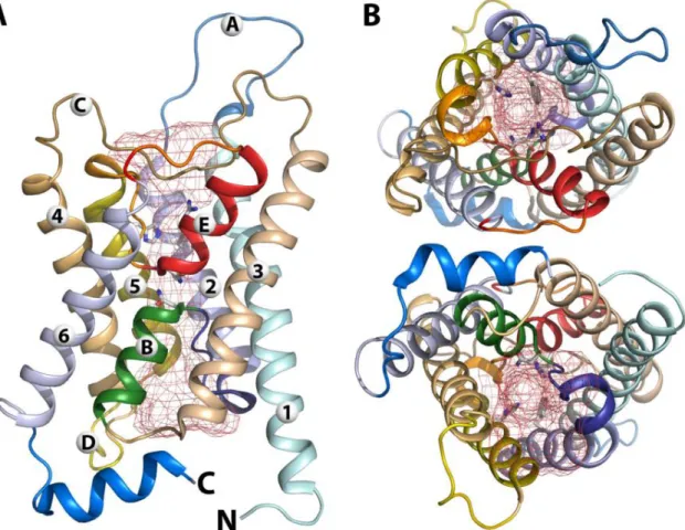
monomer is a small protein with usually fewer than 300 amino acids, organized in six transmembrane α-helices (H1-6) arranged in a right-handed helical bundle and connected by five loops (A-E), with both amino and carboxyl termini located in the cytoplasm side121,122 (Figure 7A). The angle at which the transmembrane α-helices are
oriented gives rise to funnel-shaped cytoplasmic and extracellular vestibules connected by the conduction pore. The latter is formed by two small α-helical segments, loop B, entering from exoplasmatic side and connecting H2 and H3, while loop E enters from the cytoplasmatic side and connects H5 and H6, called HB and HE respectively123.
Figure 7 - Monomeric structure of bovine AQP1. (A) side view, (B) top and bottom views. The red mesh
indicates the pore region. Reprinted with author's permission116.
Aquaporins have important characteristics (two filters) that provide selectivity for water and some small solutes, preventing protons and solutes above a certain size and charged solutes to permeate (Figure 8). The amino acid residues of the first selective filter is for most AQPs composed of two highly conserved asparagine-proline-alanine (NPA), called signature motifs, located at the end of the loops (HB and HE). These motifs are oriented 180 degrees toward each other, forming part of the surface of the aqueous pathway at the centre of the pore. The chemical/structural characteristics of this region, mostly due to the presence of these residues and the dipole behaviour of the two half
63
helices HB, generates an electric field124 that induces upon the interaction with the
asparagines, a rotation of the water molecule, thus disrupting any H-bonds between adjacent water molecules and preventing the Grottus mechanism for protons permeation102,104,109,115,125,126,127. The second important selective filter located at the
extracellular side, the aromatic/arginine (ar/R) constriction region, prevents permeation of solutes above 2.8 Å and of charged solutes109. Within this region a hydrophobic
phenylalanine residue side chain (Phe 58 in bAQP1) orients the water molecules such as to enforce strong hydrogen bonds to an arginine and a histidine (Arg 197 and His 182 in bAQP1) (Figure 8A). Besides these selective filters in the pore, the channel has in the vestibules at the entrance of either side, in the vestibules, different residues, mainly carbonyl groups that interact with water molecules82,109. Furthermore, the wall regions
of the pore have different hydrophobic/hydrophilic characteristics determining, conduction rate and the open/closed state of the channel82.
Figure 8 - Detailed view of bovine AQP1 pore region. (A) Half helices dipoles and the hydrophilic and
hydrophobic residues lining the pore are depicted in red and yellow, respectively; the ar/R and NPA selective filters are shown as well; (B) Inside view of the pore surface (hydrophilic and hydrophobic properties are painted red and yellow, respectively). Reprinted with author's permission116.
In the highly dielectric (ε = 80) bulk water, protons are well solvated by surrounding water molecules. Upon moving the proton across the AQP pore, the solvation shell needs to be removed from the proton thus creating an electrostatic barrier128,129. Inside the pore, the hydronium ion is only partially solvated by few close
water molecules, and the surrounding protein medium (ε ≈ 4) stabilizes the hydronium only to a fraction of the solvation in the bulk. Thus, a large energetic cost results for moving the hydronium from the water into the pore. Inside the pore, water molecules move in single file, so whenever and wherever bulk water-water hydrogen bonds have to
64
be ruptured to allow the water molecule to squeeze through the narrow NPA or ar/R regions (so preventing protons to permeate AQPs), the protein offers replacement interactions, which largely compensate for the energetic cost of water-water bond rupture. This remarkable complementarity to bulk water lowers the activation barrier to a large extent. Furthermore, the high permeation rates are observed both experimentally and in simulations101,109. The high permeation rate is thought to be partially due to the
hydrophobic nature of some pore regions, where water molecules have to pass rapidly, while they slowdown in hydrophilic regions upon interactions with residues. The conduction rate is so, modulated by the hydrophobicity/hydrophilic characteristics of the pore and is often referred as saltatory conduction (named after the saltatory ion conduction in axons).
3. Aquaglyceroporin structure.
The structure of Escherichia coli glycerol facilitator GlpF revealed that the AQP general fold topology is conserved among orthodox AQPs and aquaglyceroporins104,130.
Yet GlpF permeability to water is substantially less than that measured for its bacterial AQP counterpart AqpZ83,131.
The ar/R constriction region of GlpF has Trp48, Gly191, Phe200, and Arg205109
instead of Phe56, His180, Cys189 and Arg195 in AQP1. Among the water-specific AQPs, the histidine residue, which is preserved among water-specific AQPs, together with the highly conserved arginine residue providing a hydrophilic edge in juxtaposition to an aromatic residue123. On the other hand, for GlpF, the replacement of this histidine by
glycine significantly increases the size of this constriction region and sterically allows a second residue change, replacing the cysteine by a phenylalanine109,123,132. These
substitutions have two critical effects on the characteristics of the GlpF ar/R constriction region: they increase its size, making it wider than AQP1 (3.4Å and 2.8Å, respectively) and also increase its hydrophobicity109. It seems that the more hydrophobic nature of GlpF is
the primary factor responsible for its relatively reduced ability to transport water109,133.
Moreover, the ladder of aromatic residues (Phe and Trp) in GlpF constriction region forms a greasy slide that allows the efficient conduction of glycerol, small linear polyols, and urea, while making it less efficient for water passage123.
The GlpF channel lining is strongly amphipathic, with oxygens and nitrogens lined up on one side and carbons on the opposite side of the lumen surface. This amphipathic channel uniquely matches the chemical structure of glycerol, which is a composite of the polar hydroxyl group arranged on a non-polar alkyl backbone104. The glycerol permeation
through the selectivity filter of GlpF depends on the orientation of the glycerol molecule inside the pore. In the narrow ar/R selectivity filter of the channel all three hydroxyl groups of glycerol are able to form hydrogen bonds to polar protein groups or to the nearby water molecules134. Two consecutive OH groups of the glycerol molecule are
oriented with ideal geometry with respect to donor and acceptor atoms from within the channel104.