Arq Neuropsiquiatr 2007;65(4-A):1037-1039
1037
HORNER’S SYNDROME AFTER BLUNT
CERVICAL AND CHEST TRAUMA
Case report
Wellingson Silva Paiva
1, Robson Luis Oliveira De Amorim
1, Wagner Malago Tavares
1,
Eduardo Joaquim Lopes Alho
1, Brasil Ping Jeng
1, Eberval Gadelha Figueiredo
2ABSTRACT - Horner‘s syndrome is the triad of miosis, ptosis, and anhidrosis, resulting from disruption of the sympathetic pathways. This article describes an uncommon case of Horner‘s syndrome in a 22-year-old man after blunt trauma to the neck and chest without carotid artery dissection. The patient was brought to the emergency service after motorcycle fall. Neurologic examination revealed a patient presenting the score 15 at Glasgow Coma Scale. The left eyelid was 1–2 mm lower than the right. Carotid Doppler and angiotomog-raphy were undertaken and revealed no abnormalities of the carotid artery. CT disclosed a mediastinal he-matoma extending to the left apex, compressing the left sympathetic chain. The understanding of this clin-ical entity may help the surgeon to make a better differential diagnosis in trauma patients in whom prompt diagnosis is critical to stablish the correct treatment.
KEY WORDS: Horner syndrome, trauma, etiology.
Síndrome de Horner após trauma cérvico-torácico fechado: relato de caso
RESUMO - A síndrome de Horner compreende a tríade de miose, ptose e anidrose, resultado de lesão em al-gum ponto das vias simpáticas. O referido estudo apresenta um caso da referida síndrome em um jovem de 22 anos vitima de queda de moto, com escoriações no tórax e no pescoço, sem dissecção carotídea. Ao exa-me neurológico, encontrava-se com 15 pontos na Escala de Coma de Glasgow, com miose à esquerda e pto-se palpebral ipsilateral. Realizado Doppler de carótidas e angiotomografia dos vasos cérvico-cranianos não sendo evidenciadas anormalidades. A tomografia de tórax mostrou um hematoma no ápice pulmonar es-querdo, comprimindo a cadeia simpática ipsilateral. O conhecimento desta entidade clínica pode ajudar o cirurgião a fazer um diagnóstico diferencial adequado nos pacientes vítimas de traumas, nos quais o diag-nóstico correto e eficaz pode ser fundamental para a definição da conduta a ser tomada.
PALAVRAS-CHAVE: síndrome de Horner, trauma, etiologia.
1MD, Neurosurgery Resident Hospital das Clínicas University of São Paulo, São Paulo SP, Brazil; 2MD, Neurosurgeon, Chief of the Cerebrovascular Surgical Team Hospital das Clínicas University of São Paulo, São Paulo SP, Brazil;
Received 29 March 2007, received in fi nal form 18 July 2007. Accepted 25 August 2007.
Dr. Wellingson Paiva - Rua Ovidio Pires de Campos, 171 / 511 - 05403-010 São Paulo SP - Brasil. E-mail: [email protected]
Horner’s syndrome consists of a triad of symp-toms (miosis, ptosis, and anhydrosis) resulting from disruption of the sympathetic pathways somewhere between the brain and the eyes. In blunt trauma, it is usually associated with carotid artery dissection. Although the individual signs of Horner’s syndrome do not constitute an emergency, the internal carot-id artery proximity to the sympathetic ganglia makes Horner’s syndrome a potential vascular emergency1-3.
We present a case of Horner’s syndrome in a 22-year-old man after blunt trauma to the neck and head unrelated to carotid artery dissection.
CASE
A 22-year-old man was brought to the emergency room after motorcycle fall, with history of transitory loss of con-science. In the admition, he was alert and orientated, the carotid pulses were symmetric, regular in rate and rhythm, with no bruits and the chest and the abdomen had no signs of abnormalities. The patient related moderate cervical pain but no neurological defi cits were noticed except for the asy-metric pupils that measured 5 mm on the right and 2 mm on the left side, both presenting direct and consensual
re-Arq Neuropsiquiatr 2007;65(4-A)
1038
Horner’s syndrome: blunt cervical / chest trauma Paiva et al.
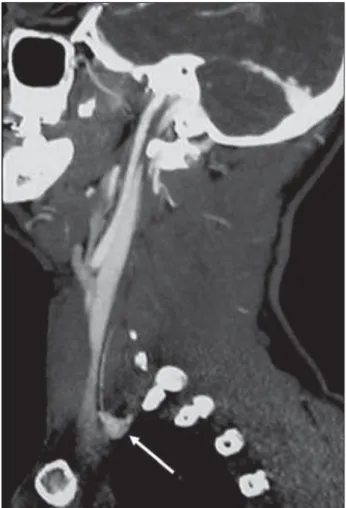
veal any rib, sternal fractures or mediastinal enlargement. Skull computed tomography (CT) showed no abnormality so as the carotid ultrasonography Doppler and the angioto-mography of the head and neck. Cervical spine CT showed a fracture of left C7 transverse process and additional investi-gation with chest CT disclosed a mediastinal hematoma ex-tending to the left lung apex, exhibiting mass effect over surrounding structures without signs of aortic dissection (Fig 2). A conservative management was adopted and the pa-tient left the hospital three days later but still with the neu-rologic signs. Follow up four weeks after discharge revealed a normal neurologic examination and no complaints.
DISCUSSION
Horner’s syndrome is an uncommon occurrence in all age groups (0.08% of blunt trauma patients)2.
Di-agnosis is namely based on clinical fi ndings, and after careful history and examination, the physician must decide whether further investigation is necessary. There is a wide variety of conditions that may cause this syndrome, postsurgical and iatrogenic causes com-prise most of the cases3-8. Penetrating neck injuries,
cervical spine dislocation and birth trauma are the major factors that lead to traumatic injury to the ocu-losympathetic pathway7,9. A history of trauma
preced-ing these fi ndings should prompt the clinician to con-sider that the carotid artery, which lies directly over the sympathetic chain in the neck, may have been in-jured, particularly if signs of head or neck trauma are present8,10,11.The investigation of choice considered by
some authors is a magnetic resonance imaging and angiography scan of the head and neck10. Therefore,
to exclude carotid injury the authors performed an ultrasonography Doppler and an angiotomography what seems to be less invasive and with a high sen-sivitity. The carotid dissection diagnosis implies an emergent condition that can lead, if misdiagnosed, to major catastrophes including massive ischemic stroke, even in a patient with minor symptoms at admission11.
In this current case further investigation showed a mediastinal and left lung apical hematoma which probably caused compression of the sympathetic
gan-glia, as the clinical fi ndings appeared in fi rst day of trauma. When it comes to blunt chest trauma, there is an association with fractures of the first rib7,9,
not seen in this patient. The fracture of the left C7 transverse process could explain the cervical pain, therefore, the correlation of this radiological fi nd-ing with the hematoma can not be affi rmed. Medi-astinal hematoma due to trauma is associated with sternal fracture, aortic dissection and extrapericardial cardiac tamponade12-14. Before the era of
multidetec-tor helicoidal CT, performed in this patient, the gold standard to investigate aortic dissection was angiog-raphy14. Asensio-Sánchez et al.15, described a case of
Horner’s syndrome due to an hematoma in the neck which was producing tracheal deformity and steno-sis. A chest hematoma causing such syndrome was described by Banks et al.16 and the signs were caused
by a tension hemothorax, a life-threatening condi-tion. In our case, the patient was hemodynamically stable and no surgical intervention was necessary. This report illustrates a condition that can be seen in the trauma emergency department and shows that a
Fig 1. Assimetric pupils and left semiptosis
Arq Neuropsiquiatr 2007;65(4-A)
1039 Horner’s syndrome: blunt cervical / chest trauma Paiva et al.
meticulous and sequenced investigation with proper complementary exams is necessary because such signs can be just the “iceberg tip”.
In conclusion, Horner’s syndrome is a very rare condition after mild neck and chest trauma. The un-derstanding of this clinical entity may help the sur-geon to make a better differential diagnosis in trau-ma patients in whom correct and prompt diagnosis can be lifesaving.
REFERENCES
1. Rudolph A. Rudolph’s pediatrics. 20.Ed. Stamford: Appleton & Lange, 1996:112-118
2. Davis JW, Holbrook TL, Hoyt DB, Mackersie RC, Field TO Jr, Shackford SR. Blunt carotid artery dissection: incidence, associated injuries, screen-ing and treatment. J Trauma 1990;30:1514-1517.
3. Kaya SO, Liman ST, Bir LS, Yuncu G, Erbay HR. Unusual Horner’s syn-drome as a complication in thoracic surgical practice. Eur J Cardiotho-rac Surg 2003;24:1025-1028
4. Pearce SH, Rees CJ, Smith RH. Horner’s syndrome: an unusual iatro-genic complication of pneumothorax. Br J Clin Pract 1995;49:48.
5. Shen SY, Liang BC. Horner’s syndrome following chest drain migration in the treatment of pneumothorax. Eye 2003;17:785-788.
6. Campbell P, Neil T, Wake PN. Horner’s syndrome caused by an inter-costal chest drain. Thorax 1989;44:305-306.
7. Bell LR, Atweh N, Ivy M, Possenti P. Traumatic and iatrogenic Horner’s syn-drome: case report and review of the literature. J Trauma 2001;51:400-404. 8. Thanvi B, Munshi SK, Dawson SL, Robinson TG. Carotid and vertebral
artery dissection syndromes. Postgrad Med J 2005;81:383-388. 9. Harris GJ, Soper RT. Pediatric fi rst rib fractures. J Trauma 1990;30:343-345.
10. Chan CC, Paine M, O’Day. Carotid dissection: a common cause of Horner’s syndrome. J Clin Exp Ophthalmol 2001;29:411-415. 11. Liu WP, Ng KC, Hung JJ. Carotid artery injury with cerebral infarction
following head and neck blunt trauma: report of a case. Yale J Biol Med 2005;78:151-156.
12. Coleman GM, Fischer R, Fuentes F. Blunt chest trauma: extrapericardial cardiac tamponade by a mediastinal hematoma. Chest 1989;95:922-924. 13. Sia AT. Cardiac tamponade: an unlikely cause of unexplained hypoten-sion in an isolated “minor” blunt chest injury. Singapore Med J 1997; 38:35-36.
14. Geusens E, Pans S, Prinsloo J, Fourneau I. The widened mediastinum in trauma patients. Eur J Emerg Med 2005;12:179-184.
15. Asensio-Sánchez VM, Sánchez-Rodríguez JC, Macías-Pascual J, Mar-tínez-Rodríguez M. Traumatic Horner’s syndrome. Arch Soc Esp Ot al-mol 2007;82:171-173.