CORPUS CALLOSUM INDEX
A practical method for long-term
follow-up in multiple sclerosis
Fernando Faria Andrade Figueira
1, Valeria Silva dos Santos
2,
Gustavo Medeiros Andrade Figueira
3, Ângela Correa Marques da Silva
4ABSTRACT - Rather than acute inflammation, long-standing multiple sclerosis (MS) course is hallmarked by relentless axonal loss and brain atrophy, both with subtle clinical expression and scarcely visible on conven-tional MRI studies. Brain atrophy imaging has sophisticated methodological requirements, not always prac-tical and accessible to most centers. Corpus callosum (CC) is a major inter-hemispheric white matter bun-dle, grossly affected by long term MS and easily assessed by MRI. To determine whether a practical imag-ing method can reliably follow presumed axonal loss in patients with progressive MS, we designed a 5-year prospective open label study, enrolling 128 consecutive patients (75 relapsing-remitting (RR) and 53 second-ary-progressive (SP)), on regular immunomodulatory therapy compared to control group, formed by 23 pa-tients with MRI considered normal. On a conventional best mid-saggital T1W, CC index (CCI) was obtained by measuring anterior, medium and posterior segments of CC, normalized to its greatest anteroposterior diameter using an orthogonal semi-automated linear system. CCI was measured at baseline and at least once yearly. Results were plotted intra-individually; baseline values were used as reference. At baseline, CCI was able to distinguish SP patients from RR and controls, and on follow-up, despite some overlap, demon-strated a progressive reduction from baseline on both RR and SP groups compared to controls. From the third year on, difference between SP and RR patients reached statistical significance, which did not corre-lated with disability measured by EDSS. So, a corpus callosum index proved practical and feasible to longi-tudinally demonstrate morphometric callosal changes with potential to be used as a tool for long-term fol-low-up, mostly in SP patients.
KEY WORDS: multiple sclerosis, MRI, corpus callosum.
Índice de corpo caloso: um instrumento prático para o seguimento a longo prazo de pacientes com escle-rose múltipla
RESUMO - Mais do que inflamação aguda, a perda celular e conseqüente atrofia cerebral são os fatos pa-tofisiológicos mais marcantes na fase progressiva da esclerose múltipla (EM). No entanto, correlatos clínicos e de imagem por ressonância magnética (IRM) destes eventos, requerem sofisticada tecnologia, nem sem-pre prática e quase nunca acessível à maioria dos centros de tratamento. Deste modo, considerando a hi-pótese de que esta perda celular compromete fibras associativas que compõem o corpo caloso (CC), estru-tura facilmente acessível à IRM convencional, nosso grupo elaborou um estudo prospectivo aberto, atual-mente com 5 anos de duração, e envolvendo 128 pacientes consecutivos, todos em acompanhamento regu-lar em nosso centro de tratamento para a EM. A aquisição do índice de CC se deu através de um “melhor” corte meio-sagital em estudo convencional de imagem ponderada por T1, utilizando um sistema linear or-togonal semi-automatizado. Este índice foi obtido no início do seguimento e sua evolução foi acompanha-da anualmente. A partir do terceiro ano deste seguimento, sua diferença entre os pacientes com a forma surto-remissiva e aqueles com a forma secundariamente progressiva alcançou significância estatística. Esta diferença não mostrou correlação com o grau de incapacidade medido pelo EDSS. Assim, um índice de CC mostrou-se uma medida prática para o seguimento de alterações morfométricas do corpo caloso, provan-do assim potencial para ser utilizaprovan-do no acompanhamento em longo prazo de pacientes com EM, em espe-cial aqueles com a forma progressiva.
PALAVRAS-CHAVE: esclerose múltipla, IRM, corpo caloso.
1Head, Dept. Neurology, Hospital da Penitência, Rio de Janeiro RJ, Brazil; 2Assistant, Dept. Neurology, Hospital da Penitência, Rio de Janeiro RJ, Brazil; 3Post-Graduate, Federal University of State of Rio de Janeiro, Rio de Janeiro RJ, Brazil; 4Speech Therapist, Dept. Neu-rology, Hospital da Penitência, Rio de Janeiro RJ, Brazil. Dr. Figueira received eventual support and honoraria for scientifi c consulting and speaking from Abbott Laboratories, Schering do Brasil, Serono and TRB Pharma. This manuscript has no fi nancial disclosure. Received 28 February 2007, received in fi nal form 4 June 2007. Accepted 3 August 2007.
Multiple sclerosis (MS) is a systemic autoimmune disease with exclusive expression over the central ner-vous system (CNS), characterized, at least at presenta-tion, in about 85% of cases, by a relapsing-remitting (RR) course. This RR course is the clinical expression of a temporal and spatial dissemination of a focal
in-fl ammatory process, clinically represented by relaps-es, most of them followed by a more or less complete and spontaneous remission1. Despite this classical
con-cept, current evidence points to a silent, more wide-spread and ongoing infl ammation, diffusely involving CNS, besides axonal loss, which has been demonstrat-ed to occur early in the course of the disease and to be a key factor for progression of disability2-5. So, as
time goes by and infl ammation tends to slow down, relapses become rarer, giving place to a more relent-less secondary progressive (SP) phase, hallmarked by axonal loss and brain atrophy. At this point, conven-tional measures used as endpoints in most pivotal short-term follow-up MS studies, such as number of relapses and magnetic resonance imaging (MRI) ac-tivity, characterized by T1 weighted (T1W) gadolini-um enhancement (Gd+) and refl ecting infl ammation, lose sensitivity. Axonal loss in MS has scarce expres-sion. Clinically, it leads to progressive and insidious changes in cognitive functions, such as memory and speed of mental processing, which impacts daily liv-ing activities but lacks specifi city and is rarely taken into account on therapeutic decisions. On convention-al imaging, its paradigm is based on morphologicconvention-al changes, as intracellular iron deposition, expressed by T2W hypointense sign at subcortical gray matter nuclei6, and volume reduction, as formally expressed
by changes in brain parenchymal fraction (BPF)7, both
too complicated to be used on daily practice. Other-wise, as it occurs widely over the CNS, clinical-imag-ing correlates for axonal loss are poorly specifi c and require sophisticated methodology, being impracti-cal to be used on bedside basis and inaccessible to most centers8,9.
Corpus callosum (CC) is a major white matter bun-dle which plays an important role in functional inter-hemispheric integration, communicating cognitive information through homotopic, and some hetero-topic, interconnections between the hemispheres10,11.
The CC is usually grossly affected by long-term dis-ease12 and, because of its functional relevance to
in-ter-hemispheric information transfer, it might be one of the components of the complex pathological pro-cess that leads to cognitive changes in MS. Further-more, it is one of the few white matter tracts that can
be discretely identifi ed by conventional MRI, having sharply demarcated two-dimensional limits on a mid-saggital T1W imaging. So, as a consequence of these anatomic and functional properties, it is reasonable to assume that CC morphometrics might be a possible marker for the integrity of these associative fi bers.
Involvement of associative fibers leads to mor-phometric changes in CC, affecting inter-hemispher-ic transfer of information and it is a major patho-logical substrate for slowing of mental processing on long-term MS9.
METHOD
Sample – One-hundred and twenty eight consecutive non-selected patients, with diagnosis of clinically defi ned MS according to the International Panel criteria13 and in-cluded in our institutional MS treatment program12, were followed for fi ve years, in an open-label trial. Sample de-mographics are shown in Table. All patients were on fol-low-up according to the standard protocol of our reference center, regularly using one of the available immunomodula-tory drugs for at least two years and had no relapses on pri-or 12-month period. A control group was fpri-ormed by twen-ty-three patients, submitted to a normal MRI study, for a non-infl ammatory neurological condition, such as epilep-sy and headache.
Imaging – Patients were submitted to the same con-ventional brain MRI scanning protocol on baseline using a 1.5T Sigma Magneton scanner (Siemens AG, Germany), according to the Consortium of Multiple Sclerosis Centers Magnetic Resonance Imaging working group standard rec-ommendations14, including an axial pre and post Gd T1W, saggital T1W and FLAIR, axial T2W and FLAIR. Serial imag-ing was repeated at least annually for a 5-year period, but Gd sequences were used only if clinically indicated. All MRI were analyzed by the same examiner (FF), and, at baseline study, data were compared to those of a blind radiologist, to achieve reproducibility.
Corpus callosum index (CCI) was obtained on a conven-tional best mid-saggital T1W image, using a simple orthog-onal semi-automated system, by drawing a straight line at greatest anteroposterior diameter of CC and a perpendicu-lar at its midline, owing to points a, b and c (Fig 1). Anterior (aa´), posterior (bb´) and medium (cc´) segments of CC were
Table. Demographic data. RR MS (N=75)
SP MS (N=53)
Control (N=23)
Gender (M/F) 29/46 19/34 9/14
measured and normalized to its greatest anteroposterior di-ameter (ab). Studies were done at baseline and yearly, and results plotted intra-individually for follow-up, matched to baseline values used as a reference standard15. For compar-ison and validation purposes only, at the beginning of fol-low-up we obtained brain parenchymal fraction in all ex-ams, using methodology previously described7.
Clinical evaluation – After a complete baseline study and fi nal diagnosis of MS, patients were introduced to our treatment center standard protocol, with an every three-month routine clinical follow-up, including disability es-timates by expanded disability status scale (EDSS)16, done at each consultation. A brief neuropsychological test bat-tery, described elsewhere9, including memory, verbal
fl u-ency, depression task and a paced auditory serial addiction Fig 1. Determination of corpus callosum index, using a “best”
midsagittal slice on a T1W brain MRI.
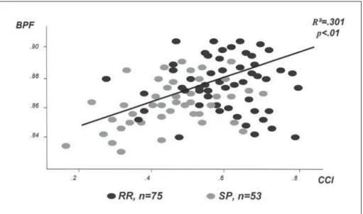
Fig 2. Correlation of corpus callosum index and brain parenchymal fraction, at baseline of the study.
test (PASAT), with rounds of 3, 2.5 and 2 seconds interval, was applied as part of baseline evaluation, always by the same examiner (AS). Scoring on PASAT was obtained by ar-ithmetical mean of the decimal fraction number of correct answers at each round.
Statistical analysis – Demographic data values are pre-sented as mean ± standard deviation (SD). Comparison of categorical variables was assessed by Pearson chi-square or the Fisher exact test, as appropriate. A p value of 0.05 or less was considered for statistical purposes of signifi cance.
The study was designed to enroll patients on regular clinical follow-up and treatment. Protocol of the study was analyzed and approved by Ethical Committee of our Hospi-tal and all patients intended to be involved gave informed consent to it.
RESULTS
Baseline – On baseline, when compared to scores of a blind radiologist, CCI determination showed a interobserver disagreement of 0.92% (SD=.32; p= 0.003)12. At this point, CCI was compared to brain
pa-renchymal fraction (BPF) that, despite being too com-plicated to be used in daily practice is a widely accept-ed standardizaccept-ed measure for total brain atrophy (Fig 2). CCI showed a linear correlation with BPF on both groups of patients (R2=.301; p<.01), and was able to
distinguish SP patients from RR and controls (p=.014 and p=.003, respectively)12. Then CCI was normalized
to its mean value separately in RR and SP patient groups to study its correlation to PASAT, as a clinical scorer for speed of inter-hemispheric transfer of in-formation, a function at least theoretically linked to callosal fi ber integrity. Results showed a linear corre-lation between PASAT score and normalized CCI
mea-sure both on RR (R2=.364; p<.01) and SP (R2=.431; p<
.001) groups of patients (Fig 3)12.
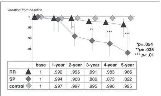
Follow-up – The sample was then prospectively followed for a 5-year period, and measures where taken at least yearly. There was a slow progressive reduction in mean CCI, observed on both group of MS patients, markedly in SP group, but also among RR patients, when compared to control. These scores were completely independent from EDSS. After 3 years, the difference between RR and SP patients scores reached statistical signifi cance (p=.035), and after 5 years, there was a clear difference between the groups (p<.01), which was, still, independent of disability status rated by EDSS (Fig 4).
DISCUSSION
As stated above, the progressive phase of MS is clinically marked by motor disability and cognitive dysfunction, resulting from axonal loss and brain atrophy. Most current tools for imaging and clinical follow-up rely on acute infl ammation paradigm, as well as most end-points of pivotal immunomodula-tory drug studies. Nevertheless, brain atrophy mea-sures might prove to be important markers of disease progression, as they refl ect the irreversible pathologi-cal process of cell damage17.
The aim of our present study is a search for a mea-sure for follow-up of progressive MS patients, easy to be applied on a bedside basis, feasible to be repro-duced by different observers, with some evidence of a clinical correlate to prove its anatomical and func-tional reliability.
Our present series is composed by 128 patients on regular treatment with any of the available im-munomodulatory drugs and has a 5-year prospective ambulatory follow-up. Conventional brain MRI was obtained annually, as part of a routine treatment program. A simple, semi-automated index, that could also be manually obtained, was calculated from a best mid-saggital T1W slice and showed a fair reproduc-ibility on baseline, as matched to a scored examina-tion of a blind expert radiologist. In spite of the low number of cases, CCI showed good anatomical and functional correlates. Anatomical correlate by match-ing to BPF, a well recognized and validated measure of whole brain atrophy, and functional by its linear relation to an also well validated cognitive function-al measure, PASAT. At this point, CCI was sensitive enough to segregate RR from SP patients, despite the signifi cant overlap between these two populations.
There was considerable overlap between groups, on follow-up, but inter-group CCI scoring analysis showed two distinctive patterns: a slowly progressive reduction, mostly among RR patients group, and a pronounced reduction, more common on SP patients. Initially a trend, after a 3-year follow-up this differ-ence became statistically signifi cant, allowing segre-gation between the groups.
More impressive information was obtained by in-tra-patient CCI scoring variation over time: a progres-sive reduction was observed on both groups, much more expressive among SP patients, from the early beginning of our study.
A particular group of 8 RR patients raises a very in-triguing question as they showed what can be called a clinical-imaging mismatch: in spite of being clinically stable, with no relapses and no disability progression after a 5-year period, a relentlessly progressive cal-losal atrophy is evident, quite a similar behavior to SP patients. This small group might simply be on a transitional stage of the continuum between RR and SP phases. On the other hand, they may represent a “look beyond the clinics”, anticipating at image what can be, later, expressed clinically as a progressive de-terioration. In this last case, CCI could have a unique predictive value for evolution, impacting on progno-sis and treatment strategies. We hope that the long-term follow-up of these patients would help answer these questions.
In conclusion, current paradigms for optimizing primary care in MS patients emphasizes not only re-duction of infl ammation, leading to a reduction in re-lapses rate and MRI activity, but also a long-term effi
-cacy, impacting on disability scores, cognitive changes and brain atrophy. Acute changes are widely accessed by conventional MRI while cell loss and brain atrophy, leading to more discrete and insidious changes, are frequently dismissed. Two-dimensional methods for assessing brain atrophy in MS, either by linear or area measures are easy to be obtained and proved very sensitive to longitudinal changes8. Otherwise, it seems
reasonable to conclude that, in some way, any imag-ing protocol for long-term follow-up in MS should recommend the inclusion of at least one standardized brain atrophy scoring. In spite of the cross-sectional design of our study, the CCI was practical, with no so-phisticated software requirements, and quite sensitive to callosal atrophy. So, it proved to have a potential to be used on long-term follow-up for patients with MS.
Acknowledgement – The authors wish to thank Miss Raquel Figueira, B. Biol., for revising the manuscript.
REFERENCES
1. Lublin FD. Clinical features and diagnosis of multiple sclerosis. Neurol Clin 2005;23:1-15.
2. Lassmann H, Bruck W, Lucchinet i C. Heterogeneity of multiple scle-rosis pathogenesis: implications for diagnosis and therapy. Trends Mol Med 2001;7:115-121.
3. Prineas J. Pathology of multiple sclerosis. In Cook S (Ed.). Handbook of multiple sclerosis. Basel: Marcel Dekker, 2001:289-324.
4. Bjartmar C, Trapp BD. Axonal and neuronal degeneration in multiple sclerosis: mechanisms and functional consequences. Curr Opin Neurol 2001;14:271-278.
5. Bjartmar C, Wujek JR, Trapp BD. Axonal loss in the pathology of multi-ple sclerosis: consequences for understanding the progressive phase of the disease. J Neurol Sci 2003;206:165-171.
6. Bakshi R, Dmochowski J, Shaikh ZA, Jacobs L. Gray mat er T2 hypoin-tensity is related to plaques and atrophy in the brains of multiple scle-rosis patients. J Neurol Sci 2001;185:19-26.
7. Rudick RA, Fisher E, Lee JC, Simon J, Jacobs L. Use of the brain pa-renchymal fraction to measure whole brain atrophy in relapsing-remit-ting multiple sclerosis. Multiple Sclerosis Collaborative Research Group. Neurology 1999;53:1698-1704.
8. Bakshi R, Dandamudi, VSR, Neema M, De C, Bermel RA. Measurement of brain and spinal cord atrophy by magnetic resonance imaging as a tool to monitor multiple sclerosis. J Neuroimaging 2005;15(Suppl):S30-S45. 9. Figueira F, Santos VS, Silva FC. A cognitive imaging correlate in
multi-ple sclerosis. Rev Chil Neuropsiquiatr 2003;41:86-87.
10. Witelson SF. Hand and sex diff erences in the isthmus and genu of the human corpus callosum: a postmortem morphological study. Brain 1989; 112:799-835.
11. Hofer S, Frahma J. Topography of the human corpus callosum revisit-ed. Comprehensive fi ber tractography using diff usion tensor magnetic resonance imaging. NeuroImage 2006;32:989-994.
12. Figueira F, Santos VS, Silva FC, Silva ACM. Corpus callosum index: a practical measure for long-term follow-up in multiple sclerosis. Neurol-ogy 2006;66:S10.006, A75.
13. McDonald WI, Compston A, Edan G, and the International Panel. Rec-ommended diagnostic criteria for multiple sclerosis: guidelines from the International Panel on the Diagnosis of Multiple Sclerosis. Ann Neurol 2001;50:121-127.
14. Consortium of Multiple Sclerosis Centers. Available at ht p://www.ms-care.org/. June 2003.
15. Figueira F, Santos VS, Silva FC, Silva ACM. Corpus callosum in-dex: a prospective study on progressive MS. Arq Neuropsiquiatr 2006;64(Suppl):S164-P552.
16. Kurtzke JF. Rating neurological impairment in multiple sclerosis: an Ex-panded Disability Status Scale (EDSS). Neurology 1983;33:1444-1452. 17. Simon JH, Jacobs LD, Campion MK, and The Multiple Sclerosis