Left Atrium Measurement in Patients Suspected of Having Heart
Failure With Preserved Ejection Fraction
Antonio José Lagoeiro Jorge
1, Mario Luiz Ribeiro
1, Maria Luiza Garcia Rosa
1, Fernanda Volponi Licio
1, Luiz Cláudio
Maluhy Fernandes
1, Pedro Gemal Lanzieri
1, Bruno Afonso Lagoeiro Jorge
1, Flavia Oliveira Xavier Brito, Evandro
Tinoco Mesquita
1Universidade Federal Fluminense1, Niterói, RJ; Universidade Federal do Rio de Janeiro2, RJ, Brazil
Abstract
Background: The pathophysiological model of heart failure (HF) with preserved ejection fraction (HFPEF) focuses on the presence of diastolic dysfunction, which causes left atrial (LA) structural and functional changes. The LA size, an indicator of the chronic elevation of the left ventricular (LV) filling pressure, can be used as a marker of the presence of HFPEF, and it is easily obtained.
Objective: To estimate the accuracy of measuring the LA size by using indexed LA volume and diameter (ILAV and ILAD, respectively) for diagnosing HFPEF in ambulatory patients.
Methods: This study assessed 142 patients (mean age, 67.3 ± 11.4 years; 75% of the female sex) suspected of having HF, divided into two groups: with HFPEF (n = 35) and without HFPEF (n = 107).
Results: The diastolic function, assessed by use of Doppler echocardiography, showed a significant difference between the groups regarding the parameters assessing ventricular relaxation (E’: 6.9 ± 2.0 cm/s vs. 9.3 ± 2.5 cm/s; p < 0.0001) and LV filling pressure (E/E’ ratio: 15.2 ± 6.4 vs. 7.6 ± 2.2; p < 0.0001). The ILAV cutoff point of 35 mL/m2 best correlated with the diagnosis of HFPEF, showing sensitivity, specificity, and accuracy of 83%. The ILAD cutoff point of 2.4 cm/m2 showed sensitivity of 71%, specificity of 66%, and accuracy of 67%.
Conclusion: For diagnosing HFPEF in ambulatory patients, the ILAV proved to be a more accurate parameter than ILAD. On echocardiographic assessment, ILAV, rather than ILAD, should be routinely measured. (Arq Bras Cardiol 2012;98(2):175-181)
Keywords: Cardiac volume; left atrium; heart failure; stroke volume.
Mailing Address: Antonio José Lagoeiro Jorge •
Rua Coronel Bittencourt 66, Boa Vista – 24900-000 – Maricá, RJ, Brazil E-mail: lagoeiro@cardiol.br, lagoeiro@globo.com
Manuscript received February 12, 2011; revised manuscript received August 12, 2011; accepted September 08, 2011.
diastasis7. Atrial size is less influenced by aging; thus, alterations
in volume can associate with ventricular pathologies7.
The diagnosis of HFPEF is challenging, especially for ambulatory patients with exertional dyspnea and multiple comorbidities. To prevent low specificity in the diagnosis of HFPEF, the signs and symptoms of HF should be associated with objective measures of LV diastolic dysfunction or with the plasma levels of natriuretic peptides, according to recommendations of the guidelines published so far8,9. The use of Doppler
echocardiography is fundamental to that diagnosis; however, the measure of the transmitral flow during diastole, the most used parameter to confirm alterations in relaxation, can be altered by normal aging4. The ratio of transmitral early LV filling velocity to
early diastolic velocity of the mitral ring (E/E’ ratio) with tissue Doppler echocardiography (TDE) is the major non-invasive marker for the diagnosis of HFPEF and is associated with LV filling pressure8,9.
The LA size, a marker of chronic elevation of the LV filling pressure, can be used as one of the parameters to confirm the presence of HFPEF, and it is easily obtained10. Initially, the measure
of the anteroposterior LA diameter (LAD) on echocardiography (M mode) was the only non-invasive method available for determining the LA size. However, limitations of the method indicate an underestimation of the actual atrial size. There are no studies comparing the accuracy of measuring indexed LA
Introduction
Epidemiological studies on heart failure (HF) have confirmed that HF with preserved ejection fraction (HFPEF) is more prevalent than HF with reduced ejection fraction (HFREF)1-3.
The pathophysiological model focuses on the presence of diastolic dysfunction, subsequent to abnormalities of relaxation or increased left ventricular (LV) stiffness, which retrogradely produces an elevation in the mean left atrial (LA) pressure, causing structural and functional changes in that chamber4.
The cardiovascular system is influenced by the normal aging process, with structural alterations and worsening of ventricular relaxation5,6. Contrary to that observed in transmitral flow
measurement at early diastole, a recent study has reported that aging does not enlarge the atrium7.
volume (ILAV) and indexed LAD (ILAD) for diagnosing HFPEF in ambulatory patients.
This study aimed at estimating the accuracy of measuring the LA size by using ILAV and ILAD for diagnosing HFPEF in ambulatory patients.
Methods
This is an observational, prospective, cross-sectional study involving 142 consecutive ambulatory patients suspected of having HF, assessed from September 2008 to December 2010. The patients underwent clinical, electrocardiographic, and Doppler echocardiographic assessment, in addition to measurement of their B-type natriuretic peptide (BNP) blood levels. Heart failure with preserved ejection fraction was defined as the presence of
signs or symptoms of HF, LV ejection fraction (LVEF) ≥ 50%, and
diastolic dysfunction8,9. Patients with the following characteristics
were excluded from this study: atrial fibrillation; pacemakers; severe heart valve disease; pericardial disease; and heart surgery within the last six months.
The diagnosis of diastolic dysfunction considered the following
criteria: (1) TDE showing E/E’ ratio ≥ 15; and (2) E/E’ ratio suggestive
of diastolic dysfunction (> 8 and < 15). The confirmation of the diagnosis required other echocardiographic measurements, such as the following: indexed LV mass > 122 g/m2, for women, and
> 149 g/m2, for men; and ratio between early and late LV filing
velocity (E/A ratio) lower than 0.5, with a deceleration time of the E wave greater than 280 m/s, for individuals over 50 years of age. In addition to the echocardiographic criteria, a BNP level greater than 200 pg/mL was also used. Heart failure with preserved ejection fraction was excluded in patients with E/E’ ratio and BNP level lower than 8 and 100 pg/mL, respectively8,9.
This study was approved by the ethics committee on research in human beings of the medical school of the Universidade Federal Fluminense (UFF). All patients provided written informed consent at the time of inclusion in the study.
Doppler echocardiography was performed by VIVID 7 device (GE®, USA), and analyzed by use of the ECHOPAC software by an experienced echocardiographer, with no previous knowledge of the results of the other tests. The exam was performed according to the American Society of Echocardiography/European Association of Echocardiography recommendations for chamber quantification11.
The linear anteroposterior measurement of the LAD was obtained by use of M mode and indexed to body surface. The left atrial volume (LAV) was measured with the biplane method of discs (modified Simpson’s rule), by use of the apical four and two-chamber view at end ventricular systole, and indexed to body surface. The parameters of diastolic function were estimated by use of the mean of five consecutive beats. The early (E) and late (A) transmitral flow and the deceleration time of the E wave were measured. The velocity of myocardial relaxation at early diastole (E’) was assessed through TDE in the septal and lateral segments of the mitral ring, and the mean of those measurements was obtained. All exams were recorded in digital media for future analyses and reviews.
The BNP blood level was measured by use of Triage BNP Test
(Biosite, USA), a rapid fluoro immuno assay for the quantitative measurement of BNP, with Triage Meter reading. The BNP values were expressed as pg/mL.
Statistical analysis
Statistical analysis was performed with the SPSS® software,
version 17.0. The continuous variables with normal distribution were expressed as mean ± standard deviation, and the others as median. The Student t, Mann-Whitney, and chi-square tests were used to assess the difference of the means between continuous variables with normal distribution, between continuous variables without normal distribution, and between categorical variables, respectively. Pearson correlation was used to measure the association between ILAV, ILAD, and the clinical and echocardiographic variables. A receiver operator characteristic (ROC) curve was built to express the sensitivity and specificity of ILAV and ILAD in diagnosing HFPEF. A statistical significance level of 0.05 was adopted.
Results
The major characteristics of the patients involved in this study are shown in Table 1. Their mean age was 67.3 ± 11.4 years, and
75% of them were of the female sex. The major comorbidities were arterial hypertension (90%) and diabetes (29%). Patients with
HFPEF had lower values of estimated glomerular filtration rate as compared with patients without HFPEF (73.6 ± 35.4 vs. 94.1 ± 41.3 mL/min; p = 0.009). The major variables obtained by use of Doppler echocardiography and TDE were as follows: LVEF (73.1 ± 8.1); E’ (8.7 ± 2.6 cm/s); E/A (0.88 ± 0.47); indexed LV mass (90.3 ± 25.0 g/m2); E/E’ (9.5 ± 4.9); ILAV (31.5 ± 9.9 mL/m2);
and ILAD (2.4 ± 0.4 cm/m2).
The diastolic function assessed by use of Doppler echocardiography differed between the two groups, and the difference was significant in ventricular relaxation (E’: 6.9 ± 2.0 cm/s vs. 9.3 ± 2.5 cm/s; p < 0.0001) and LV filling pressure (E/E’ ratio: 15.2 ± 6.4 vs. 7.6 ± 2.2; p < 0.0001).
The BNP level was five times higher in patients with HFPEF (133.2 [117.0] pg/ml vs. 26.6 [16.0] pg/ml; p < 0.0001).
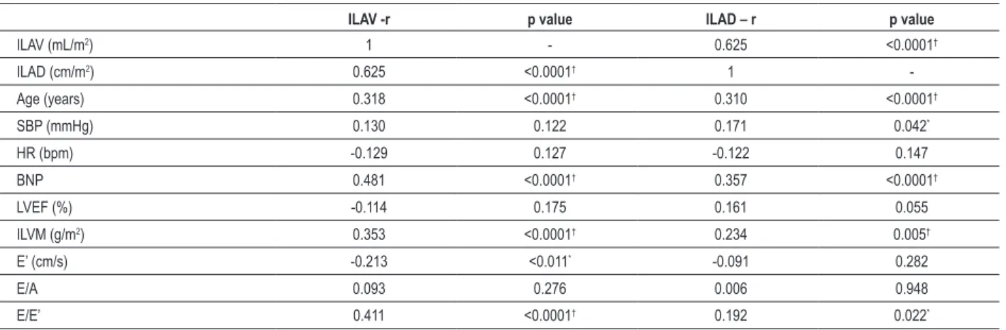
To determine the correlation of ILAV and ILAD with clinical, laboratory and echocardiographic parameters (Table 2), the patients were assessed as a single group. A significant correlation between ILAV, E/E’ ratio, and BNP was observed. The ILAV was mildly related to age, indexed LV mass, E’, and E/A ratio, while the ILAD was mildly related to age, BNP, and indexed LV mass.
In detecting HFPEF, the areas under the ROC curve were 0.89
(95% CI: 0.82 – 0.95; p < 0.0001) for ILAV, and 0.75 (95% CI:
0.65 – 0.84; p < 0.0001) for ILAD. The cutoff point of 35 mL/m2
for ILAV best correlated with the diagnosis of HFPEF, with sensitivity
of 83%, specificity of 83%, accuracy of 83%, and positive likelihood
ratio of 4.9. The cutoff point of 2.4 cm/m2 for ILAD had sensitivity of 71%, specificity of 66%, accuracy of 67%, and positive likelihood
ratio of 2.1(Figure 1).
Discussion
This study’s findings indicate that ILAV can be an important marker of diastolic dysfunction, in addition to helping in the diagnosis of HFPEF when other measures, such as BNP and TDE, are not available. This study also shows that ILAD is a less accurate method to diagnose HFPEF.
Table 1 - Characteristics of the patients with and without HFPEF
Characteristic Total (n = 142) HFPEF (n = 35) No HFPEF (n = 107) p value
Age (years) 67.3±11.4 73.6±12.0 65.3±10.5 < 0.0001
Sex (female %) 75 91 70 0.011
Hypertension (%) 90 94 89 0.343
Diabetes 27 40 22 0.042
BMI (kg/m2) 29.2±5.8 28.1±5.6 29.5±5.8 0.205
HR (bpm) 75.0±14.8 76.8±17.2 74.4±13.9 0.395
SBP (mmHg) 153±26 164±31 150±24 0.009
DBP (mmHg) 90±15 90±18 90±14 0.782
BNP (pg/mL)* 52.9(23) 133.2(117) 26.6(16) <0.0001
Hemoglobin (g/dL) 13.5±1.3 12.8±1.2 13.7±1.3 <0.0001
TC (mg/dL) 206±47 205±51 207±46 0.839
Glucose (g/dL) 105±30 112±42 102±26 0.111
HbA1C (%) 6.1±1.2 6.3±1.2 6.1±1.1 0.320
Creatinine (g/dL) 0.89±0.27 0.92±0.23 0.88±0.29 0.537
GFR (mL/min) 89.1±40.7 73.6±35.4 94.1±41.3 0.009
LVEF (%) 73.1±8.1 71.5±9.3 73.6±7.6 0.181
ILAV (mL/m2) 31.5±9.9 41.9±9.4 28.1±7.4 <0.0001
ILAD (cm/m2) 2.4±0.4 2.7±0.5 2.3±0.4 <0.0001
ILVM (g/m2) 90.3±25.0 99.8±26.9 87.2±23.7 0.009
E (cm/s) 75.3±27.4 99.1±37.2 67.6±17.6 <0.0001
A (cm/s) 92.1±30.4 108.7±49.5 86.7±18.9 <0.0001
DT (ms) 256.3±79.8 269.3±122.6 252.9±64.8 0.384
E’ (cm/s) 8.7±2.6 6.9±2.0 9.3±2.5 0.0001
E/A 0.88±0.47 1.08±0.76 0.83±0.31 0.006
E/E’ 9.5±4.9 15.2±6.4 7.6±2.2 <0.0001
HFPEF – heart failure with preserved ejection fraction; BMI – body mass index; HR – heart rate; SBP – systolic blood pressure; DBP - diastolic blood pressure; BNP –
B-type natriuretic peptide; TC - total cholesterol; HbA1C - glycated hemoglobin; GFR - estimated glomerular iltration rate; LVEF – left ventricular ejection fraction; ILAV – indexed left atrial volume; ILAD – indexed left atrial diameter; ILVM – indexed left ventricular mass; E – mitral low velocity peak at early diastole; A - mitral low velocity peak at the end of diastole; DT - initial mitral low deceleration time; E’ – mitral ring velocity at early diastole. (*) BNP showed an abnormal distribution, being, then, described as median. Other variables were expressed as mean ± standard deviation. Categorical variables were expressed as percentages. For comparison between the groups, Student t test was used for continuous variables, and chi-square test for categorical variables. Statistical signiicance for p < 0.05.
Table 2 - Correlation of ILAV and ILAD with clinical, laboratory and Doppler echocardiographic parameters
ILAV -r p value ILAD – r p value
ILAV (mL/m2) 1 - 0.625 <0.0001†
ILAD (cm/m2) 0.625 <0.0001† 1
-Age (years) 0.318 <0.0001† 0.310 <0.0001†
SBP (mmHg) 0.130 0.122 0.171 0.042*
HR (bpm) -0.129 0.127 -0.122 0.147
BNP 0.481 <0.0001† 0.357 <0.0001†
LVEF (%) -0.114 0.175 0.161 0.055
ILVM (g/m2) 0.353 <0.0001† 0.234 0.005†
E’ (cm/s) -0.213 <0.011* -0.091 0.282
E/A 0.093 0.276 0.006 0.948
E/E’ 0.411 <0.0001† 0.192 0.022*
In addition, measuring LAV is important for the prognosis of patients, independently of their associated diseases. An observational study including 6,657 patients with previous history of neither atrial fibrillation nor heart valve disease has shown that an ILAV of 34 mL/m2 is an independent predictor
of death, HF, atrial fibrillation, and ischemic stroke13.
Evaluating LAV is clinically important because there is a significant relation between LA remodeling and the echocardiographic indices of diastolic function and BNP levels, as shown in this study (Table 2). However, the velocity and time intervals measured by use of Doppler echocardiography reflect the filling pressures at the time of measurement, while LAV often reflects the cumulative effect of the filling pressures over time. Left atrial changes act, thus, as a barometer of the heart and can reflect the degree of LV diastolic dysfunction14.
A possible explanation for the importance of LAV in the diagnosis and prognosis of HFPEF is the fact that LA remodeling results from the chronic elevation in LA pressures. In patients with dyspnea and normal LVEF, the increase in LAV supports the hypothesis that the difficulty in breathing results from HFPEF. Thus, the lack of an increase in LAV helps to rule out the diagnosis of HFPEF12.
Some guidelines8,9 use the cutoff point of 40mL/m2,
which, in this study showed high sensitivity, but low specificity for the diagnosis of HFPEF. In this study, the cutoff point of 35 mL/m2, slightly lower than that of the guidelines, provided sensitivity and specificity of 83%, with 83% accuracy for confirming HFPEF in ambulatory patients
with HF symptoms.
Figure 1 – ROC curve of ILAD and ILAV for diagnosing HFPEF.
S
ens
iti
vi
ty
1-Speciicity
Area P value Conidence interval
ILAV ILAD
Indexed LAV Indexed LAD Reference line 0.886
0.746
<0.0001 <0.0001
0.820 -0.952 0.651 -0.841
1.0
0.8
0.6
0.4
0.2
0.0
Some previous studies have shown that the atrial size naturally enlarges with advancing age, leading to the hypothesis that such enlargement could alter atrial function, increasing the possibility of the occurrence of atrial arrhythmias. However, no study assessing atrial volume has confirmed that observation. A study, assessing that topic and comparing young and elderly individuals (over the age of 70 years), has reported subtle changes in the extremes of age7.Initial studies showing greater
influence of age on the LA size has used less geometrically strict methods in M mode for that assessment15. Although the
measurement of the anteroposterior LAD is universally used in clinical practice, it can have low accuracy as a surrogate for volume, because its use is based on the unlikely assumption that there is a constant relation between atrial dimensions, which was not confirmed in this study (Figure 2). The LAV measurement offers greater accuracy for assessing the actual LA size, being more sensitive to changes in size15.
In this study, although ILAD showed a significant correlation
with ILAV (r = 0.605; p < 0.0001), it had lower accuracy (67%), sensitivity (71%), and specificity (66%) as compared with ILAV,
when the cutoff point of 2.4 cm/m2 was used to diagnose HFPEF.
Conclusion
The ILAV measurement can predict the presence of diastolic dysfunction and is a more accurate method than ILAD to diagnose HFPEF in ambulatory patients with signs or symptoms of HF. When assessing HFPEF by use of Doppler echocardiography, ILAV, rather than ILAD, should be routinely measured.
Potential Conflict of Interest
No potential conflict of interest relevant to this article was reported.
Sources of Funding
There were no external funding sources for this study.
Study Association
This article is part of the thesis of master submitted by Antonio José Lagoeiro Jorge, from Universidade Federal Fluminense e Universidade Federal do Rio de Janeiro.
Figure 2 – Correlation between ILAV and ILAD in individuals with and without HFPEF.
Inde
xed l
eft a
tri
al
v
ol
um
e
Indexed left atrial diameter
R = 0.366 p < 0.0001 70.00
60.00
50.00
40.00
30.00
20.00
10.00
References
1. Owan TE, Hodge DO, Herges RM, Jacobsen SJ, Roger VL, Redfield MM. Trends in prevalence and outcome of heart failure with preserved ejection fraction. N Engl J Med. 2006;355(3):251-9.
2. Moutinho MAE, Colucci FA, Alcoforado V, Tavares LR, Rachid MB, Rosa ML, et al. Insuficiência cardíaca com fração de ejeção preservada e com disfunção sistólica na comunidade. Arq Bras Cardiol. 2008;90(2):132-7.
3. Tribouilloy C, Rusinaru D, Mahjoub H, Soulière V, Lévy F, Peltier M, et al. Prognosis of heart failure with preserved ejection fraction a 5 year prospective population-based study. Eur Heart J. 2008;29(3):339-47.
4. Borlaug BA, Paulus WJ. Heart failure with preserved ejection fraction: pathophysiology, diagnosis, and treatment. Eur Heart J. 2011;32(6):670-9.
5. Bryg RJ, Williams GA, Labovitz AJ. Effect of aging on left ventricular diastolic filling in normal subjects. Am J Cardiol. 1987;59(9):971-4.
6. We i J Y. A g e a n d t h e c a r d i o v a s c u l a r s y s t e m . N E n g l J M e d . 1992;327(24):1735-9.
7. Thomas L, Levett K, Boyd A, Leung DY, Schiller NB, Ross DL. Compensatory changes in atrial volumes with normal aging: is atrial enlargement inevitable? J Am Coll Cardiol. 2002;40(9):1630-5.
8. Paulus WJ, Tschöpe C, Sanderson JE, Rusconi C, Flachskampf FA, Rademakers FE, et al. How to diagnose diastolic heart failure: a consensus statement on the diagnosis of heart failure with normal left ventricular
ejection fraction by Heart Failure and Echocardiography Associations of the European Society of Cardiology. Eur Heart J. 2007;28(20):2539-50.
9. Bocchi EA, Marcondes Braga FG, Ayub-Ferreira SM, Rohde LE, Oliveira WA, Almeida DR, et al. Sociedade Brasileira de Cardiologia.III Diretriz brasileira de insuficiência cardíaca crônica. Arq Bras Cardiol. 2009;93(supl 1):1-71.
10. Nagueh SF, Appleton CP, Gillebert TC, Marino PN, Oh JK, Smiseth OA, et al. Recommendations for the evaluation of left ventricular diastolic function by echocardiography. J Am Soc Echocardiog. 2009;22(2):107-33.
11. Lang MR, Bierig M, Devereux RB, Flachskampf FA, Foster E, Pellikka PA, et al. Recommendations for chamber quantification. Eur J Echocardiogr. 2006;7(2):79-108.
12. Tsang TS, Barnes ME, Gersh BJ, Bailey KR, Seward JB. Risks for atrial fibrillation and congestive heart failure in patients >/=65 years of age with abnormal left ventricular diastolic relaxation. Am J Cardiol. 2004;93(1):54-8.
13. Abhayaratna WP, Seward JB, Appleton CP, Douglas PS, Oh JK, Tajik AF, et al. Left atrial size: physiologic determinants and clinical applications. J Am Coll Cardiol. 2006;47(12):2357-63.
14. Lester SJ, Tajik JA, Nishimura RA, Oh JK, Khandheria BK, Seward JB. Unlocking the mysteries of diastolic function: deciphering the Rosetta Stone 10 years later. J Am Coll Cardiol. 2008;51(7):679-89.