DOI: 10.5935/2359-4802.20180047
REVIEW ARTICLE
Mailing Address: Debora Carvalho Grion
Avenida Marques do Paraná, 349, apto. 810. Postal Code: 24030-215, Centro, Niterói, RJ - Brazil. E-mail: [email protected], [email protected]
Phenotype Mapping of Heart Failure with Preserved Ejection Fraction
Evandro Tinoco Mesquita, Debora Carvalho Grion, Miguel Camargo Kubrusly, Bernardo Barcelos Fernandes Fumagalli Silva, Érico Araújo Reis Santos
Universidade Federal Fluminense (UFF), Niterói, RJ - Brazil
Manuscript received September 20, 2017, revised manuscript January 03, 2018, accepted January 16, 2018.
Heart Failure / physiopathology; Stroke Volume; Phenotype; Machine Learning; Artificial Intelligence.
Keywords
Abstract
Heart failure with preserved ejection fraction (HFPEF) has become the main phenotypic model of heart failure (HF) in community and referral patients in Brazil and in the world. Despite advances in the development of new drugs for HF treatment, there has been no significant improvement in mortality of this condition.
According to many studies, this can be explained by the heterogeneous nature of HF physiopathology, whose basic mechanisms may result in different clinical presentations, culminating in the emerging of different phenogroups in this syndrome. In this context, phenotype mapping of HFPEF has emerged as a possible solution, since it enables the development of clinical trials that establish specific therapeutic strategies for each phenotypic profile.
New technologies in the field of artificial intelligence have enabled the assessment of a large volume of data and infer intrinsic patterns and different outcomes. Thereby, it is possible to obtain mutually exclusive categories of HFPEF, with a phenotype mapping of the syndrome and grouping of patients according to their phenotypic features. Besides, other diseases can have the same clinical phenotype but different pathophysiological basis, the so called “phenocopies”.
These tools enable the analysis and categorization of the wide spectrum of heart failure, contributing to solve the dilemmas of the treatment of this syndrome.
Introduction
Heart failure with preserved ejection fraction (HFPEF) has become the main phenotypic model of heart failure
(HF) in community and referral patients in Brazil and in the world.1,2
Only two forms of clinical presentations of HFPEF used to be recognized – first, in the outpatient setting, elderly women patients, intolerant to exercise, usually
with no clinical evidence of congestion;3,4 and second,
patients admitted to emergency departments with hypertensive crisis, acute atrial fibrillation and acute
pulmonary edema.5
Clinical profiles of HFPEF have been gradually identified. For example, HFPEF has been associated with pulmonary arterial hypertension and valve diseases – aortic stenosis, mitral stenosis – and deposition diseases, such as senile amyloidosis.6,7
In the last decades, progresses have been made in the understanding of pathophysiological mechanisms involved in HFPEF and the influence of comorbidities in the development and progression of the disease. In addition to diastolic dysfunction, abnormal chronotropic response, left atrial dysfunction, and altered physiology of coronary endothelium and systemic and pulmonary microcirculation have been reported. Molecular changes related to oxidative stress and a proinflammatory state have been also described, and seem to be associated with aging, hypertension, obesity and other cardiovascular
and non-cardiovascular diseases.8,9
D e s p i t e a d v a n c e s i n t h e s t u d y o f H F P E F pathophysiology and development of new drugs, there has been no significant improvement in mortality or clinical outcome of this condition.10
medicine, a new paradigm that has been successfully used in oncology. This approach considers genetic variability, environment, and lifestyle of each patient, allowing an individualized approach for the treatment and prevention of diseases.11-13
The aim of the present study was to present a narrative review of the literature to describe the clinical phenotypes of HFPEF and its potential impact on the management of patients and on clinical research.
Methods
Bibliographic review
We conducted a narrative review, from a clinical perspective, of studies published in MEDLINE using the PubMed search engine. The following MeSH (Medical Subject Heading) terms were used - (heart failure with preserved ejection fraction [tiab] OR diastolic heart failure [tiab] OR hfnef [tiab] OR hfpef [tiab]) AND (phenoc* [tiab] OR phenotype* [tiab]).
The search was carried out in February 2017, and 136 articles published in the period from 1990 and 2017 were identified. Thirty articles were independently selected by four investigators for detailed analysis. Additional articles were selected from the reference lists of the retrieved articles.
Pathophysiology of HFPEF
Heart failure (HF) is a complex clinical syndrome characterized by symptoms and signs caused by abnormal cardiac function and/or structure that leads to decreased cardiac output and/or increased intracardiac pressures.
HF patients can have different phenotypes according to morphofunctional characteristics of the disease, and
receive different therapeutic approaches.14,15 Based on
this, patients are usually classified into patients with HF with reduced ejection fraction (HFrEF), marked by left ventricular ejection fraction (LVEF) lower than 40% - and HF with preserved ejection fraction (HFpEF), characterized by LVEF greater than 50%. Recently, the European Society of Cardiology has proposed a new phenotype – “HF with midrange ejection fraction” – with intermediate ejection fraction (LVEF between 40 and 49%)
and a clinical profile different from HFeEF and HFpEF.16
The main diagnostic criteria of HFPEF – the focus of this study – are the clinical profile of LVEF equal to or greater than 50%, increased levels of brain natriuretic peptide (BNP) (greater than 35pg/mL or NT-proBNP
greater than 125 pg/mL) and at least one of these two criteria – important structural cardiac disease (left ventricular hypertrophy and/or increased left atrium)
and diastolic dysfunction.16
HFPEF is characterized by reduced end-diastolic volume, left ventricular hypertrophy, and increased left atrial volume and left ventricular filling pressure. These pathophysiological abnormalities are associated with increased left ventricular stiffness, decreased left ventricular relaxation, cardiomyocyte hypertrophy, myocardial interstitial fibrosis and reduced intramyocardial capillaries.17-19 In addition, a proportion
of patients with HFpEF present atrial fibrillation, which
further aggravates cardiac function.20
The classical presentations - HFeEF and HFpEF – used to be distinguished only by the remodeling pattern of cardiac chambers and extension of myocardial dysfunction, culminating in different therapeutic responses. However, it is known today that morphofunctional changes are also based on molecular alterations, which are also different
between these conditions.10
Left ventricular diastolic dysfunction, an important diagnostic criterion for HFPEF, may be explained by increased myocardial stiffness, resulting from changes in
extracellular matrix and/or cardiomyocytes.10 There are
evidence that extracellular matrix stiffness results mainly from collagen metabolism. Excess deposition of type I collagen, the subtype with the highest stiffness property, is explained by increased synthesis and/or decreased degradation of this compound. Type I collagen synthesis can be measured by procollagen type I carboxy-terminal propeptide, which derives from type I procollagen and acts as a biomarker. Decreased degradation of type I collagen is caused by downregulation of matrix metalloproteinases (MMPs) and/or upregulation of tissue inhibitors of metalloproteinases (TIMPs). TIMP-1 plasma levels have also been suggested as promising
biomarkers in HFPEF.9,10
Excess collagen is found in only one third of patients with HFPEF, even in the presence of ventricular stiffness, which usually results from an intrinsic cardiomyocyte condition, and may be related to the protein structure
and/or to the disruption of the sarcomere structure.9,10
- N2B (stiffer) and N2BA (more compliant). Changes in the ratio of one isoform to the other and phosphorylation of the fibers, as well as oxidative stress can have an impact on myocardial compliance, leading to stiffness.9,18
Disruption of sarcomere structure is the mechanical factor of ventricular relaxation. It is an energy-consuming reaction, and, for this reason, the lack of energy stores impairs a normal left ventricular relaxation. Recent studies have demonstrated a decreased phosphate creatinine/adenosine ratio in patients with HFPEF, which is consistent with a decline
in myocardial energy store.21-23
In addition to interstitial (collagen-related changes) and structural (regulation of constituent proteins) changes, unbalanced levels of chemical mediators, especially of monophosphate cyclic guanine (cGMP), may also explain myocardial stiffness in HEPEF. Activation of protein kinase G (PKG) by cGMP results in phosphorylation cascade of proteins important for cardiomyocyte integrity – phosphorylation of titins inhibits cardiac hypertrophy and increases myocardial compliance, phosphorylation of potassium channels inhibits tissue ischemia, and phosphorylation of troponin I increases left ventricular relaxation. Also, PKG activation by cGMP increases
calcium reuptake by sarcoplasmic reticulum.9
Low BNP, microvascular inflammation and oxidative stress, which are common in several conditions, such as obesity and insulin resistance, suppresses GMPc synthesis pathways. This, in turn, inhibits PKG phosphorylation cascade and culminates in myocardial stiffness, characteristic of HFPEF.8,9
Although HFPEF is commonly referred as diastolic HF, the disease is not limited to ventricular relaxation
problems. A study24 demonstrated that myocardial
contractility may be decreased in HFPEF, even if the end-systolic elastance (ESE) – used to measure myocardial contractility – is increased.24 This apparent contradiction
may be explained by the influence of cardiac chamber geometry on ESE. Concentric hypertrophy, characteristic of HFPEF, independently increases ESE, even with reduced left ventricular contractility.21
In HFPEF, vascular stiffness is generalized, resulting in elevated pressure, which aggravates ventricular stiffness and attenuates vascular dilation in exercise, thereby decreasing blood supply to musculoskeletal system. Increased vessel stiffness, associated with elevated left heart pressure, increases pulmonary pressure and
consequently the mortality of these patients.21
Defects in diastolic, systolic, vascular and chronothropic functions elucidate the heterogeneous nature and complexity of HFPEF. Its multiple pathophysiological factors indicate the need of phenotyping of these patients, and identification of specific causes of the worsening of each phenotype. This strategy has become increasingly possible with biomolecular advances in medicine and will possibly guide therapeutic decisions based on specific pathophysiological changes.
Modulation of HFPEF phenotypes by epigenetics – a new frontier
Epigenetics is an emerging science involving the study of changes in the regulation of genes and their expression, regardless of their sequences. Environmental factors can affect intracellular signaling pathways in a way that can affect chromatin structure, resulting in the passage of altered gene expression patterns to the offspring by epigenetic memory, affecting the
phenotypes of the diseases.25,26
New evidence suggests the involvement of epigenetic regulation in target cells related to cardiovascular
pathogenesis, including HF and its different phenotypes.27
Cardiomyocytes, for example, can adapt to environmental stress by epigenetic regulation. This dysregulation in genetic expression provides information about the pathogenesis of cardiac and vascular remodeling, dysfunction of progenitor cells and endogenous repair system, inflammation, fibrosis and cardiac dysfunction.28,29
Four epigenetic mechanisms in cardiovascular diseases have been identified – DNA methylation, chromatin remodeling by adenosine triphosphate (ATP)-dependent enzymes, histone modification and microRNA-dependent
mechanisms.30-32 Recent findings have associated these
mechanisms with HFPEF-related diseases; however, evidence on the role of epigenetics in changes in cardiac function and structure, and clinical trials corroborating theories involving both epigenetics and cardiovascular disease are still lacking. Advances in studies on this field should contribute to HF prevention and provide enough evidence for the stratification of HF phenotypes.
Clinical phenotypes and phenotypic mapping
by machine learning algorithms. Machine learning is a field of artificial intelligence, in which a computer is programmed to learn the relationship between the objects of study by data processing and accumulate experience with previous problem-solving approaches. Machine learning algorithms are classified into supervised and unsupervised. While supervised learning is focused on outcome prevention, unsupervised learning aims to infer intrinsic structures of the data.
Therefore, in this approach, a large volume of data can be analyzed and mutually exclusive categories of HFPEF can be obtained by phenotype mapping of the syndrome and grouping of patients in subgroups according to phenotypic characteristics. Phenotypic classification of patients with HFPEF would be helpful to the development of clinical trials on therapeutic strategies specific to each phenotypic profile.12
A recent study was the first to identify phenogroups of a heart disease, and the first to use the machine learning technique as an approach to solve the heterogeneity of a
cardiovascular syndrome by phenotype analysis.33
The data analyzed for patients’ classification using the machine learning approach included clinical variables, physical features, laboratory data and electrocardiogram and echocardiogram parameters.
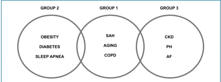
Although the patients shared many clinical characteristics, they were classified in three subgroups with distinct characteristics and prognosis:33
• Group 1, composed of younger patients, with
moderate diastolic function and relatively normal BNP levels. These patients have the mildest myocardial remodeling, electrical dysfunction and hemodynamic change, although 65% of them had moderate diastolic dysfunction and elevated pulmonary capillary pressure (PCP) and pulmonary artery systolic pressure.
• Group 2 involved obese, diabetic patients, with
a high prevalence of sleep apnea and impaired left ventricular relaxation. This group showed the highest PCP and highest pulmonary vascular resistance.
• Group 3 was composed of older patients, with
significant chronic kidney disease and pulmonary hypertension. In this phenogroup, a more severe myocardial remodeling and electric dysfunction was observed, with a longer QRS-T interval, higher relative thickness of cardiac walls, higher left ventricular mass index, higher E/e’ ratio and worse right ventricular function.33
In addition, different phenogroups had different clinical course and outcomes, and distinct risk stratification.
Prognosis was divided into the following categories: death, hospitalization for non-cardiac causes, hospitalization for cardiac causes, and hospitalization for HF. In group 1, the most frequent prognostic factors were hospitalization for cardiovascular and hospitalization for non-cardiovascular diseases; in group 2, hospitalization for non-cardiovascular causes and HF, and in group 3, the most prevalent outcome
was death, followed by hospitalization for HF.33
However, although ideally the subgroups should be mutually excluding, some patients had overlapping clinical features, especially in the analysis of group 1 patients (Figure 1). Even so, this was a pioneer study in
the phenotyping of complex cardiovascular syndromes.33
In light of the above, one may infer that the use of the machine learning tool in international centers would provide new, essential information on HFPEF epidemiology.
Considering that this study was conducted in a North American setting,33 it is expected that results observed in
the subgroups are different from those in South America. Therefore, application of the technique in Latin American prevalence studies is paramount for future phenotype mapping of HFPEF in Brazil.
Treatment
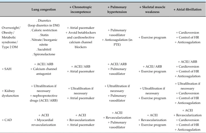
Classical therapeutic approach of HFPEF has not reduced mortality and morbidity rates of these patients. Thus, considerable differences between the phenogroups indicate the importance of a specific therapeutic approach, since advances in therapies have been so far hampered by such phenotypic complexity. To deal with that, new therapies that have a direct effect on signaling cascades involved in the pathophysiology of the HFPEF
have been proposed.34,35 Today, these therapies varied
from signaling pathways of systemic inflammation to myocardial elasticity, and additional therapies to different comorbidities associated with the same pattern of phenotypic predisposition to the disease.
Aging, obesity, systemic hypertension, type 2 diabetes mellitus, kidney failure, and sleep apnea can trigger a chronic systemic inflammation that affects the myocardium and other organs. The patient may have pulmonary hypertension, sodium retention and impaired oxygen extraction by skeletal muscles.
Figure 1 - Phenogroups of heart failure with preserved ejection fraction. SAH: systemic arterial hypertension; COPD: chronic obstructive pulmonary disease; CKD: chronic kidney disease; PH: pulmonary hypertension; AF: atrial fibrillation.
GROUP 2 GROUP 1 GROUP 3
OBESITY
DIABETES
SLEEP APNEA
SAH
AGING
COPD
CKD
PH
AF
For hypertension phenotype, anti-hypertensive are the most recommended treatment, such as angiotensin converting enzyme (ACE) inhibitors, angiotensin II receptor blockers and calcium channel blockers, highlighting the importance of this treatment to HF prevention, to vascular conditions not related to HF (such as stroke and myocardial infarction) and to improve the quality of life of HFPEF patients. The CHARM-Preserved, PEP-CHF and TOPCAT studies demonstrated a reduction in hospitalization rates of patients with HFPEF by blockage of the renin-angiotensin-aldosterone system. These studies used, respectively, an angiotensin II receptor blocker, ACE inhibitors and an aldosterone antagonist (spironolactone).36-38 Inhibition of the
renin-angiotensin-aldosterone system would be of benefit to the patients due to the association of neurohormonal
activation with hypertension and volume retention.39,40
Control of heart rate is mediated by activation of the sympathetic system, which has a direct effect on adverse outcomes in patients with HFPEF. A study derived from the I-Preserve study showed an association between increased heart rate and higher incidence of death for cardiovascular causes and hospitalizations in patients
in sinus rhythm.41 Therefore, heart rate control would
be an effective treatment target.
In patients with pulmonary hypertension, the use of dobutamine improved pulmonary vascular function, and studies on new pulmonary vasodilators targeting
GMPc, endothelin and nitric oxide (NO)32 have also been
developed (Table 1). In patients with kidney dysfunction, sildenafil had no significant effects in the RELAX clinical
trial.42 Due to the high prevalence of patients with HFPEF
and pulmonary hypertension, and its intrinsic relationship with morbidity and mortality, pulmonary vasodilation is
paramount in the treatment of these patients.43
Based on the studies reviewed, we conclude that phenotype mapping in HFPEF has enabled the development of a new generation of clinical trials aimed at new therapeutic approaches (Figure 2).
HFPEF accounts for nearly half of HF patients on treatment and its prevalence has increased. Cardiovascular disease phenotypes are complex, with many influencing factors. Systemic inflammatory reaction and microvascular endothelial dysfunction lead to ventricular remodeling and dysfunction. Specific therapeutic interventions should be multifaceted and focused on stages of these signaling cascades. New therapeutic approaches should encompass metabolic control, modulation of inflammatory response, control of pulmonary hypertension, prevention of muscle weakness, and reduction of sodium and water retention. Due to the wide range of interventions, phenotype mapping becomes an essential tool for future investigations and clinical trials (to confirm the results). Possibly, there will be more significant changes as new genetic, cellular, molecular and immunologic biomarkers are incorporated and used to discriminate treatment groups in a clear and objective manner.
Cardiac diseases that simulate HFPEF – Phenocopies
Table 1 - Example of therapeutic strategies for different phenotypes of heart failure with preserved ejection fraction
Lung congestion + Chronotropic incompetence
+ Pulmonary hypertension
+ Skeletal muscle
weakness + Atrial fibrillation
Overweight/ Obesity/ Metabolic syndrome/ Type 2 DM
. Diuretics (loop diuretics in DM)
. Caloric restriction . Statin . Nitrate/Inorganic
nitrite . Sacubitril . Spironolactone
+ Atrial pacemaker + Avoid betablockers
and cardioselective calcium channel
blockers
+ Pulmonary vasodilator + Anticoagulation (in
PTE)
+ Exercise program
+ Cardioversion + Control of HR + Anticoagulation
+ SAH
+ ACEI/ARB + Calcium channel
antagonist
+ ACEI/ARB + Atrial pacemaker
+ ACEI/ARB + Pulmonary vasodilator
+ ACEI/ARB + Exercise program
+ ACEI/ARB + Cardioversion + Control of HR + Anticoagulation
+ Kidney dysfunction
+ Ultrafiltration if necessary + nephroprotective drugs (ACEI/ARB)
+ Ultrafiltration if necessary + Atrial pacemaker
+ Ultrafiltration if necessary + Pulmonary
vasodilator
+ Ultrafiltration if necessary + Exercise program
+ Ultrafiltration if necessary + Cardioversion + Control of HR + Anticoagulation + CAD + ACEI + Myocardial revascularization + ACEI + Revascularization
+ Atrial pacemaker
+ ACEI + Revascularization + Pulmonary vasodilator + ACEI + Revascularization + Exercise program
+ ACEI + Revascularization
+ Cardioversion + Control of HR + Anticoagulation
Adapted from: Shah SJ, Kitzman DW, Borlaug BA, van Heerebeek L, Zile MR, Kass DA, Paulus WJ. Phenotype-specific treatment of heart failure with preserved ejection fraction: a multiorgan roadmap. Circulation. 2016;134(1):73-90. DM: diabetes mellitus; SAH: systemic arterial hypertension; CAD: chronic artery disease; ACEI: angiotensin converting enzyme inhibitor; ARB: angiotensin II receptor blocker; PTE: pulmonary thromboembolism; HR: heart rate
Figure 2 - The process towards effective therapies of heart failure with preserved ejection fraction: HFPEF: heart failure with preserved ejection fraction.
HFPEF: a complex syndrome Understanding its pathophysiology Subdividing the syndrome into clinical phenotypes Inferring new therapeutic targets New clinical trials Individualized therapeutic approach
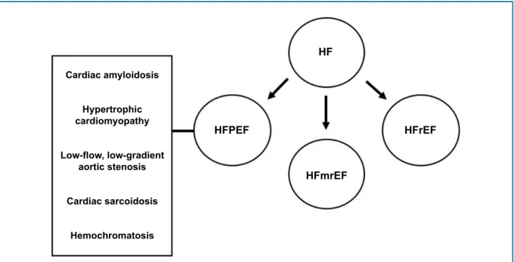
diseases was found to mimic their phenotypic features. We found that this concept may be extended to HFPEF. Due to the heterogeneity nature of HFPEF, other diseases may have the same clinical phenotype and thereby be considered their “phenocopies”. Although both therapeutic intervention and prognosis of the diseases are different, their similar clinical presentation hampers the differential diagnosis. One pertinent example of a disease that mimics the clinical pattern of HFPEF is cardiac amyloidosis.44,45
Figure 3 - Phenocopies of heart failure with preserved ejection fraction. HF: heart failure; HFPEF: heart failure with preserved ejection fraction; HFmrEF: heart failure with mid-range ejection fraction; HFrEF: heart failure with reduced ejection fraction.
Cardiac amyloidosis
Hypertrophic cardiomyopathy
Low-flow, low-gradient aortic stenosis
Cardiac sarcoidosis
Hemochromatosis
HFPEF
HFmrEF
HFrEF HF
hypertrophy, with late diagnosis due to its gradual progression. Nevertheless, it is worth pointing out that individuals with HFPEF usually have other comorbidities that independently contribute to diastolic dysfunction.46-48
In addition to cardiac amyloidosis, “phenocopies” include other diseases such as hypertrophic
cardiomyopathy, cirrhotic cardiomyopathy,49 low-flow,
low-gradient aortic stenosis, cardiac sarcoidosis and hemochromatosis (Figure 3).
The identification of “phenocopies” in HFPEF may enable an individualized approach to molecular targets and functional abnormalities, such as the use of certain drugs in senile amyloidosis, and betablockers and/or calcium channel antagonists in hypertrophic cardiomyopathy. Besides, the chance of diagnostic errors may decrease and that of early diagnosis of other diseases may increase when the presence of diseases that mimic HFPEF is considered.
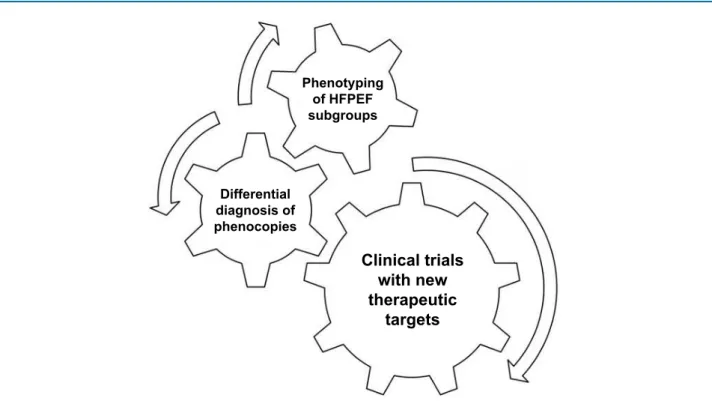
The use of the machine learning technique for patients’ grouping by phenotypes allows the analysis of a wide variety of variables and relationship between them, and to classify them in mutually exclusive phenogroups. In addition to allowing a phenotype categorization and to contribute to a therapeutic revolution, the identification of possible “phenocopies” is crucial for the differential diagnosis of a HFPEF model (Figure 4).
Conclusions
HFPEF is a common syndrome, whose prevalence will increase in the community. However, classification of phenogroups and results of therapeutic approaches are still incipient.
Today, the concept of adopting a phenotype network to explain HFPEF disrupts the Cartesian model in suggesting a complex approach of these patients, who may have many morphofunctional patterns. These distinct patterns may be related to abnormal signaling processes in the myocardium and associated with systemic inflammation, which is increased in patients with HFPEF and comorbidities.
Phenotype mapping of heterogeneous clinical syndromes, such as HFPEF, enables the categorization of patients, and can serve as a basis for the development of clinical trials and identification of new therapeutic approaches.
Author contributions
1. Owan TE, Hodge DO, Herges RM, Jacobsen SJ, Roger VL, Redfield MM. Trends in prevalence and outcome of heart failure with preserved ejection fraction. N Engl J Med. 2006;355(3):251-9.
2. Jorge AL, Rosa ML, Martins WA, Correia DM, Fernandes LC, Costa JA, et al. The prevalence of stages of heart failure in primary care: a population-based study. J Card Fail. 2016;22(2):153-7.
3. Borlaug BA, Melenovsky V, Russell SD, Kessler K, Pacak K, Becker LC, et al. Impaired chronotropic and vasodilator reserves limit exercise capacity in patients with heart failure and a preserved ejection fraction. Circulation. 2006;114(20):2138-47.
4. Ennezat PV, Lefetz Y, Marechaux S, Six-Carpentier M, Deklunder G, Montaigne D, et al. Left ventricular abnormal response during dynamic
exercise in patients with heart failure and preserved left ventricular ejection fraction at rest. J Card Fail. 2008;14(6):475-80.
5. Shah AM, Solomon SD. Phenotypic and pathophysiological heterogeneity in heart failure with preserved ejection fraction. Eur Heart J. 2012;33(14):1716-7.
6. Samson R, Jaiswal A, Ennezat PV, Cassidy M, Le Jemtel TH. Clinical phenotypes in heart failure with preserved ejection fraction. J Am Heart Assoc. 2016;5(1). pii: e002477.
7. Pedrotty DM, Jessup M. "Frailty, thy name is woman": syndrome of women with heart failure with preserved ejection fraction. Circ Cardiovasc Qual Outcomes. 2015;8(2 Suppl 1):S48-51.
References
Figure 4 - Future management of heart failure with preserved ejection fraction. HFPEF: heart failure with preserved ejection fraction. Phenotyping
of HFPEF
subgroups
Differential diagnosis of phenocopies
Clinical trials
with new
therapeutic
targets
interpretation of the data: Mesquita ET, Grion DC, Kubrusly MC, Silva BBFF, Santos EAR. Writing of the manuscript: Mesquita ET, Grion DC, Kubrusly MC, Silva BBFF, Santos EAR. Critical revision of the manuscript for intellectual content: Mesquita ET, Grion DC, Kubrusly MC, Silva BBFF, Santos EAR.
Potential Conflict of Interest
No potential conflict of interest relevant to this article was reported.
Sources of Funding
There were no external funding sources for this study.
Study Association
This study is not associated with any thesis or dissertation work.
Ethics approval and consent to participate
8. Lim SL, Lam CS. Breakthrough in heart failure with preserved ejection fraction: are we there yet? Korean J Intern Med. 2016;31(1):1-14.
9. van Heerebeek L, Paulus WJ. Understanding heart failure with preserved ejection fraction: where are we today? Neth Heart J. 2016;24(4):227-36.
10. Borlaug BA, Redfield MM. Diastolic and systolic heart failure are distinct phenotypes within the heart failure syndrome. Circulation. 2011;123(18):2006-14.
11. Mesquita ET, Jorge AJ, Souza Junior CV, Cassino JP. Systems biology applied to heart failure with normal ejection fraction. Arq Bras Cardiol. 2014;102(5):510-7.
12. Shah SJ, Katz DH, Deo RC. Phenotypic spectrum of heart failure with preserved ejection fraction. Heart Fail Clin. 2014;10(3):407-18.
13. Joyner MJ. Precision medicine, cardiovascular disease and hunting elephants. Prog Cardiovasc Dis. 2016;58(6):651-60.
14. Borlaug BA, Olson TP, Lam CS, Flood KS, Lerman A, Johnson BD, et al. Global cardiovascular reserve dysfunction in heart failure with preserved ejection fraction. J Am Coll Cardiol. 2010;56(11):845-54.
15. Tan YT, Wenzelburger F, Lee E, Heatlie G, Leyva F, Patel K, et al. The pathophysiology of heart failure with normal ejection fraction: exercise echocardiography reveals complex abnormalities of both systolic and diastolic ventricular function involving torsion, untwist, and longitudinal motion. J Am Coll Cardiol. 2009;54(1):36-46.
16. Ponikowski P, Voors AA, Anker SD, Bueno H, Cleland JG, Coats AJ, et al; Authors/Task Force Members; Document Reviewers. 2016 ESC Guidelines for the diagnosis and treatment of acute and chronic heart failure: The Task Force for the diagnosis and treatment of acute and chronic heart failure of the European Society of Cardiology (ESC). Developed with the special contribution of the Heart Failure Association (HFA) of the ESC. Eur J Heart Fail. 2016;18(8):891-975.
17. Little WC. Diastolic dysfunction beyond distensibility: adverse effects of ventricular dilatation. Circulation. 2005;112(19):2888-90.
18. Zile MR, Baicu CF, Ikonomidis JS, Stroud RE, Nietert PJ, Bradshaw AD, et al. Myocardial stiffness in patients with heart failure and a preserved ejection fraction: contributions of collagen and titin. Circulation. 2015;131(14):1247-59.
19. Borlaug BA. The pathophysiology of heart failure with preserved ejection fraction. Nat Rev Cardiol. 2014;11(9):507-15.
20. Zakeri R, Chamberlain AM, Roger VL, Redfield MM. Temporal relationship and prognostic significance of atrial fibrillation in heart failure patients with preserved ejection fraction: a community-based study. Circulation. 2013;128(10):1085-93. Erratum in: Circulation. 2013;128(24):e465.
21. Borlaug BA, Paulus WJ. Heart failure with preserved ejection fraction: pathophysiology, diagnosis, and treatment. Eur Heart J. 2011;32(6):670-9.
22. Smith CS, Bottomley PA, Schulman SP, Gerstenblith G, Weiss RG. Altered creatine kinase adenosine triphosphate kinetics in failing hypertrophied human myocardium. Circulation. 2006;114(11):1151-8.
23. Phan TT, Abozguia K, Nallur Shivu G, Mahadevan G, Ahmed I, Williams L, et al. Heart failure with preserved ejection fraction is characterized by dynamic impairment of active relaxation and contraction of the left ventricle on exercise and associated with myocardial energy deficiency. J Am Coll Cardiol. 2009;54(5):402-9.
24. Baicu CF, Zile MR, Aurigemma GP, Gaasch WH. Left ventricular systolic performance, function, and contractility in patients with diastolic heart failure. Circulation. 2005;111(18):2306-12.
25. Napoli C, Grimaldi V, De Pascale MR, Sommese L, Infante T, Soricelli A. Novel epigenetic-based therapies useful in cardiovascular medicine. World J Cardiol. 2016;8(2):211-9.
26. Berezin A. Epigenetics in heart failure phenotypes. BBA Clin. 2016 May 30;6:31-7.
27. Marín-García J, Akhmedov AT. Epigenetics of the failing heart. Heart Fail Rev. 2015;20(4):435-59.
28. Schiano C, Vietri MT, Grimaldi V, Picascia A, De Pascale MR, Napoli C. Epigenetic-related therapeutic challenges in cardiovascular disease. Trends Pharmacol Sci. 2015;36(4):226-35.
29. Berezin AE. Circulating cell-free mitochondrial DNA as biomarker of cardiovascular risk: new challenges of old findings. Angiol. 2015;3(4):161-3.
30. Kunkel GH, Chaturvedi P, Tyagi SC. Resuscitation of a dead cardiomyocyte. Heart Fail Rev. 2015;20(6):709-19.
31. Xiao D, Dasgupta C, Chen M, Zhang K, Buchholz J, Xu Z, et al. Inhibition of DNA methylation reverses norepinephrine-induced cardiac hypertrophy in rats. Cardiovasc Res. 2014;101(3):373-82.
32. Sayed D, Hong C, Chen IY, Lypowy J, Abdellatif M. MicroRNAs play an essential role in the development of cardiac hypertrophy. Circ Res. 2007;100(3):416-24.
33. Shah SJ, Katz DH, Selvaraj S, Burke MA, Yancy CW, Gheorghiade M, et al. Phenomapping for novel classification of heart failure with preserved ejection fraction. Circulation. 2015;131(3):269-79.
34. Shah SJ, Kitzman DW, Borlaug BA, van Heerebeek L, Zile MR, Kass DA, et al. Phenotype-specific treatment of heart failure with preserved ejection fraction: a multiorgan roadmap. Circulation. 2016;134(1):73-90.
35. Senni M, Paulus WJ, Gavazzi A, Fraser AG, Díez J, Solomon SD, et al. New strategies for heart failure with preserved ejection fraction: the importance of targeted therapies for heart failure phenotypes. Eur Heart J. 2014;35(40):2797-815.
36. Yusuf S, Pfeffer MA, Swedberg K, Granger CB, Held P, McMurray JJ, et al; CHARM Investigators and Committees. Effects of candesartan in patients with chronic heart failure and preserved left-ventricular ejection fraction: the CHARM-Preserved Trial. Lancet. 2003;362(9386):777-81.
37. Cleland JG, Tendera M, Adamus J, Freemantle N, Polonski L, Taylor J; PEP-CHF Investigators. The Perindopril in elderly people with chronic heart failure (PEP-CHF) study. Eur Heart J. 2006;27(19):2338-45.
38. Pitt B, Pfeffer MA, Assmann SF, Boineau R, Anand IS, Claggett B, et al; TOPCAT Investigators. Spironolactone for heart failure with preserved ejection fraction. N Engl J Med. 2014;370(15):1383-92.
39. Kanwar M, Walter C, Clarke M, Patarroyo-Aponte M. Targeting heart failure with preserved ejection fraction: current status and future prospects. Vasc Health Risk Manag. 2016 Apr 15;12:129-41.
40. Biolo A, Rohde LE. O impacto dos polimorfismos genéticos e da farmacogenética na avaliação e manejo da insuficiência cardíaca. Revista da Sociedade de Cardiologia do Rio Grande do Sul. 2004;13(3):1-5.
41. Bohm M, Perez AC, Jhund PS, Reil JC, Komajda M, Zile MR, et al; I-Preserve Committees and Investigators. Relationship between heart rate and mortality and morbidity in the Irbesartan patients with heart failure and preserved systolic function trial (I-Preserve) Eur J Heart Fail. 2014;16(7):778-87.
42. Lindman BR, Dávila-Román VG, Mann DL, McNulty S, Semigran MJ, Lewis GD, et al. Cardiovascular phenotype in HFpEF patients with or without diabetes: a RELAX trial ancillary study. J Am Coll Cardiol. 2014;64(6):541-9.
43. Shah AM, Claggett B, Sweitzer NK, Shah SJ, Anand IS, O'Meara E, et al. Cardiac structure and function and prognosis in heart failure with a preserved ejection fraction: findings from the echocardiographic study of the Treating Preserved Cardiac Function Heart Failure with an Aldosterone Antagonist (TOPCAT) Trial. Circ Heart Fail. 2014;7(5):740-51.
44. Morner S, Hellman U, Suhr OB, Kazzam E, Waldenström A. Amyloid heart disease mimicking hypertrophic cardiomyopathy. J Intern Med. 2005;258(3):225-30.
45. Rapezzi C, Longhi S, Milandri A, Lorenzini M, Gagliardi C, Gallelli I, et al. Cardiac involvement in hereditary-transthyretin related amyloidosis. Amyloid. 2012;19 Suppl 1:16-21.
in a patient with hyper-immunoglobulin E-emia. Am J Case Rep. 2016 Apr 11;17:235-40.
47. Banypersad SM, Moon JC, Whelan C, Hawkins PN, Wechalekar AD. Updates in cardiac amyloidosis: a review. J Am Heart Assoc. 2012;1(2):e000364.
48. Ton VK, Mukherjee M, Judge DP. Transthyretin cardiac amyloidosis: pathogenesis, treatments, and emerging role in heart failure with preserved ejection fraction. Clin Med Insights Cardiol. 2015;8(Suppl 1):39-44.
49. Bicca J, Jarske LP, Silva TO, Gismondi R, Mocarzel LO, Lanzieri PG. Cirrhotic Cardiomyopathy. Int J Cardiovasc Sci. 2016;29(2):139-48.