Antonio José Lagoeiro Jorge
1, Eduardo Nani da Silva
1, Luiz Cláudio Maluhy Fernandes
1, Mário Luiz Ribeiro
1,
Evandro Tinoco Mesquita
1, Fernanda Volponi Licio
1,2Universidade Federal Fluminense1, Niterói, RJ; Universidade Federal do Rio de Janeiro2, Rio de Janeiro, RJ - Brazil
Mailing address: Antonio Jose Lagoeiro Jorge •
Rua Coronel Bittencourt 66 - Boa Vista - 24.900-000 - Maricá, RJ - Brazil E-mail: [email protected], [email protected]
Manuscript received July 12, 2009; revised manuscript received October 04, 2009, manuscript accepted October 15, 2009.
Abstract
Background: Heart failure with normal ejection fraction (HFNEF) is now the most prevailing model of HF in different epidemiological studies, and abnormalities in mild systolic function (subclinical) have been observed in those patients when the left ventricular contractility (LV) is evaluated in the longitudinal axis (S`) by tissue Doppler echocardiography (TDE), even in normal LV ejection fraction.
Objective: To evaluate whether patients with HFNEF, according to the new criteria set out by the European Society of Cardiology, present changes in systolic function detected by S’ measurement when compared with patients whose HFNEF has not been confirmed.
Methods: One hundred eighteen patients with signs or symptoms of HF underwent BNP measurement and TDE with measurements of longitudinal axis velocity during systole (S’) and diastole (E’) and measures of transmitral flow during diastole (E, A).
Results: HFNEF was confirmed in 38 patients (32.2%). Peak myocardial velocity during systole (S’) and myocardial velocity in early diastole (E’) were significantly reduced in patients with HFNEF compared to patients whose HFNEF was deleted (7.8 ± 2.3 cm/s vs 9.4 ± 2.5 cm/s p=0.002 - 7.7 ± 2.6 cm/s vs 9.4 ± 2.5 cm/s - p=0.001). Mean BNP values were higher in patients with HFNEF (140.5 ± 122.4 pg/ml vs 23.1 ± 25.0 pg/ml p <0.0001). S’ correlated significantly with E’ (r=0.457 - p<0.0001) revealing a strong link between ventricular contraction and relaxation.
Conclusion: Our findings show a reduction of systolic function, as measured by S ’in HFNEF and a linear correlation between the systolic (E / E ‘and E’) and diastolic dysfunction degree. (Arq Bras Cardiol. 2010; [online]. ahead print, PP.0-0)
Key words: Heart failure; stroke volume; ventricular dysfunction; echocardiography, Doppler.
outpatients with signs or symptoms of HF. This document set diagnostic criteria including left ventricular ejection fraction (LVEF) ≥ 50%, end-diastolic volume index (EDV-I) ≤ 97 mL/ m2 and diastolic dysfunction that may be assessed by invasive
cardiac catheterization through measurements obtained by TDE or by natriuretic peptide blood test9.
LVEF is the most widely used index to assess cardiac function in both clinical and experimental studies10. This is
especially due to the lack of an ideal measure of cardiac contractility. As its measurement and understanding are relatively easy, LVEF has remained as the most commonly used index. Although LVEF measurement has some prognostic value in certain situations, it is influenced by preload, postload, heart rate, myocardial contractility and dyssynchrony10.
The distribution of myocardial fibers is not uniform throughout the LV wall. The bundles of subendocardial and subepicardial muscles are arranged longitudinally, while the fibers located in the middle of the wall are aligned circumferentially. This group of muscle fibers is primarily responsible for LV radial axis contraction. As the fibers of the longitudinal axis at the heart base correspond to the atrioventricular ring (AR), changes in the longitudinal axis can
Introduction
The accelerated aging of the population and high prevalence of hypertension have caused an increase in heart failure with normal ejection fraction (HFNEF)1-3.Recent
epidemiological studies with data from primary care in Brazil4
confirm that HFNEF is already more prevalent than HF with reduced ejection fraction (HFREF)5,6.
The pathophysiological model of HFNEF focuses on the presence of diastolic dysfunction due to abnormalities of relaxation and/or increased of left ventricular stiffness. Diastolic changes retrogressively produce left atrial average diastolic pressure increase and pulmonary venous hypertension, and consequently the onset of exertional dyspnea in these patients7,8.
be measured by the movement of the AR ring through TDE (S’), and a cutoff value greater than 7.5 cm/s had a sensitivity of 79% and a specificity of 88% in predicting normal global function of LV11.
Studies8,12-15 have shown that HFNEF with normal systolic
function is a very rare clinical situation and the most common one is the association of HFNEF with mild abnormalities (subclinical) of the systolic function, which can be observed by measuring the longitudinal axis shortening velocity (S’) through TDE. Normal LVEF in patients with HFNEF is directly related to the development of left ventricular hypertrophy (LVH), and in the presence of LVH, the ejection fraction is maintained within normal range despite a significant reduction in systolic volume8. Indeed, the apparent preservation of systolic function
is more a reflection of LVEF constraints, and evidence of systolic dysfunction may be obtained through TDE16.
We studied patients with clinical suspicion of HFNEF to assess whether those whose diagnosis was confirmed, according to new criteria set out by the SEC, would present alterations in systolic function detected by S’ measurement when compared with patients whose HFNEF was excluded.
Methodology
A prospective observational study evaluated 118 patients (mean age 68.8 ± 12.0 and 72.9% female) with clinically suspected acute HF with LVEF ≥ 50%. We excluded patients with severe valve disease, cardiac pacemakers, patients undergoing cardiac surgery in the past six months and patients with severe pulmonary disease. Patients were divided into two groups, one without HFNEF (n = 80, mean age 66.0 ± 10.8) and another with HFNEF confirmed (n = 38, mean age 75.0 ± 12.1) according to the criteria of the European Society of Cardiology to diagnose or exclude HFNEF9. All patients
signed an informed consent, and the research was approved by the Ethics Committee on Research of the Medical School of Universidade Federal Fluminense (n.00410.258.000-08).
Doppler echocardiogram was performed through VIVID GE 7 and analyzed by the software EchoPAC by an experienced echocardiographer without prior knowledge of other test findings. All examinations were reviewed by a second echocardiographer, who issued a first examiner independent report. The images were obtained through a parasternal window and an apical window for two and four cameras. Ventricle and left atrium sizes were measured at M mode in the parasternal window. Ejection fraction was calculated through the modified Simpson’s method. All valves and their flow patterns were inspected for valve diseases. Diastolic function parameters were estimated by averaging five consecutive heartbeats. Early transmitral flow (E), late transmitral flow (A), the ratio between them (E/A), and early transmitral flow deceleration time were measured (DT). Myocardial relaxation rate in early diastole (E’) was measured in septal, lateral, anterior and posterior segments of the mitral annulus. The mean of these measures was determined. Systolic function was globally measured through left ventricle ejection fraction and measurements of longitudinal axis stretch during systole (S’) through TDE, and the normal pattern of myocardial velocity during systole measured by TDE includes a positive
initial deflection that corresponds to isovolumetric contraction (S1) and a second positive wave (S2), usually called S’, which corresponds to the LV ejection.
All tests are recorded and filed in digital media for future analyses or reviews.
All patients underwent BNP measurement through Triage BNP Test, which is a rapid test through fluorimmunoanalysis for quantitative measurement of B-type natriuretic peptide using whole blood or plasma anticoagulated with EDTA, with readings by Triage Meter.BNP levels are expressed in pg/ml.
Patients underwent resting electrocardiogram with 12 leads. The exams were evaluated by two cardiologists. Chest radiograph and lateral radiograph was performed in all patients.
Patients who had systolic BP ≥ 140 mmHg and/or diastolic BP ≥ 90 mmHg or were taking antihypertensive drugs were classified as hypertensive.
The program SPSS (Version 15.0 SPSS Inc. Chicago) was used to perform statistical analyses. All data were presented using summarized descriptive tables. Continuous variables with normal distribution were expressed as means ± standard deviation. Categorical variables were expressed in absolute numbers or percentages. Categorical variables were compared using the Qui-square test. Comparisons between groups were performed using T test for independent samples. The sample size was estimated in 30 patients with HFNEF, which could provide a power of 99% for the paired T test and 89% for the unpaired T test.
Results
Using the algorithm of the European Society of Cardiology, HFNEF could be confirmed in 38 patients (32.2%) and excluded in 80 patients (67.8%).
The main demographic characteristics of patients with and without HFNEF are presented in Table 1. In patients with HFNEF we observed an older age group (75.0 ± 12.1 vs 66.0 ± 10.8 - p <0.0001). No significant difference regarding sex (78.9%
vs 70.0% - p = 0.307) and hypertension was found (97.4% vs
86.3% - p = 0.062). Concerning renal dysfunction and atrial fibrillation, a significant difference in patients with HFNEF was found as compared to patients without HFNEF (52.6% vs 15.0% and 26.3% vs 2.5% p-value < 0.0001, respectively).
Heart rate was significantly higher in patients with HFNEF (84.9 ± 21.3 bpm vs 73.5 ± 12.1 bpm - p <0.0001). There was no significant difference between groups for diastolic blood pressure (90.6 ± 16.7 mmHg vs 88.3 ± 11.9 mmHg, p = 0.397), although systolic blood pressure was significantly different between groups ( 158.9 ± 30.1 mmHg vs 146.4 ± 22.5 mmHg, p = 0.013).
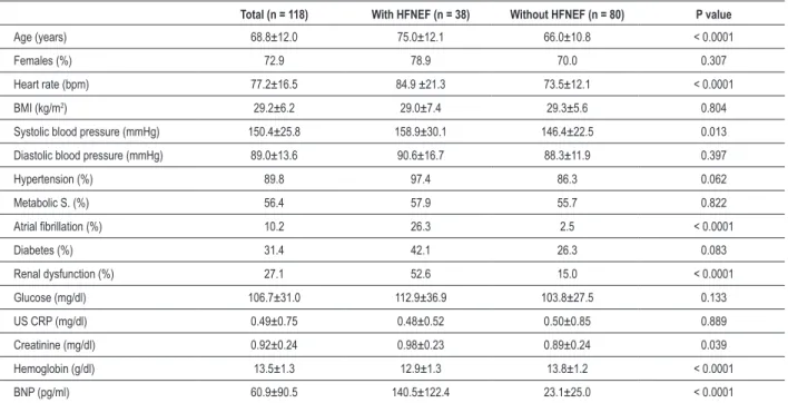
Natriuretic peptide levels were significantly different among patients with and without HFNEF (140.5 ± 122.4 pg/ml vs
23.1 ± 25.0 pg/ml p <0.0001) (fig. 1).
Table 2 displays the TDE parameters. LVEF was similar in both groups, while the other measures (E, E/A LAV-I, S’, E’, E/E’, and LVMI) were significantly different between groups.
Table 1 - Clinical and laboratory characteristics
Total (n = 118) With HFNEF (n = 38) Without HFNEF (n = 80) P value
Age (years) 68.8±12.0 75.0±12.1 66.0±10.8 < 0.0001
Females (%) 72.9 78.9 70.0 0.307
Heart rate (bpm) 77.2±16.5 84.9 ±21.3 73.5±12.1 < 0.0001
BMI (kg/m2) 29.2±6.2 29.0±7.4 29.3±5.6 0.804
Systolic blood pressure (mmHg) 150.4±25.8 158.9±30.1 146.4±22.5 0.013
Diastolic blood pressure (mmHg) 89.0±13.6 90.6±16.7 88.3±11.9 0.397
Hypertension (%) 89.8 97.4 86.3 0.062
Metabolic S. (%) 56.4 57.9 55.7 0.822
Atrial ibrillation (%) 10.2 26.3 2.5 < 0.0001
Diabetes (%) 31.4 42.1 26.3 0.083
Renal dysfunction (%) 27.1 52.6 15.0 < 0.0001
Glucose (mg/dl) 106.7±31.0 112.9±36.9 103.8±27.5 0.133
US CRP (mg/dl) 0.49±0.75 0.48±0.52 0.50±0.85 0.889
Creatinine (mg/dl) 0.92±0.24 0.98±0.23 0.89±0.24 0.039
Hemoglobin (g/dl) 13.5±1.3 12.9±1.3 13.8±1.2 < 0.0001
BNP (pg/ml) 60.9±90.5 140.5±122.4 23.1±25.0 < 0.0001
BMI - body mass index, US CRP - ultrasensitive C-reactive protein, BNP - [B-type natriuretic peptide]; Categorical variables = Pearson chi-square; Numeric variables = T Test. Signiicant differences between groups for p < 0.05.
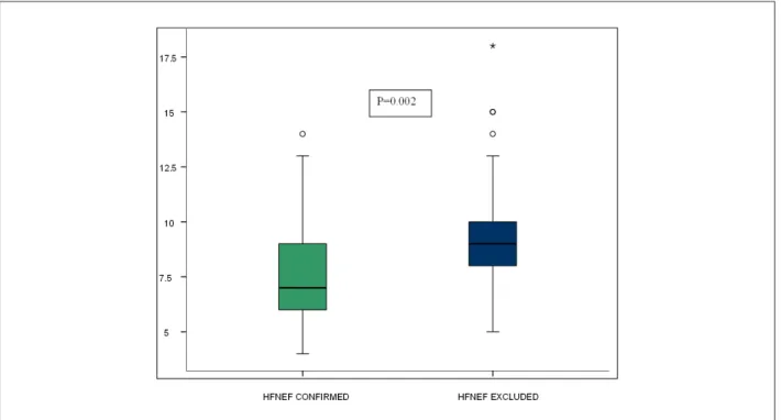
function was assessed in the radial axis by measuring the left ventricle ejection fraction (71.0 ± 9.5% vs 72.9 ± 7.9% - p = 0.229). However, assessment of LV systolic function in the longitudinal axis (S’) through TDE was significantly different between the two groups (7.8 ± 2.3 cm/s vs 9.4 ± 2.5 cm/s - p = 0.002) (fig. 2).
Parameters such as left atrial volume index (LAV-I) and E/A ratio for the assessment of diastolic function revealed a significant difference between patients with and without HFNEF (42.5 ± 15.3 ml/m2vs 28.4 ± 8.3 ml/m2 p < 0.0001
and 1.04 ± 0.70 vs 0.81 ± 0.29 p = 0.021).
LV diastolic function evaluated by TDE was significantly different for the parameters that assess LV relaxation (E’- 7.7 ± 2.6 cm/s vs 9.4 ± 2.5 cm/s - p = 0.001 ) and the pressure of LV filling (E/E’ - 14.3 ± 6.5 vs 7.5 ± 2.0 - p < 0.0001).
A correlation of S’ with other Doppler echocardiography parameters was found (Table 3) as the E/E’ ratio, E’ and E/A ratio (r = (-) 0.435, r = 0.457 p <0.0001, r = (-) 0.240, p = 0.014) and also with the LVMI (r = (-) 0.229 - p = 0.014), although BNP-I and LAV-I values were not correlated (r = (-) 0.125 p = 0.183, r = (-) 0.122 p = 0.194).
Discussion
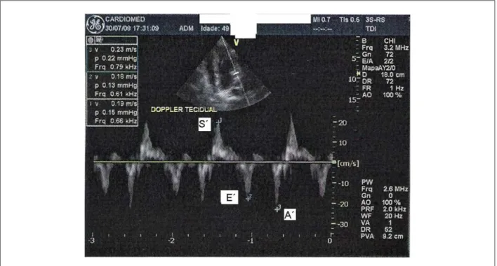
This study demonstrated that patients with HFNEF have a worse systolic function when assessed by measuring the mitral annulus velocity in the longitudinal axis (S’) (fig. 3). Systolic function assessed in the radial axis is preserved in
Table 2 - Characteristics of systolic and diastolic function by Doppler echocardiography and tissue Doppler echocardiography
Total (n = 118) With HFNEF (n = 38) Without HFNEF (n = 80) P value
LVEF (%) 72.3±8.4 71.0±9.5 72.9±7.9 0.229
LVMI (g/m2) 93.9±24.6 100.5±25.9 90.7±23.5 0.044
E (cm/s) 78±30 101±38 67±19 < 0.0001
A (cm/s) 93±32 110±52 87±19 0.001
E/A ratio 0.87±0.44 1.04±0.70 0.81±0.29 0.021
DT (ms) 257±86 269±126 253±68 0.455
LAV-I (ml/m2) 32.9±12.8 42.5±15.3 28.4±8.3 < 0.0001
S’ (cm/s) 8.9±2.4 7.8±2.3 9.4±2.5 0.002
E’ (cm/s) 8.8±2.6 7.7±2.6 9.4±2.5 0.001
E/E’ 9.7±5.1 14.3±6.5 7.5±2.0 < 0.0001
LVEF - left ventricle ejection fraction; LAV-I - left atrial volume index; TD - deceleration time, LVMI - left ventricular mass index; Numeric variables - T Test. Signiicant differences between groups for p <0.05.
Figure 2 - Comparison between S’ values in patients with and without HFNEF, 7.8 ± 2.3cm/s vs 9.4 ± 2.5 cms p = 0.002.
this population, as noted by the LV ejection fraction above 50%. Our results are similar to those of Tan et al17, which
showed that patients with HFNEF have abnormalities of LV systolic and diastolic function and that HFNEF is not an isolated disorder of diastole17.
For many years, the HFNEF pathophysiology has been related to abnormal diastolic relaxation and/or LV stiffness. Discussions of systolic function have been undervalued due to be normal LVEF in these patients18. However, LVEF as a
systolic function index has significant limitations because of the dependence on pre-and post-load, suboptimal reproducibility and low sensitivity in detecting small reductions in LV systolic function.
New mechanistic discussions involving the systolic and diastolic function by means of imaging methods that assess mechanisms of LV contraction and relaxation allow a better understanding of the link between systole and diastole in the normal heart and HFNEF19. Due to the orientation of its muscle
fibers, when the LV contracts, it promotes a rotational motion that builds energy, and during diastole, the energy stored is released, promoting a diastolic suction that contributes to the LV filling in the next cardiac cycle. Even small changes in systolic function, not detected by LVEF measurement, may change ventricular contraction leading to a decreased relaxation and loss of suction that affects quick LV filling.
Figure 3 - Evaluation of systolic (S’) and diastolic function (E’ and A’) by tissue Doppler echocardiography.
Table 3 - Correlation between systolic function evaluated in the longitudinal axis (S’) with TDE and BNP variables
Variables Pearson - r P value
E’(cm/s) 0.457 < 0.0001
E/E’ (-)0.435 < 0.0001
E/A (-)0.240 0.014
LVMI (g/m2) (-)0.229 0.014
LAV-I (ml/m2) (-)0.122 0.194
BNP (pg/ml) (-)0.125 0.183
LAV-I - left atrial volume index; LVMI - left ventricular mass index, BNP - B-type natriuretic peptide. Correlation is signiicant up to p = 0.05.
dP/dT20. S’ has the advantage of simple measurement, which is
easily detectable in most patients and has high reproducibility. Most studies point out to the relative independence of pre-and post-load in S’21 values. Some of S’ limitations include the
fact that we cannot discriminate between active and passive movements due to wall or heart movement as a whole and that this measure is angle-dependent. Patients’ movements, breathing and heart rate may also interfere with this measure21.
S’ is a marker of early systolic dysfunction in many pathological conditions that are characterized by conservation of conventional systolic indices, including hypertrophic cardiomyopathy, coronary disease, obesity and hypertension22.
High blood pressure promotes a gradual diastolic dysfunction along with the progress of concentric remodeling and hypertrophy. Advanced diastolic dysfunction (E / E ’> 15) is associated with the presence of impairment of longitudinal systolic function in hypertensive patients in a symptomatic
(HFNEF) or asymptomatic way.
This study is the first in the literature to prospectively use new diastology criteria in characterizing patients with HFNEF. Therefore, it was observed that patients with criteria for HFNEF presented a significant increase in pulmonary venous pressure measured indirectly by E/E’ ratio, lower E’ and S’ values, suggesting reduced ventricular relaxation and contractility, as well as a BNP six times higher. Our data are quite similar to those found in other studies that have shown a significant relationship between systole peak (S’) and annular velocity in early diastole (E’)14,17,23, demonstrating a strong
connection between LV contraction and relaxation where a modified LV systolic function has a significant impact on ventricular relaxation.
A limitation in our study is that systolic function was not measured by other techniques that can be used to assess it.
Another limitation is that the LV function, measured by longitudinal axis shortening, is sensitive to the effects of hypertension, ischemia, diabetes and age, which are situations that may precede HFNEF. However, in our study, except in relation to age, other variables showed no statistically significant difference between the two groups.
Conclusion
by S’ and relaxation assessed by E’ support the contemporary view of the connection of systolic and diastolic abnormalities in these patients.
Potential Conflict of Interest
No potential conflict of interest relevant to this article was reported.
Sources of Funding
There were no external funding sources for this study.
Study Association
This article is part of the thesis of master submitted by Antonio José Lagoeiro Jorge, from Universidade Federal Fluminense.
References
1. Vasan R, Benjamin E, Levy D. Prevalence, clinical features and prognosis of diastolic heart failure: an epidemiologic perspective. J Am Coll Cardiol. 1995; 26 (7): 1565-74.
2. Pernenkil R, Vinson J, Shah A, Beckham V, Wittenberg C, Rich M. Course and prognosis in patients or 70 years of age with congestive heart failure and normal versus abnormal left ventricular ejection fraction. Am J Cardiol. 1997;79 (2): 216-9.
3. Smith GL, Masoudi FA, Vaccarino V, Radford MJ, Krumholz HM. Outcomes in heart failure patients with preserved ejection fraction. J Am Coll Cardiol. 2003; 41 (9): 1510-8.
4. Moutinho MAE, Colucci FA, Alcoforado V, Tavares LR, Rachid MB, Rosa ML, et al. Insuficiência cardíaca com fração de ejeção preservada e com disfunção sistólica na comunidade. Arq Bras Cardiol. 2008; 90 (2): 145-50. 5. Owan TE, Hodge DO, Herges RM, Jacobsen SJ, Roger VL, Redfield MM.
Trends in prevalence and outcome of heart failure with preserved ejection fraction N Engl J Med. 2006; 355 (3): 251-9.
6. Tribouilloy C, Rusinaru D, Mahjoub H, Soulière V, Levy F, Peltier M, et al. Prognosis of heart failure with preserved ejection fraction a 5 year prospective population-based study. Eur Heart J. 2008; 29 (3): 339-47.
7. Lester SJ, Tajik AJ, Nishimura RA, Oh JK, Khanderia BK, Seward JB. Unlocking the mysteries of diastolic function deciphering the Rosetta stone 10 years later. J Am Coll Cardiol. 2008; 51 (7): 679-89.
8. Maciver DH, Townsend M. A novel mechanism of heart failure with normal ejection fraction. Heart. 2008; 94 (4): 446-9.
9. Paulus WJ, Tschöpe C, Sanderson JE, Rusconi C, Flachskampf FA, Rademakers FE, et al. How to diagnose diastolic heart failure: a consensus statement on the diagnosis of heart failure with normal left ventricular ejection fraction by heart failure and echocardiography associations of the European Society of Cardiology. Eur Heart J. 2007; 28 (20): 2539-50.
10. Sanderson JE, Fraser AG. Systolic dysfunction in heart failure with a normal ejection fraction: echo-Doppler measurements. Progr Cardiovasc Dis. 2006; 49 (3): 196-206.
11. Alam M, Wardell J, Andersson E, Samad BA, Nordlander R. Effects of first myocardial infarction on left ventricular systolic and diastolic function with the use of mitral annular velocity determined by pulse-wave Doppler tissue imaging. J Am Soc Echocardiogr. 2000; 13 (5): 343-52.
12. Vinereanu D, Nicolaides E, Tweddel AC, Fraser AG. “Pure” diastolic
dysfunction is associated with long-axis systolic dysfunction: implications for the diagnosis and classification of heart failure. Eur J Heart Fail. 2005; 7 (5): 820-8.
13. Yu C, Lin H, Yang H, Kong S, Zhang Q, Lee S. Progression of systolic abnormalities in patients with “isolated” diastolic heart failure and diastolic dysfunction. Circulation. 2002; 105 (10): 1195-201.
14. Garcia EH, Perna ER, Farias EF, Obregon RO, Macin SM, Parras JI, et al. Reduced systolic performance by tissue Doppler in patients with preserved and abnormal ejection fraction: new insights in chronic heart failure. Int J Cardiol. 2006; 108 (2): 181-8.
15. Nishikage T, Nakai H, Lang RM, Takeuchi M. Subclinical left ventricular longitudinal systolic dysfunction in hypertension with no evidence of heart failure. Circ J. 2008; 72 (2): 189-94.
16. Brutsaert DL. Cardiac dysfunction in heart failure: the cardiologist’s love affair with time. Prog Cardiovasc Dis. 2006; 49 (3): 157-81.
17. Tan YT, Wenzelburger F, Lee E, Heatlie G, Leyva F, Patel K, et al. Exercise echocardiography reveals complex abnormalities of both systolic and diastolic ventricular function involving torsion, untwist, and longitudinal motion. J Am Coll Cardiol. 2009; 54 (1): 36-46.
18. Zile MR, Baicu CF, Gaasch WH. Diastolic heart failure - abnormalities in active relaxation and passive stiffness of the left ventricle. N Engl J Med. 2004; 350 (19): 1953-9.
19. Marwick TH. The deconvolution of diastole. J Am Coll Cardiol. 2009; 54 (1): 47-8.
20. Hsiao SH, Huang WC, Sy CL, Lin SK, Lee TY, Liu CP. Doppler tissue imaging and color M-mode flow propagation velocity: are they really preload independent? J Am Soc Echocardiogr. 2005; 18 (12): 1277-84.
21. Borges MC, Colombo RC, Gonçalves JG, Ferreira J de O, Franchini KG. Longitudinal mitral annulus velocities are reduced in hypertensive subjects with or without left ventricle hypertrophy. Hypertension. 2006; 47 (5): 854-60. 22. Yamada H, Oki T, Tabata T, Iuchi A, Ito S. Assessment of left ventricular systolic
wall motion velocity with pulsed tissue Doppler imaging: comparison with peak dP/dt of the left ventricular pressure curve. J Am Soc Echocardiogr. 1998; 11 (5): 442-9.