INTRODUCTION
The mortality rate of patients with cirrhosis ad mitted to an intensive care unit (ICU) due to organ dysfunction ranges from 34% to 69% depending on the reason for admission, the presence of organ failure (OF) and the severity of the underlying liver disease. Over the last ten years it has markedly decreased from around 90%100% to 41%50% in some reports(25, 74).
Besides wellrecognized complications of chro nic liver disease, such as ascites, infections, variceal bleeding (VB) and hepatic encephalopathy (HE), intensive care physicians now face different clinical scenarios. These include acuteonchronic liver failure (ACLF), VB with requirement for early transjugular intrahepatic portacaval shunt (TIPS) placement and nosocomial and healthcare associated (HCA) infec tions, particularly spontaneous bacterial peritonitis (SBP) due to multiresistant bacteria(26, 53, 73).
Management of decompensated cirrhosis in
INTENSIVE CARE MANAGEMENT OF
PATIENTS WITH LIVER DISEASE:
proceedings of a single-topic conference
sponsored by the Brazilian Society of
Hepatology
Paulo Lisboa
BITTENCOURT
1,2, Carlos
TERRA
3, Edison Roberto
PARISE
4,
Alberto Queiroz
FARIAS
5and Members of the Panel in the 1st Single-Topic Conference
of Intensive Care Management of Patients with Liver Disease*
ABSTRACT– Survival rates of critically ill patients with liver disease has sharply increased in recent years due to several improvements in the management of decompensated cirrhosis and acute liver failure. This is ascribed to the incorporation of evidencebased strategies from clinical trials aiming to reduce mortality. In order to discuss the cuttingedge evidence regarding critical care of patients with liver disease, a joint single topic conference was recently sponsored by the Brazilian Society of Hepatology in cooperation with the Brazilian Society of Intensive Care Medicine and the Brazilian Association for Organ Transplantation. This paper summarizes the proceedings of the aforementioned meeting and it is intended to guide intensive care physicians, gastroenterologists and hepatologists in the care management of patients with liver disease.
HEADINGS – Liver cirrhosis. Acute liver failure. Intensive care medicine. Acute on chronic liver failure. Complications.
Declared conflict of interest of all authors: none Disclosure of funding: no funding received
1 Hospital Português, Salvador, BA; 2 Hospital Universitário Professor Edgard Santos, Universidade Federal da Bahia, Salvador, BA; 3 Hospital Universitário Pedro Ernesto,
Universidade do Estado do Rio de Janeiro, RJ; 4 Universidade Federal de São Paulo, SP; 5 Departamento de Gastroenterologia da Faculdade de Medicina da
Universi-dade de São Paulo, SP; 6 Liver Unit, IMDiM, Hospital Clınic, Universidad de Barcelona, IDIBAPS and CIBERehd, 7 Departamento de Gastroenterologia, Hospital Federal
do Bonsucesso, Rio de Janeiro, RJ; 8 Universidade Federal de Ciências da Saúde de Porto Alegre e Irmandade da Santa Casa de Misericórdia de Porto Alegre, RS.
*Vincent Arroyo6, Javier Fernandez6, Gustavo Pereira7, Luiz Marcelo Maubouisson5, Guilherme Marques Andrade5, Fernando Gomes de Barros Costa,5 Liana Codes1,2,
Antônio Ricardo Andrade1,2, Angelo Matos8, André Torres, 3 Fernanda Couto, 7 Ivan Zyngier7.
Correspondence: Paulo Lisboa Bittencourt. Rua Prof. Clementino Fraga, 220, 1901 – CEP: 40170-050 – Salvador, BA, Brasil. E-mail: [email protected]
ICU has changed due to the emergence of new evi dencebased treatments associated with improved sur vival, tailoring of intensive care measures, availability of artiicial and bioartiicial liver support systems, as well as the widespread use of liver transplantation (LT) for criticallyill patients, due to the MELD allocation policy, which is based on severity of liver disease(13, 62, 74).
In order to discuss recent advances in this emerging ield, the Brazilian Society of Hepatology in coope ration with the Brazilian Society of Intensive Care Medicine and the Brazilian Association for Organ Transplantation sponsored a joint singletopic con ference on the critical care management of patients with liver disease, which was held in Rio de Janeiro on May 5th 2014.
PART I. MANAGEMENT AND TREATMENT OF PATIENTS WITH CIRRHOSIS IN THE ICU
1) Hyponatremia
Clinically relevant hyponatremia in cirrhosis is deined as a reduction in serum sodium to below 130 mmol/L(4, 8, 44).
Recent studies have shown that hyponatremia is an important prognosis marker both before and after liver transplantation (LT)(50, 54, 67). Moreover, hyponatremia has gained attention
because of the discovery of the vaptans, which improve solutefree water excretion by counteracting the effects of arginine vasopressin (AVP) in the renal tubules(85). These
drugs are currently being assessed for the management of hyponatremia associated with cardiac failure, inappropriate antidiuretic hormone secretion as well as cirrhosis. In clinical practice, hyponatremia is classiied into three types: hypo volemic, euvolemic and hypervolemic, with some patients exhibiting a mixture of conditions.
With the exception of a few circumstances of hyponatre mia with hypovolemia due to diuretic use or gastrointestinal losses, most of the cases of hyponatremia in cirrhosis result from increased extracellular luid volume (dilutional hypona tremia)(44). Conditions such as hypotonic luid administration,
heart failure and renal failure frequently seen in the ICUs should be ruled out in the differential diagnosis of dilutional hyponatremia. In cirrhosis, total body water is increased, yet effective arterial volume is decreased (“relative central hypovolemia”). The reduction in effective arterial volume is a consequence of the increased intrahepatic resistance and splanchnic arterial vasodilation, which is caused by the excessive release of nitric oxide and other compounds such as endotoxin, substance P and endocannabinoids. This pro cess leads to sodium avidity in the proximal portion of the nephron, by activation of the renin–angiotensin–aldosterone axis and excessive ADHmediated free water reabsorption in the collecting tubule.
Arterial baroreceptors, found in the left ventricle and the carotid sinus, have been shown to be a potent regulator of ADH secretion. Their activation counteracts the suppressive effects of hypoosmolality.
In patients with cirrhosis and ascites, the nonosmotic release of ADH from the posterior pituitary becomes the dominant force of water retention, resulting in impaired free water excretion and dilutional hyponatremia(44).
The action of ADH on the kidney occurs predominantly in the principle cells of the collecting tubule. The stimu lation of the vasopressin receptor, V2, by ADH leads to downstream activation of a cyclic AMPbased pathway and subsequent upregulation of the aquaporin channel AQP2 in the apical membrane of the principle cell. This allows the free low of water from the tubular luid back into circulation(85).
Low serum sodium levels (<135 mmol/L) are prevalent in both inpatients and outpatients, and are associated with severe ascites, frequent use of largevolume paracentesis, impaired renal function, higher frequencies of HE, SBP, hepatorenal syndrome (HRS), higher rates of inhospital mortality and poor shortterm prognosis(4).
Several lines of evidence show that serum sodium con centration is an independent predictor of survival among liver transplantation candidates(50, 54, 67).
In cases of hyponatremia, water moves into the cells to maintain the osmotic balance, causing cell swelling. Increases of cell volume are particularly important in the brain, as the skull restricts brain enlargement. For this reason, brain cells have defensive mechanisms to limit cerebral edema, which is the extrusion of intracellular solutes to decrease intracellu lar osmolality, until it matches that of plasma. In the early stages of the development of hyponatremia, there is a rapid loss of intracellular electrolytes, particularly potassium, usually within the irst 24 hours. Subsequently, there is a loss of lowmolecular weight organic compounds, known as organic osmolytes, including myoinositol, glutamine, choline, and taurine.
The combined losses of both electrolytes and organic osmolytes from the brain cells enable effective regulation of brain volume during hyponatremia. The effectiveness of this mechanism in preventing lethal edema depends, among other factors, on the severity of hyponatremia and rate of reduction of the serum sodium concentration. Adaptation is more ef icient in chronic hyponatremia than in acute hyponatremia. There is evidence that such cerebral adaptation to hypo natremia is also present in cirrhosis. Of similar importance to the central nervous system are the changes that occur after recovery of hyponatremia. When the serum sodium concen tration returns to normal, there is restoration of electrolyte and osmolyte levels in brain cells; electrolytes are restored quickly, whereas correction of organic osmolytes is slow, particularly if the duration of hyponatremia has been long. This is a major clinical concern because rapid correction of hyponatremia may lead to severe brain damage, because of a lack of adequate brain adaptation to the normalized os molality of the extracellular luid. This is known as osmotic demyelination syndrome(50).
Studies speciically assessing neurological symptoms in cirrhosis with hyponatremia are lacking. However, the clinical experience indicates that signiicant neurological manifes tations such as headache, focal motor deicits, seizures, and cerebral herniation are very uncommon. It is likely that the relatively low incidence of neurological manifestations of hypervolemic hyponatremia in patients with cirrhosis is rela ted to the fact that in most patients hyponatremia is chronic rather than acute. This allows suficient time for the brain to adjust to the hypoosmolality of the extracellular luid.
Apart from hyponatremia, it is believed that lowgrade cerebral edema is one of the factors leading to HE, as dis cussed in section 5 of this manuscript. In patients with HE and lowgrade cerebral edema, hyponatremia may represent a second osmotic hit to astrocytes, causing further depletion of osmotic counteractive systems. In this situation, cells would probably not tolerate additional changes in volume, and HE would develop or persist even in the absence of any stimuli(50).
Hyponatremia before LT has also been associated with an increased risk of renal failure and infectious complications, higher use of blood products, longer duration of hospital stay, and, most importantly, increased shortterm mortality rates after LT(50, 54, 67).
Conventional treatment of ascites in cirrhosis includes sodium restriction, diuretic therapy, and largevolume paracentesis. Fluid restriction in combination with orally administered aldosterone antagonists and loop diuretics is currently the primary approach for treatment of hypervole mic hyponatremia in cirrhosis.
However, in recent years, a number of vasopressin recep tor antagonist agents, which inhibit the effects of AVP and increase free water excretion, have been assessed for treatment of hyponatremia in cirrhosis(4, 84).
The oral selective vasopressin V2receptor antagonist tolvaptan is approved for treating hypervolemic and euvole mic hyponatremia, including that caused by cirrhosis. It has proved to be effective, safe, and improves shortterm quality of life (one month) in cirrhosis.
However, there are no longterm data on eficacy and safety, nor are there data on other outcomes such as HE or survival. Tolvaptan is a high cost drug, approved in USA for the management severe hypervolemic hyponatremia (<125 mmol/L) in cirrhosis, cardiac failure and SIADH, while in Europe it is approved only for the management of SIADH(85).
Patients who can beneit the most from treatment with tolvaptan are those with severe hyponatremia awaiting liver transplantation. Treatment with tolvaptan should be started in the hospital with low doses and serum sodium should be closely monitored to avoid rapid correction of hyponatremia to less than 10 mmol/day. This is relevant because patients with very low sodium concentration are at greatest risk of neurological complications. Patients with type1 hepatorenal syndrome and hyponatremia should be treated with terlip ressin or other vasoconstrictors and albumin(85).
2) Circulatory failure
Diuretics, antibiotics, and human serum albumin (HSA) are the most frequently used treatments for the management of patients with cirrhosis. According to the CANONIC study database(71), a prospective European investigation of
1348 patients with decompensated cirrhosis, HSA was pres cribed for 60% of the patients during hospital admission. Prevention of paracentesisinduced circulatory dysfunction, prevention of type1 HRS associated with bacterial infections and treatment of type1 HRS are the main intended aims of therapy(78, 79). In these cases, treatment with HSA is associated
with improved survival(88).
Decompensated cirrhosis is a condition associated with systemic inlammation, which plays an important role in the pathogenesis of organ failure. Although, the beneicial effects of HSA have been traditionally attributed to plasma volume expansion, they could also relate to its effects modulating systemic and organ inlammation(7, 35).
Three major features characterize decompensated cir rhosis. The irst is multiorgan dysfunction. The second is
a systemic inlammatory reaction with increased plasma and ascitic luid concentration of cytokines and Creactive protein (CRP). The third is an increased systemic oxidative stress with high levels of oxidized HSA and other markers of oxidative stress(7). Systemic inflammation, oxidative
stress, and organ dysfunction are moderate in patients with decompensated cirrhosis and severe in patients with acute onchronic liver failure (ACLF)(71). Translocation of bacterial
products (i.e., lipopolysaccharide, bacterial DNA) or of viable organisms from the intestinal lumen to the circulation due to quantitative and qualitative changes in gut microbiota, impairment in intestinal mucosal barrier, increased epithelial permeability, and impaired intestinal immunity are important mechanisms of systemic inlammation in cirrhosis(7). Sys
temic inlammatory response can be triggered by bacterial antigens (PathogensAssociated Molecular Patterns, PAMPs) or by intrinsic factors released into the circulation as a result of trauma or cell injury (Damaged Associated Molecular Pat terns, DAMPs). Specialized receptors of the innate immune system recognize these factors and release inlammatory mediators such as cytokines, and reactive oxygen (ROS) and nitrogen species (RNS). However, systemic inlammation may also occur in response to acute liver damage (i.e., acute alcoholic hepatitis) or other mechanisms(7).
Close interactions exist between bacterial translocation, local inlammation, and cardiovascular dysfunction in de compensated cirrhosis. Activation of the intestinal immune system by bacterial translocation causes local release of NO and other vasodilators, leading to the characteristic hyper dynamic circulation of cirrhosis.
At more advanced stages, there is effective hypovolemia, activation of the reninangiotensin system (RAS), sympa thetic nervous system (SNS), antidiuretic hormone (ADH) and ascites formation. The activated sympathetic nervous system induces changes in the gut microbiota and impairs intestinal immunity, thus producing a vicious circle promot ing the progression of cardiovascular dysfunction(7). A slow
but progressive impairment of left ventricular function and cardiac output also develops in decompensated cirrhosis and contributes to circulatory dysfunction. Recent experimental data suggest that impairment in cardiac function in cirrho sis is related to inlammation, tumornecrosis factor alpha (TNFalpha)related activation of inducible NOsynthase and oxidative stress in cardiac tissue(7). ACLF is characterized
by acute organ failure(s) (liver, renal, brain, coagulation, circulation, and respiration) in patients with compensated or decompensated cirrhosis. ACLF develops in the setting of severe systemic inlammatory reaction due to bacterial infection, acute alcoholic hepatitis or other precipitating factors. The frequency of organ failure correlates directly with the degree of systemic inflammation(71). Therefore,
of systemic inlammation to organs, leading to abnormal distribution of bloodlow within the microcirculation and cell dysfunction related to mitochondrial oxidative stress(7, 71).
Bacterial infection is a frequent precipitating factor of HE. Peripheral inlammation may affect cerebral function through afferent vagal nerves activated by cytokines at the site of inlammation, by lipopolysaccharide or cytokines which interact with the brain in areas lacking the bloodbrainbar rier or by diffusion to the brain of endothelial mediators. Activation of microglia and synthesis of proinlammatory cytokines in the brain have also been demonstrated in experi mental models of liver failure. Circulatory dysfunction in patients with systemic inlammation reduces brain perfusion. Systemic inlammation increases the inhibitory effect of ammonia in brain function. There are marked differences in HE between patients with decompensated cirrhosis and patients with ACLF. In the irst group HE is of low severity and diuretics are the most common precipitating cause. By contrast, HE in ACLF is severe and bacterial infection or acute liver injury are the main precipitating factors. Organ dysfunction in cirrhosis therefore varies according to the mechanism and degree of systemic inlammation(7, 24, 71).
Considering the potential role of systemic inlamma tion in cirrhosis and the effect of HSA in innate immune response and oxidative stress, it is reasonable to suggest that some effects of HSA (prevention and treatment of type1 HRS, treatment of SBP) might be related to these properties(7). In patients with cirrhosis and SBP, treatment
with intravenous albumin in addition to an antibiotic re duces the incidence of renal impairment and mortality in comparison to treatment with an antibiotic alone(88). It is
known that in patients with HRS type 1, the association of albumin with analogues of vasopressin improves renal func tion and survival(68, 81). In this regard, albumin infusion was
reported to be superior to hydroxyethyl starch in improving hemodynamics, including left ventricular function, cardiac output, and peripheral vascular resistance in patients with SBP without complications(35).
Circulatory dysfunction in cirrhosis is related to systemic inlammation leading to arterial vasodilation and impairment of left ventricular function. The differences in circulatory dysfunction between patients with type1 HRS and those with decompensated cirrhosis and/or type2 HRS are related to differences in timecourse and grade of systemic inlam mation. In type1 HRS, systemic inlammation is acute and severe. In decompensated cirrhosis and/or type2 HRS it is moderate and prolonged. Albumin is probably effective in the prevention and treatment of HRS by regulating the systemic inlammatory reaction(7).
3) Renal failure and hepatorenal failure
Renal failure (RF) is seen in 39% to 49% of the patients with cirrhosis admitted to the ICU(19, 23). The most common
causes of RF are hypovolemia, bacterial infection, paren chymal kidney disease and hepatorenal syndrome (HRS). They are identiied as causes of RF in 40%, 32%, 15% and 12% of the cases, respectively(19). Hepatorenal syndrome is
usually seen in patients with advanced cirrhosis and ascites. It is characterized by an intense renal vasoconstriction, which leads to very low renal perfusion and glomerular iltration rate (GFR)(46, 80). The development of portal hypertension
in cirrhosis is associated with arterial vasodilation in the splanchnic circulation due to the local release of nitric oxide and other vasodilatory substances. These circulatory changes induce arterial hypotension that is compensated by the development of a hyperdynamic circulation (increased heart rate and cardiac output). As the disease progresses, arterial vasodilation increases, leading to activation of high pressure baroreceptors, relex stimulation of the reninangio tensin and sympathetic nervous systems, increase in arterial pressure to normal or near normal levels, sodium and water retention and ascites formation. Splanchnic circulation is resistant to the effect of angiotensinII, noradrenaline and vasopressin, due to the local release of nitric oxide and other vasodilators. The maintenance of arterial pressure is due to vasoconstriction of extrasplanchnic vascular territories such as the kidneys. HRS develops in the inal phase of the disease, when there is extreme deterioration in effective ar terial blood volume, severe arterial hypotension and intense renal vasoconstriction. Several lines of evidence suggest that cardiac dysfunction may also play a role in the pathogenesis of this syndrome(58, 78).
There are two different types of HRS. Type2 HRS de velops in nonazotemic patients with cirrhosis and refractory ascites with moderate and relatively steady renal failure. By contrast, Type1 HRS is characterized by increasing serum creatinine levels, reaching a value greater than 2,5 mg/dL in less than two weeks(80). Type1 HRS frequently occurs
in a closed relationship with a precipitating factor such as bacterial infection, mainly SBP, gastrointestinal hemorrhage, a major surgical procedure or an acute hepatitis lare in a patient with cirrhosis.
The diagnosis of HRS is based on the exclusion of other types of renal failure that may occur in patients with cirrhosis. The criteria required for diagnosis are reported below(80):
Cirrhosis with ascites.
Serum creatinine >133 mmol/L (1.5 mg/dL).
No improvement of serum creatinine (decrease to a level of ≤133 mmol/L) after at least 2 days of diuretic withdrawal and volume expansion with albumin. The recommended dose of albumin is 1 g/kg of body weight per day up to a maximum of 100 g/day.
Absence of shock.
No current or recent treatment with nephrotoxic drugs. Absence of parenchymal kidney disease as indicated
by proteinuria >500 mg/day, microhaematuria (>50 red blood cells per high power ield) and/or abnormal renal ultrasonography.
HRS is the complication of cirrhosis associated with the worst prognosis. The median survival time for type1 HRS is 15 days. Patients with type2 HRS have a median survival time about 6 months.
because it allows both the liver disease and renal failure to be treated. The longterm survival of patients with HRS who undergo liver transplantation is good, with a threeyear probability of survival higher than 60%. This survival rate is only slightly reduced compared with that of transplantation in patients without HRS (which ranges between 70% and 80%). The main problem of liver transplantation in type 1 HRS is its applicability. Due to their extremely short survival time, most patients die before transplantation.
Vasoconstrictors improve circulatory function by induc ing vasoconstriction of the splanchnic arterial bed, thereby suppressing the activity of endogenous vasoconstrictor systems and improving renal perfusion. In combination with intravenous albumin, this pharmacological approach may reverse HRS in approximately 40%50% of patients(68, 81, 87).
Different vasoconstrictors have been used, including terli pressin, midodrine and noradrenaline. Although data on other vasoconstrictors are promising, terlipressin is the most studied, and should be used in progressive dosage starting with 0.5 mg / 4 hours. If serum creatinine does not decrease by more than 30% in 3 days, the dose should be doubled. Albumin should be administered starting with a priming dose of 1g/kg of body weight followed by 2040 g/day. Midodrine and noradrenaline, which have been shown to be effective and safe, can also be used. A recent randomized study including fortysix patients with HRS type1, evaluated the safety and eficacy of terlipressin and noradrenaline (starting with 0.5 mg/h) in the treatment of HRS. The main conclusion of this study was that noradrenaline is as safe and effective as terlipressin, but less expensive in the treatment of HRS(87).
These conclusions were conirmed by a recent metaanalysis of four studies comprising 154 patients(72).
The treatment of HRS with vasoconstrictors increases the risk of potentially serious adverse events, such as myocardial infarction. Assessment of potential contraindications and close monitoring of adverse events is essential.
Currently, few patients with HRS have been treated by transjugular intrahepatic portacaval shunt (TIPS).
Published data suggest that TIPS is effective in normal izing serum creatinine in a signiicant proportion of patients with HRS, rendering it an alternative treatment of type1 HRS. The applicability of transplantation is relatively low, since it is usually contraindicated in patients with severe liver failure or severe HE. At the time of writing, no studies had compared TIPS and vasoconstrictors in the treatment of HRS.
The use of MARS in patients with type 1 HRS was not associated with beneicial effects and therefore should not be given outside clinical trials(80).
4) Adrenal failure
Adrenal insuficiency (AI) is the clinical manifestation of deicient production or action of glucocorticoids. Cor tisol is the major endogenous glucocorticoid secreted by the adrenal cortex and has several biological effects. Glu cocorticoids modulate immune response by stimulation of antiinlammatory cytokine production and inhibition
of proinflammatory cytokine production, inflammatory cell migration, and expression of inlammatory mediators. They are also responsible for maintenance of myocardial contractility and vascular tone by modulating reactivity to reninangiotensinaldosterone and sympathetic nervous systems, regulating vascular permeability and decreasing production of vasodilators. The synthesis and secretion of cortisol is regulated by the hypothalamicpituitaryadrenal axis’ production of adrenocorticotropic hormone (ACTH) and corticotropinreleasing hormone (CRH).
In patients with decompensated cirrhosis, increased production of cytokines due to bacterial translocation, hy poperfusion of the adrenal gland and reduced serum levels of cholesterol (the main precursor of adrenal steroids), may lead to reduced cortisol synthesis. These indings may explain the increased frequency of AI observed in patients with cirrhosis when compared to the general population.
Diagnosis of AI is most commonly established by the ACTH stimulation test. The test uses 250μg of synthetic ACTH given IV, with cortisol measured at baseline and after 60 minutes (peak cortisol). The difference between baseline and peak cortisol is called delta cortisol. Most studies use a delta cortisol level lower than 9 μg/dL as the diagnostic criterion. In critically ill patients, a baseline serum cortisol lower than 15 μg/dL is also considered indicative of AI.
In patients with decompensated cirrhosis, the prevalence of AI ranges between 26% and 39%. There is no clear rela tionship between liver and adrenal function, as evidenced by the equal proportion of patients with ChildPugh C score and comparable values of MELD scores between patients with and without AI(94). Nevertheless, lower serum levels of
cholesterol (but not triglycerides) were reported in patients with AI. Adrenal insufficiency was also associated with greater impairment of circulatory and renal functions, as evidenced by lower mean arterial pressure and serum sodium, and higher values of BUN and plasma renin activity.
Interestingly, patients with AI had a higher probability of sepsis and type1 HRS and lower survival rates. These data indicate that, in patients with decompensated cirrhosis, AI is a common complication that develops independently of the degree of liver function and is associated with greater morbidity and mortality when compared with patients with normal adrenal function. In patients with cirrhosis and septic shock, the prevalence of AI ranges between 51% and 76%, a frequency almost double the one observed in patients with decompensated cirrhosis and almost 50% greater than the one observed in noncirrhotic subjects with septic shock.
insuficiency was shown to be an important prognostic factor and an independent predictor of hospital mortality(5, 33, 94).
Previous studies have shown that administration of low doses of hydrocortisone improves shock reversal and survival in septic shock. Two studies have speciically addressed this issue in patients with cirrhosis(5, 33). In a prospective study, 25
patients with septic shock had adrenal function evaluated, and those who had AI received 50 mg IV hydrocortisone every 6 hours. Results were compared with a retrospective cohort of 50 patients with septic shock not treated with steroids. Shock resolution was more frequent and faster in the group treated with hydrocortisone. This group also showed a lower frequency of renal failure in the ICU, with a comparable incidence of new infections and GI bleeding. Survival was signiicantly higher for the group treated with supplementary steroids, indicating a beneicial effect of the treatment of AI in patients with cirrhosis and septic shock.
Recently, a randomized, double blind trial evaluated the effects of low doses of hydrocortisone versus placebo in a group of 75 patients with cirrhosis. The authors showed a similar beneicial effect on shock reversal, but a higher frequency of gastrointestinal bleeding was observed in the hydrocortisone group. Both groups showed similar survival rates after 28 days(5).
In summary, adrenal insuficiency is common in patients with cirrhosis and is correlated with higher morbidity and mortality. However, more studies are needed to better clarify possible role of stressdose corticosteroid supplementation in the management of this condition.
5) Cerebral dysfunction and hepatic encephalopathy Hepatic encephalopathy is an important multifactorial neurological syndrome that can occur in patients with chronic liver disease and acute liver failure (ALF), related or unrelat ed to portosystemic shunts(1). It represents a progressive but
potentially reversible cause of cerebral dysfunction with a wide array of neuropsychiatric, cognitive and motor symp toms, ranging from minor signs of altered brain function to deep coma. Hepatic encephalopathy was shown to increase mortality and to diminish the quality of life of patients with cirrhosis(1, 15). The probability of transplantfree survival after
the irst episode of acute HE is only 42% at 1 year and 23% at 3 years(89). Those with severe HE in the ICU carry, respec
tively, a 35% and 54% inhospital and 1year mortality(38).
The underlying mechanisms involved in the pathogenesis of HE are not completely understood, but it is speculated that the failure to detoxify nitrogenderived products (especially ammonia) predominantly found in the intestine is involved(76).
This may be related both to impaired hepatic clearance and portosystemic shunting. Hepatic encephalopathy is classiied as type A when associated with ALF, type B when related to portosystemic bypass or shunting and type C when associa ted with cirrhosis. Type C is further divided into episodic, persistent and minimal HE. Episodic HE can be triggered by recognized risk factors; spontaneous and recurrent HE (more than two episodes in a year) in the absence of these triggering events. Persistent HE is deined by the presence
of chronic neuropsychiatric signs and symptoms, usually graded as mild, severe or controlled only with drug therapy. Minimal HE is a preclinical syndrome that can be diagnosed only with neuropsychological or complex neurophysiological tests. All types are associated with some degree of cerebral dysfunction due to astrocyte ammonia swelling, impaired glial and neuronal function, mainly due to hyperammonemia, which may disrupt synapsis and lead to HE symptoms(1). In
contrast to ALF, HE in cirrhosis occurs without cerebral edema and intracranial hypertension and is usually triggered by some precipitant factors(15). The West Haven classiication
is commonly used to assess severity of HE in four grades. However, recently the International Society for Hepatic En cephalopathy and Nitrogen Metabolism proposed another classiication named SONIC that encompasses minimal and grade I HE as covert HE and grade II to IV HE as clinical
ly apparent HE(9). Common signs and symptoms include
somnolence, confusion, bradykinesia, asterixis, dysarthria, ataxia, progressive alterations in muscular relexes and coma. Seizures, transient focal deicits and nystagmus are uncom
mon(24). Myelopathy as well as extrapyramidal symptoms
may be predominant in some patients(15, 56).
The diagnosis of HE is usually straightforward in subjects with cirrhosis, but it should be pointed out that it is important to exclude other causes of brain dysfunction, particularly in patients that failed to recover promptly post treatment. Blood ammonia may be useful in those patients, but one should keep in mind that its concentration was shown to vary in individual patients and can be in the normal range of 10% of patients with overt HE. Computed tomography scans may be useful to rule out intracranial hemorrhage, infarction, abscess or tumors. Furthermore, magnetic resonance imaging of the brain can depict typical abnormalities seen in subjects with HE, such as deposition of paramagnetic substances in the basal ganglia, decrease in brain size, increase in brain water and changes in organic osmolytes. Electroencephalogram triphasic waves are very common but not speciic for HE(24).
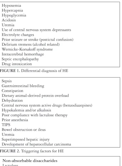
It should be pointed out that most patients with HE could recover only with the withdrawal of triggering factors (Figure 2). According to clinical judgment, it is important to rule out infection; discontinue diuretics and administer intravenous luids for those who are volume depleted, correct electrolyte disturbances, stop offending drugs such as ben zodiazepines and narcotics, restore or enhance bowel move ments in those patients with obstipation or gastrointestinal hemorrhage. Equally important but generally neglected, are general supportive measures, such as adequate care to protect those patients from selfinlicted injury and/or aspiration pneumonia, to provide nutrition in order to avoid hypogly cemia as well as malnutrition. In this regard, it is important to guarantee a minimal caloric intake of 3540 Kcal/Kg/ day. Protein restriction does not have an apparent beneit in episodic HE. With cirrhosis, despite the fact that protein could contribute to HE, patients should receive 1,21,5g/Kg/ day of protein. This is important in order to prevent further depletion of muscle mass in an already malnourished patient. For those with severe HE, solutions containing branched chain amino acids (BCAAs) and reduced amounts of aromat ic amino acids seem to improve neurological symptoms(56).
The management of HE in ALF is discussed part III section 1 of this manuscript.
PART II: INFECTIONS, SEPSIS, SEPTIC SHOCK AND SURVIVAL SEPSIS CAMPAIGN GUIDELINES (SSCG) IN
PATIENTS WITH CIRRHOSIS
1) Infections
The prevalence of infections in cirrhosis is reported to be 25%35% in hospitalized patients and as high as 59% in subjects admitted to the ICU(32, 36, 47). In decreasing order
of frequency, intraabdominal infections, particularly SBP; urinary tract infections (UTI); pneumonia; bacteremia and cellulitis are the most frequent types of infections seen in patients with cirrhosis(6). However, in the ICU, pneumonia
is the most common infection observed in patients with cirrhosis. Isolation of causative organs is possible in 50% to 70% of those patients(47). Gramnegative bacilli (GNB)
and Grampositive cocci (GPC) are the cause of commu nityacquired infections in, respectively, 60% and 30%35% of the patients, whereas CPC is seen in 60% of nosocomial infections and usually associated with invasive clinical proce dures(32, 36, 47). Multiresistant bacterial infections are reported
with higher frequency in health care associated (HCA) and nosocomial acquired infections with increasing prevalence from 10% to 23% over the last decade. Multiresistant strains are now isolated from 20% of HCA and 39% of nosocomial infections(32, 36). The presence of infections due to multiresis
tant bacteria is associated with a twofold increase in mor tality(32). Fungal infections are also particularly common in
ICU(32, 36, 48). When compared to ICU patients without chronic
liver diseases, subjects with cirrhosis and infections have a signiicantly higher incidence of septic shock and organ fail ure, require renal replacement therapy (RRT) more often and suffer decreased survival rates(47). Mortality of patients with Sepsis
Gastrointestinal bleeding Constipation
Dietary animal-derived protein overload Dehydration
Central nervous system active drugs (benzodiazepines) Hypokalemia and/or alkalosis
Poor compliance with lactulose therapy Prior anesthesia
TIPS
Bowel obstruction or ileus Uremia
Superimposed hepatic injury
Development of hepatocellular carcinoma
FIGURE 2. Triggering factors for HE
Hypoxemia Hypercapnia Hypoglycemia Acidosis Uremia
Use of central nervous system depressants Electrolyte changes
Prior seizure or stroke (postictal confusion) Delirium tremens (alcohol related) Wernicke-Korsakoff syndrome Intracerebral hemorrhage Septic encephalopathy Drug intoxication
FIGURE 1. Differential diagnosis of HE
Non-absorbable disaccharides
Lactulose
• rectal enemas: 300-500 mL in 1 liter of warm water (retained for 1 hour) two to three times a day
Lactulose or Lactitol
• oral or nasoenteric dose of 15-40 mL two to three times a day
Antibiotics
Rifaximin
• oral or nasoenteric dose of 550mg twice daily
Neomycin (abandoned due to nefrotoxicity and/or ototoxicity • oral dose of 500mg four times daily
Metronidazole
• oral or nasoenteric dose of 4000 mg two times daily (use only in the short-term due to the risk of polyneuropathy)
Flumazenil
• Intravenous injection of 1-3mg (potentially effective, but very short duration of action)
L-ornitine L-aspartate (LOLA)
• 20-30g, IV, over 4 hours, once daily for 3-7 days • 3 g oral twice daily
FIGURE 3. Pharmacological agents used to treat HE
effective for treatment of overt HE as well as for prevention of HE recurrence. In this respect, it has been shown to be superior to NAD in several controlled trials(57). Flumazenil
has no signiicant effect on recovery or survival, but may be useful for HE triggered by benzodiazepines(15). Other drugs,
cirrhosis and septic shock admitted to the ICU is reported to be 76%. Delay in administration and/or inappropriate use of antibiotics, as well as initial employment of a single antibiotic agent as empirical therapy, were all associated with decreased survival rates(6).
The frequency of infection due to multiresistant bacteria and its impact on survival has led to changes in infection preventative measures and a modiication of antibiotic guide lines(3, 34). Empirical antibiotic therapy should be selected
by considering the type of infection, the site of acquisition (nosocomial, HCA or communityacquired), the severity of infection (sepsis, severe sepsis or septic shock) and local epidemiological pattern of resistant bacteria.. It is important to emphasize the importance of antibiotic prophylaxis in subjects with VB in order to prevent subsequent infectious complications and the employment of albumin to prevent HRS in order to improve shortterm survival in patients with SBP(34).
In summary, bacterial infections in patients with cirrhosis admitted to the ICU are very prevalent and associated with poor prognoses. Prompt and appropriate antibiotic treatment is essential in the management of infected patients with cir rhosis admitted to the ICU. Thirdgeneration cephalosporins continue to be the goldstandard antibiotic treatment of many of the infections acquired in the community. Empirical treatment of nosocomial and possibly some HCA infections should be adapted to the local epidemiological pattern of antibiotic resistance and should be also deined according to the severity of the infection.
2) Fluids and vassopressors
In order to improve survival(27), the survival sepsis cam
paign guidelines (SSCG) recommend administration of broadspectrum antibiotics in patients with severe sepsis and septic shock (after blood cultures collection) within the irst hour after the recognition of sepsis. Early resuscitation should be carried out in the irst 6 hours with crystalloids (at least 30 mL/kg) in subjects with hypovolemia or tissue hypoperfusion, to achieve hemodynamic improvement based on either dynamic or static variables including mean arterial pressure (MAP) higher than 65 mmHg, central venous pressure of 812 mmHg or 1215 mmHg in mechani callyventilated subjects, urinary output >0.5 mL/Kg/hour, central venous oxygen saturation (ScvO2) >70% and lactate restoration to normal levels. It is notable, however, that in patients with cirrhosis ScvO2 levels higher than 70% could occur in hypovolemic subjects due to the presence of hyperdy namic circulation, and slowed lactate clearance is commonly ascribed to the reduction in its excretion by the liver.
Fluid challenge with albumin can also be considered in patients who continue to require substantial amounts of crystalloid to maintain adequate mean arterial pressure. Its use, in association with crystalloids, was associated with a signiicant higher mean arterial MAP and lower heart rate (HR) when compared to crystalloid resuscitation alone, but no improvement in survival was observed(17). In addition to
providing volume expansion, albumin also acts as a potent
antioxidant and detoxifying substance capable of restoring endothelial function in cirrhosis. Albumin use in subjects with cirrhosis and SBP was shown to prevent HRS and to improve systemic hemodynamics and survival(88). The use
of hydroxyethyl starch, was shown to be detrimental in the treatment of septic shock and should be avoided.
Norepinephrine is considered the irstchoice vasopressor. Vasopressin (0.03 U/min) can be added to norepinephrine to raise MAP to desired levels, but its use should not be recom mended as initial therapy. Dobutamine infusion can be added to norepinephrine in the presence of myocardial dysfunction or ongoing signs of hypoperfusion despite resuscitation.
3) Corticosteroids and glicemic control
Cortisol is known to be responsible for vascular tonus, endothelial integrity, vascular permeability, total corporal water distribution and also for decreasing the levels of cyto kines such as interleukin1, interleukin6 and tumor necrosis factoralpha(2, 93).Patients with cirrhosis are predisposed to
develop adrenal insuficiency (AI) due to low levels of cho lesterol synthesis (which is the substrate for cortisol produc tion), and to the high frequency of concurrent endotoxemia and coagulopathy that can induce adrenal hemorrhage or infarction(2, 93). Relative AI is deined as inadequate cellular
response to corticosteroid activity in critically ill patients. Its prevalence in patients with cirrhosis varies from 10% to 87% according to the presence of severe sepsis or shock septic, severe upper gastrointestinal bleeding and liver transplan tation. Moreover, AI is associated with conditions such as liver and renal failure, refractory septic shock and hospital mortality(2, 93).
These discrepancies in the frequency of AI in cirrhosis are caused by different laboratorial diagnostic criteria en countered to establish AI. An International Task Force has standardized the AI deinition in the ICU as a delta (peak minus basal) cortisol level less than 250 nmol/L (9 µg/dL) after standard ACTH test or random serum total cortisol less than 276 nmol/L (<10µg/dL)(2, 93).
Latest surviving sepsis campaign guidelines suggest the use of intravenous hydrocortisone (200 mg/day) for refractory septic shock with an evidence grade of 2C(27). In this respect,
two previous trials have reported distinct results regarding the use of corticosteroids in septic shock with either reduction in or with no effect on mortality(27). Up to 8% of the patients in
both trials had liver disease. Recently, Tsai et al.(94) enrolled
101 critically ill patients with cirrhosis and severe sepsis or septic shock. They found that the group with AI had a higher hospital mortality rate when compared with patients without AI (81% compared with 37%, respectively). The trial also showed that independent factors that predicted AI were mean arterial pressure, serum bilirubin, vasopressor dependency, and bacteremia.Fernández et al.(33) retrospectively evaluated
75 critically ill patients with cirrhosis and found that 68% of them had AI. Administration of low doses of hydrocortisone was associated with a signiicant increase in reversal of shock and increased hospital survival. Arabi et al.(5) conducted a
cortisone in septic shock, enrolling 75 patients with cirrhosis within 24 to 48 hours of shock. This study was stopped for futility even in the presence of improvement of hemody namic status in the treated group of patients due to the fact that they had also higher rates of severe hyperglycemia and gastrointestinal bleeding. Shock relapse was also frequently reported after weaning of corticosteroid in the treated group of subjects. In summary, more largescale trials are needed to settle the potential beneits of corticosteroids in patients with cirrhosis and AI.
Hyperglycemia leads to impairment of neutrophil func tion, apoptosis and may be a procoagulant. The NICESUG AR trial randomized 6,030 critically ill patients in intensive (with goals for glucose levels between 81108 mg/dL) and conventional (with goals for glucose levels between 140180 mg/dL) insulin therapy. Even though 30% of those patients had some liver dysfunction, it was unclear how many were diagnosed with cirrhosis. The study showed that the intensive group had a higher mortality rate and incidence of hypogly cemia when compared to conventional therapy(39). As there
are no trials reported to address this question in patients with cirrhosis, most papers suggest a less strict glucose target (140180 mg/dL) in critically ill patients with cirrhosis(45).
PART III: CONTROVERSIES IN THE MANAGEMENT OF PATIENTS WITH LIVER DISEASE IN THE ICU
1) Acute liver failure
Acute liver failure (ALF) is a lifethreatening critical ill ness that occurs in patients without previously known liver disease. It is characterized by the sudden onset of jaundice followed by HE and signs and symptoms of liver dysfunc tion. It is potentially reversible but carries a high mortality rate(13). Trey and Davidson(92) have deined ALF, also known
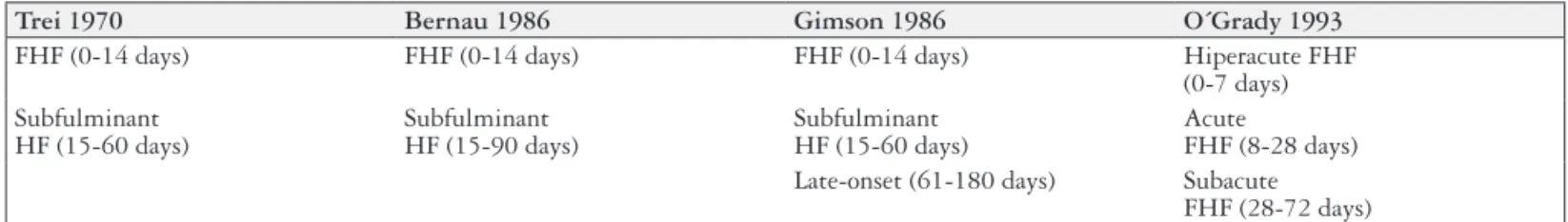
as fulminant hepatic failure, as onset of HE within 8 weeks of the irst symptoms, usually jaundice, in the absence of preexisting liver disease. Others have suggested different time frames between disease onset and the development of HE, as well as different terminology (Figure 4). The American Association for the Study of Liver Diseases deines ALF as the presence of HE and coagulopathy expressed as an INR higher than 1.5, in the absence of preexisting cirrhosis within 26 weeks of the onset of the irst symptoms(62).
The main etiologies of ALF are summarized in Figu re 5(52). There is a striking geographical heterogeneity in the
epidemiology of ALF worldwide. Hepatotoxicity due to acetaminophen is the most common cause of ALF in the United States of America and United Kingdom, followed by idiosyncratic hepatotoxicity due to other drugs and inde terminate etiology. Viral hepatitis is uncommon in the West. Hepatitis A, B and E are the most common causes of ALF in Asia and Africa(13, 52).
To determine the etiology of ALF it is important to ob tain a comprehensive clinical history and a detailed physical examination to guide laboratory and imaging evaluation, including: INR, sodium, potassium, chloride, bicarbonate, calcium, magnesium, phosphate, glucose, AST, ALT, alkaline phosphatase, GGT, total bilirubin, albumin, urea, creatinine, arterial ammonia, amylase, lipase, arterial blood gas, arterial lactate, complete blood count, blood type, toxicology screen with acetaminophen level, viral serologies (antiHAV IgM, HBsAg, antiHBc IgM, antiHEV, antiHCV, HCV RNA, Herpes simplex virus IgM, Varicella zoster virus, HIV), ce ruloplasmin levels, antinuclear antibody, antismooth muscle antibody and antiliver kidney microssome type 1 antibody, abdominal ultrasound and or computed tomography scan or magnetic resonance imaging if appropriate by clinical judgment(62).
Trei 1970 Bernau 1986 Gimson 1986 O´Grady 1993
FHF (0-14 days) FHF (0-14 days) FHF (0-14 days) Hiperacute FHF (0-7 days) Subfulminant
HF (15-60 days)
Subfulminant HF (15-90 days)
Subfulminant HF (15-60 days)
Acute
FHF (8-28 days) Late-onset (61-180 days) Subacute
FHF (28-72 days)
FIGURE 4. Deinitions of Acute Liver Failure
FHF Fulminant hepatic failure, HF: Hepatic failure.
Viral hepatitis Hepatitis A, B*, C, E and delta
Other Viruses Herpes simplex virus*, Varicella zoster virus*, Epstein Barr virus, Cytomegalovirus, Paracetamol*
Other drugs Isoniaziad, NSAIDs, Valproic acid, etc Amanita phalloides*
Vascular disorders* Budd-Chiari syndrome, ischemic hepatitis, heart failure Pregnancy disorders* Acute fatty liver of pregnancy, HELLP syndrome, eclampsia Autoimmune hepatitis* Acute fatty liver of pregnancy, HELLP syndrome, eclampsia Malignant involvement of the liver* breast, lung, lymphoma, melanoma
FIGURE 5. The main causes of acute liver failure
Differential diagnosis of the cause of ALF is important to rule out chronic liver disease, acute on chronic liver failure, to evaluate therapy directed for the underlying disorder and to assess prognosis. In this regard, patients with either hepatitis A or paracetamol induced ALF carry a transplantfree sur vival rate higher than 50%, whereas their counterparts with nonparacetamol druginduced or Wilson disease ALF have mortality rates as high as 90% to 100%.
It is important to consider treatment of the conirmed or presumed cause of ALF, including cessation of possible offending drugs. Nacetylcysteine should be considered in paracetamol overdose and in the initial phase of nonparace tamol ALF, when HE is grade I or II(61). Immediate delivery
should be recommended in acute fatty liver of pregnancy. In cases of suspected autoimmune hepatitis AIH or Wilson dis ease, a trial of corticosteroids or Dpenicillamine respectively is recommended. In case of Amanita Phalloides intoxication penicillin is prescribed, and in ALF due to herpes virus or hepatitis B antivirals are recommended. Other diverse mea sures such as hemodynamic support, chemotherapy or TIPS may be required for ischemic hepatitis, massive neoplastic in volvement of the liver and acute BuddChiari syndrome(13, 62).
The causes of ALF that require evaluation for treatment are highlighted in Figure 5.
Besides etiology, other prognostic factors include age, time interval between EH and jaundice, pH, INR, factor V, creatinine and bilirubin, MELD score, serum lactate, serum phosphate, alpha 1fetoprotein, galactose elimination capac ity, C13 methacetin breath test, GC globulin, liver volume by CT scan and indocyanine green clearance. Several of those aforementioned parameters were combined into prognostic systems, like Kings College and the Clichy criteria (Figures 6 and 7) in order to identify those patients with higher mortal ity without LT. The most employed worldwide and in Brazil is the Kings College system, whose speciicity is good, but is somewhat limited in its sensitivity(13, 62).
Once the diagnosis of ALF has been established, the patient should be referred to a liver unit to allow a deinite etiological diagnosis, to initiate speciic treatments and eva luate the criteria for possible OLT”
Systemic complications of ALF include intracranial hy pertension (ICH) due to cerebral edema, particularly seen in mechanically ventilated subjects with grades III or IV HE, circulatory and renal failure, metabolic and hydroelectrolitic abnormalities, coagulopathy, infections, sepsis and septic shock(60, 90).
In order to prevent cerebral edema, it is important to maintain ICP lower than 20 mmHg and cerebral perfusion pressure equal or higher than 50 mmHg with SaO2 >95%. It is advisable in subjects with grades III or IV HE to keep the head elevated, to avoid stimulation, hypotension, fever, hyponatremia and hypoglycemia. To protect airways, tracheal intubation and mechanical ventilation may be warranted. Placement of ICP monitoring devices can be useful for early detection of ICH and to guide therapy. However, it should be pointed out that their use was not associated with enhanced survival. The frequency of bleeding adverse events associated with the placement of ICP devices is reported to be around 10% and occur more often with the insertion of intracerebral or subdural ICP catheters. ICP monitoring is useful in the presence of urgent requirement for liver trans plantation according to Kings College criteria, seizures or pupillary abnormalities, more than two criteria for SIRS, ammonia levels higher than 150 mmol/L, hyponatremia, need for vasopressors or evidence of highly decreased or highly increased cerebral blood low by jugular venous O2 saturation or transcranial Doppler. Whenever possible, it is preferable to use extradural ICP transducers(13, 95).
Whenever clinically suspected or detected by ICP moni toring, ICH should be aggressively treated with intravenous mannitol. Shortterm hyperventilation may be useful in cases of impending brainstem herniation. Prophylaxis of seizures with phenytoin is no longer recommended but treatment should be aggressive whenever ICH is present due to its undesired effects in ICP. Appropriate sedation, whenever possible with propofol is recommended, Hypertonic saline is of value to maintain sodium levels between 145155 mmol/L. Moderate hypothermia around 3334o C can also lower ICP with no improvement in survival rates(60).
Sepsis is common in the course of ALF and may lead to impaired hepatic regeneration and ICH due to SRIS. Surveillance for infection is required as well as prompt an timicrobial treatment whenever it is suspected. Antibiotic prophylaxis is possibly helpful but not proven. Circulatory failure must be treated cautiously with intravenous luids and vasopressors, preferably noradrenaline, to maintain adequate organ perfusion, particularly CPP. Electrolyte and metabolic disturbances are common and must be avoided or corrected. It is advisable to start enteral nutrition as early as possible. Coagulation abnormalities should not be corrected in the absence of bleeding and invasive procedures. Continuous venousvenous hemodialysis is the best approach for renal replacement therapy in those subjects with renal failure(13).
Non-paracetamol ALF
Prothrombin time higher than 100 seg (INR higher than 6.5) independently of the grade of HE or at least three out of four parameters: age less than 10 or higher than 40 years, drug-induced or unknown etiology, time interval between EH and jaundice higher than 7 days, bilirrubin higher than 300 mmol/L and INR higher than >3.5
Paracetamol ALF
Arterial pH<7.3 after resuscitation and or
Combination of grade of HE equal or higher than 3, creatinine equal or higher than 300 mmol/L and INR higher han 6.5
FIGURE 6. King’s College prognostic system
Presence of grade III or IV of HE and
Age less than 30 years and factor V lower than 20% Age higher than 30 years and factor V lower than 30%
The evaluation for LT remains a major dilemma. The most common criteria used worldwide for identiication of those ALF subjects with a survival beneit with LT are the Kings College and the Clichy prognostic systems. In Barce lona, patients with the HE grade III or IV, sub fulminant or sub acute course or lack of improvement with conventional treatment are regarded as appropriate candidates for LT. In most programmes, a pragmatic casebycase evaluation of the risk beneit proile is employed. Other strategies besides cadaveric LT include auxiliary heterotopic LT, livingdonor LT and artiicial and bioartiicial support systems. Auxiliary heterotopic LT and livingrelated LT are usually infeasible in critically ill subjects with ALF. Data concerning liver support systems are presented elsewhere in this manuscript. Their use is controversial, and there is no undisputed evidence that they can improve survival, but instead they may play a temporary role providing a bridge for LT(13, 62).
In summary, ALF is a life threatening illness usually as sociated with a dismal prognosis without LT. It is important to search carefully for its cause in order to establish prognosis and to evaluate introduction of speciic therapy for the un derlying disorder. All patients with ALF should be referred to an ICU for advanced life support in a tertiary care center for prompt evaluation for LT.
2) Prognostic scores: Child-Pugh Score (CPS), Model for End-stage Liver Disease (MELD), sofa and clif-sofa
The occurrence of the irst episode of liver decompen sation marks a change in the prognosis of the liver disease. Variceal bleeding, SBP, HRS and sepsis are the main compli cations requiring admission to the ICU. A major proportion of these patients develop organ failure. The mortality rate of critically ill patients with cirrhosis is very high due to poor prognosis related to endstage liver disease, late referral for organ support, lack of knowledge of the management of cirrhosis, absence of a liver transplantation program, as well as reduced availability of ICU beds and the high cost of treatment(45).
The main objectives of prognostic scores in patients with cirrhosis are to estimate the probability of mortality within a given time interval, to determine which therapeutic option is the most appropriate with respect to the patient’s condition, whether a patient has an acceptable chance of survival after a given treatment and whether a resourcespending treatment such as OLT is justiied.
Various prognostic models have been developed and ap plied to patients with cirrhosis, including ICU scores such as SOFA, RFH score, SAPS II, APACHE II, which were shown to predict outcomes better in the ICU, when compared to conventional liver scores such as MELD and ChildPugh scores(3, 22). MELD variants that included Na were not shown
to predict ICU mortality better than MELD alone in such patients(63). Some authors found that a modiied SOFA for
cirrhosis, named CLIFSOFA and leukocyte counts were independent predictors of mortality in patients with acute onchronic liver failure (ACLF)(71).
Cutoff levels are essential for determining outcomes. A French study showed that mortality was best correlated with SOFA ≥10.5 and MELD ≥28.5 after the irst day of ICU ad mission(63). Mortality of patients with cirrhosis has reportedly
decreased on the ICU in recent years. This improvement in survival may be a consequence of new therapies such as terlipressin or TIPS(22, 45). An aggressive treatment initiated
at an earlier stage may also account for improving results. Early referral to the ICU would reduce the risk of disease worsening and maybe improve the survival rate.
Although outcomes have improved over time, mortality rates for critically ill patients with cirrhosis remain high. Studies showed that patients admitted to the ICU with VB or HE had better survival rates when compared to patients admitted with sepsis. The presence of infection is associated with poor survival at 2 months. Fungal infection signiicantly impacts on ICU mortality. Bilirubin at admission, infection at admission or acquired during ICU stay, mechanical venti lation and vasopressor therapy are all independently related to mortality(63, 64).
There is a correlation between the number of organs re quiring support and ICU mortality. Patients have a mortality rate of over 90% if they experience failure of three or more organs or if they require more than three types of organ support or replacement therapy (mechanical ventilation, vasopressor therapy, renal replacement therapy, MARS), unlike the mortality rate, estimated at 2%, for patients with cirrhosis but without organ failure(63).
Some authors suggest that all critically ill patients with cirrhosis should be admitted to the ICU at an earlier stage of decompensation, to optimize their management. Reassessment of these patients with ICU scores should take place 2 to 4 days thereafter. The persistence of three or more organ failures and the need for three or more organ supports may lead to consideration of the limitations of invasive treatments, as unfavorable results are almost certain in this setting(63, 64).
3) Artificial and bioartificial liver support systems The artiicial and bioartiicial support systems were designed to enhance shortterm survival of patients with acute liver failure and acute on chronic liver failure in order to provide a bridge to liver transplantation or liver regene ration in the case of ALF(62). The Molecular Adsorbent
Recirculating System (MARS), the fractionated plasma separation and adsorption (Prometheus), the singlepass albumin dialysis and the Hepa Wash procedure are artii cial liver support systems that are commercially available and reported to be safe(97). The rationale for their use is the
removal of albuminbound toxins. Their beneit is limited to secondary endpoints in randomized clinical trials such as improvement of HE and mean arterial pressure, increased cerebral blood perfusion, reduction in ICP and cerebral edema, removal of proinlammatory cytokines in ALF, but without any effect on mortality either in ALF or in ACLF(11, 62, 59, 97). Furthermore, several other bioartiicial
tAssist, the extracorporeal liver support device (ELAD), the modular liver support system (MELS), the bioartiicial liver support system (BLSS) and the Amsterdam Medical Center bioartiicial liver (AMCBAL). When compared to the arti icial liver support systems, they appear to be less effective and much more complex and expensive. At present, their use has not been associated with improved transplantfree survival, and the use of either liver support system is not recommended outside clinical trials(13, 62).
4) Limiting intensive care support in patients with cirrhosis
It is a challenge to limit intensive care support in pa tients with liver disease in light of the recent improvement in patient survival in ICUs, the availability of liver support systems and liver transplantation (LT). In this regard, the survival beneit of LT is seen most markedly in patients with MELD scores higher than 15(83). However, critically ill
subjects with cirrhosis and organ dysfunction usually have a poor prognosis. In those patients, the presence of more than two organ failures, deined by Sequential Organ Failure Assessment (SOFA) score, is associated with a mortality risk of 50% that approaches almost 100% in the presence of more than three organ failures(45, 71). The requirement for inotropic
support, mechanical ventilation or RRT was also associated with adverse prognosis. In the presence of acute on chronic liver failure (ACLF), CLIFSOFA was also reported to be a reliable prognostic parameter with better performance when compared to MELD and ChildPugh score, showing that extrahepatic organ failure is more important than liver failure in prediction of overall mortality. In this respect, patients with ACLF grades 2 and 3 have a 28day mortality, respectively, of 32% and 77%(71).
5) Sedoanalgesia
The management of pain and analgesia of patients with cirrhosis is concerning even for hepatologists, because side effects of analgesics and narcotics can be severe in subjects with cirrhosis. The most common complications include HE, AKI and gastrointestinal bleeding. There is a paucity of highquality, prospective data regarding the pharmacology, the proiles of many analgesics and their adverse effects in patients with endstage liver disease.
The International Association for the Study of Pain de ines pain as a disagreeable emotional and sensory experience associated with actual or potential tissue damage. The use of scales to monitor pain improves the clinical outcome of those patients in distress, including reduction of their time on me chanical ventilation and shorter length of stay in an ICU(12).
Treatment of non-neuropathic pain
The main cause of ALF in the US is the acetaminophen. Due to this fact, the use of this drug is usually incorrectly considered unsafe in patients with cirrhosis. However, the
Food and Drugs Administration (FDA) recommends the use acetaminophen in subjects with liver diseases, in doses between 23 g/day, without concurrent consumption of more than three alcoholic drinks per day(20). Hepatotoxicity due
to nonsteroidal antiinlammatory drugs (NSAID)is well
documented. Besides, they also inhibit prostaglandin synthe sis, which may precipitate HRS. Opiates also offer risks of toxicity. Tramadol, fentanyl, oxycodone and hydromorphone are the best options and may be used in reduced dosages in cirrhosis. The association with laxatives is recommended in order to avoid constipation and HE.
Treatment of the neuropathic pain
Patients with cirrhosis may feel pain due to neuropathies caused by diabetes, alcoholism, thiamine deiciency and/or cryoglobulinemia, needing adjuvant drugs for treatment of pain(29). Anticonvulsants, such as gabapentin and pregabalin
are suitable drugs, since they are neither metabolized in the liver, nor do they bind to plasma proteins. Use in conjunction with laxatives is recommended.
Sedation
Frequently, agitation in the ICU is treated with sedatives by mistake. Instead, it is advisable to immediately identify and treat the causes of the agitation, such as delirium, HE, pain, hypoxemia, hypotension, hypoglycemia, alcohol or drug abstinence. When they are needed, the sedatives must be used to induce only mild sedation, and the use of the monitoring sedation scales and light sedation protocols are associated with improvement in the clinical outcomes of the ICU. The most reliable sedation scales are The Richmond AgitationSedation Scale (RASS) and the SedationAgita tion Scale (SAS), the target for the sedation being between 2 and 0 on the RASS or 34 on the SAS, or consciousness during the day.
Benzodiazepine is metabolized in the liver and in those with cirrhosis it may trigger HE. Nonbenzodiazepine sed atives are recommended to improve the clinical results of those in mechanical ventilation and to reduce the incidence of delirium. Propofol is useful to provide lowlevel sedation with quick awakening after drug interruption, but smaller dosages are usually required. Dexmedetomidine is the only sedative permitted for patients that are not intubated in ICU in the United States, and its infusion does not need to be dis continued during weaning for extubation. Its metabolism is hepatic and those with severe hepatic dysfunction have their clearance reduced, therefore smaller dosages are required as stated for propofol.