Hospital São Vicente de Paulo - Universidade de Passo Fundo
Mailing address: Luís Sérgio Moura Fragomeni – Rua Teixeira Soares, 777 - S/702 99010-080 - Passo Fundo, RS, Brazil - E-mail: [email protected] English version by Stela Maris C. e Gandour
Arq Bras Cardiol, volume 80 (nº 4), 431-7, 2003
Luís Sérgio de Moura Fragomeni, Fabiano Fernandes Vieira, Júlio Cesar de Mello Bajerski, Roque Paulo Falleiro, Gustavo Hoppen, Iselso Sartori
Passo Fundo, RS - Brazil
Infective Endocarditis. Surgical Therapy
Despite all diagnostic and therapeutic advances, acti-ve intracardiac infection, so-called infectiacti-ve endocarditis, is still related to a high risk of morbidity and mortality in Brazil and in other countries 1-4 . In a continental and
heteroge-neous country like ours, these advances frequently have a compromised application: patients initially managed in community hospitals with no laboratory resources or ima-ging techniques, such as ultrasonography, tomography, or magnetic resonance imaging, are frequently referred to spe-cialized centers in an already advanced stage of the disease. In addition, regional centers, despite the increasing surgical demand, may still lack a large volume of surgeries, having, therefore, less experience in managing this type of patient. On the other hand, it would be impossible to imagine the entire national demand absorbed by 3 or 4 centers here con-sidered to be excellent. Undoubtedly, early recognition, mainly stimulated by a high degree of suspicion of the di-sease and aggressive therapy, is the major factor for a suc-cessful strategy. Age, left ventricular function, presence of a cardiac valvular prosthesis, renal failure, diabetes, embo-lic compembo-lications, and the infecting agent are the major fac-tors that determine the course of infective endocarditis 5-7.
In a retrospective study, we analyzed the major causes of morbidity and mortality in patients with infective endocar-ditis in a regional cardiology center and what can be done to improve the prognosis of this special group of patients.
Methods
From May 1986 to April 2001, 34 patients underwent cardiac surgery for infective endocarditis in the Service of Cardiothoracic Surgery of the Hospital São Vicente de Pau-lo, in the city of Passo Fundo, in the Brazilian state of Rio Grande do Sul. That is a university-affiliated general hospi-tal with 640 beds, where 40 to 50 cardiac surgeries with extracorporeal circulation are performed every month. Its area of influence encompasses 300 municipalities in the states of Rio Grande do Sul and Santa Catarina, involving a population of 1,050,000 inhabitants. The hospital is located in the city of Passo Fundo (170 thousand inhabitants) in the
Objective - To assess the major causes of surgical morbidity and mortality in patients with infective endo-carditis operated upon in a regional cardiology center.
Methods - Thirty-four patients underwent surgical treatment for infective endocarditis. Their ages ranged from 20 to 68 years (mean of 40.6) and 79% were males. Their NYHA functional classes were as follows: IV - 19 (55.8%) patients; III - 12 (35.2%) patients; II - 3 (8.8%) patients. Blood cultures were positive in only 32% of the cases. Eight patients had already undergone previous cardiac surgery, whose major indication (82.3%) was heart failure refractory to clinical treatment.
Results - Four (11.7%) patients died at the hospital. Follow-up was complete in 26 (86%) patients. Five (14.7%) patients died later, 12, 36, 48, 60, and 89 months after hospital discharge. Of the 21 patients being currently followed up, 1 is in NYHA functional class III, and 5 in NYHA functional class II.
Conclusion - A high degree of clinical suspicion, at an early diagnosis, and indication of surgical treatment prior to deterioration of left ventricular function and installation of generalized sepsis may improve prognosis.
northern region of the state of Rio Grande do Sul. Following a defined protocol, the medical records of those patients were reviewed and their clinical evolution analyzed.
The male sex prevailed, corresponding to 79.4% of the patients; their ages ranged from 20 to 68 years (mean age of 40.6 years). According to the NYHA functional class classi-fication of heart failure, 19 (55.8%) patients were class IV; 12 (35.2%) were class III; and 3 (8.8%) were class II. Blood cul-tures were performed in all patients, and microorganisms were identified in only 11 (32.3%) patients. Streptococcus viridans (5 cases) and Staphylococcus aureus (3 cases) were the microorganisms most frequently found. Klebsiella
and Escherichia coli were identified in 2 other patients. All cases were confirmed as infective endocarditis by surgical findings. The patients with negative blood cultures also had negative tissue cultures.
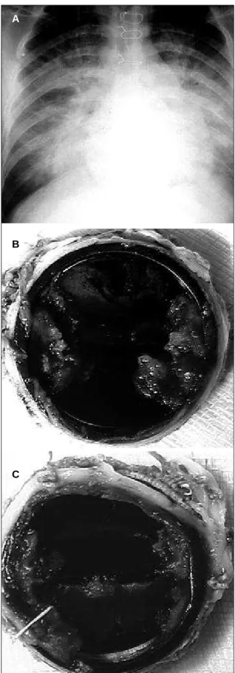
Clinical, laboratory, and radiological findings sugges-ting the diagnosis of infective endocarditis were in accor-dance with those of 2-dimensional Doppler echocardiogra-phy performed in all patients. In addition, 4 patients also underwent cine coronary angiography. Clinical signs com-patible with cerebral embolism were identified in 2 patients. Their complementary study included computerized tomo-graphy of the brain in both patients and angiotomo-graphy of the internal carotid artery in 1 patient. In the latter, a cerebral abscess was drained and cardiac surgery was performed within 4 weeks. These patients neither underwent previous surgery nor were taking oral anticoagulant agents. Antibio-tic therapy was performed from the beginning of the symp-toms until the surgical procedure for a period ranging from 2 to 120 days (mean of 28.1 days). Eight patients had already undergone the following cardiac surgeries: 7 patients underwent aortic valve replacement (4 biological and 3 me-chanical prostheses) and 1 underwent mitral valve replace-ment (mechanical prosthesis) (fig. 1a, 1b, 1c). In patients without previous surgical treatment, the following lesions were identified: mitral valve prolapse (4), mitral stenosis (1), congenital mitral insufficiency (1), and congenital aortic stenosis (1).
Of the patients undergoing surgical treatment, 19 (55.8%) had clinical signs of severe heart failure due to val-vular lesions, but the systemic infection was considered under control. The remaining, in addition to the indication for valvular prosthesis implantation due to severe insuffi-ciency, had their clinical picture impaired by the presence of sepsis, cardiogenic shock, abscess, aneurysm, or fistula. One patient was under mechanical ventilatory support. In 3 patients, the source of endocarditis was previous hemodia-lysis due to acute renal failure that developed after blood dyscrasia because of infection from lizard toxin (Lonomia obliqua). Hemodialysis due to chronic renal failure and aortic valve calcification were indications in 2 other patients.
All patients were operated upon with extracorporeal circulation in moderate systemic hypothermia (28oC),
myo-cardial protection with topical cooling of the cardiac surfa-ce, and anterograde intracoronary infusion of a crystalloid
Fig. 1 – 1a) Implantation of double leaflet mitral prosthesis 14 months before. Active infective endocarditis; 1b) Atrial face of the prosthesis with vegetations; 1c) Ventricular face of the prosthesis blocked by vegetations.
A
B
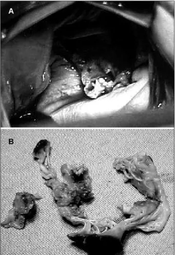
cardioplegic solution. Due to annular fragility and aiming at strengthening the suture, the stitches were usually ancho-red in a Teflon pillow. When the aortic ring requiancho-red wide-ning or the fistulas required closing, a flap of bovine pericar-dium preserved in glutaraldehyde was used. The most frequently impaired valve was the aortic valve (17 patients -50%), followed by the mitral valve (11 patients – 32.3%) (fig. 2a, 2b), and the following associations: mitral-aortic in 3 (8.8%), mitral-tricuspid in 2 (5.8%), and aortic-tricuspid in 1 patient (3.4%). Two patients requiring mitral valve replace-ment by a mechanical prosthesis due to destruction of the valvular apparatus by infective endocarditis had a signifi-cant associated tricuspid insufficiency that required correc-tion of the DeVega annuloplasty type, but no signs of infec-tive endocarditis were observed in that valve. One patient (2.9%) had impairment of the tricuspid valve by infective endocarditis; the 4 patients treated with mitral valvuloplas-ty had vegetations characteristic of infective endocarditis in the valvular apparatus. Annuloplasty techniques 8-10 with
simple sutures (1) or placement of a Carpentier ring (3), par-tial resections of the impaired leaflets and repaired with direct sutures, when adequate (1), or repaired with bovine pericardium flap (3) were used in these patients. These con-servative techniques were used only in situations in which the partial resection of the impaired leaflets did not prevent
complete resection of the vegetations. Five patients had total atrioventricular block at the time of the surgical deci-sion. The surgical procedures are specified in table I.
Results
The major indication for surgery was the presence of refractory heart failure in 28 (82.3%) patients. Third-degree atrioventricular block, central embolism, and periannular aortic and mitral abscess with the formation of aortic/right atrium/left atrium fistula complete the remaining indications. Duration of aortic clamping ranged from 21 to 130 minutes (mean of 67.8 minutes). The mean length of stay in the intensive care unit was 5.2 days, and the mean postopera-tive hospital stay was 21 days. Of the 5 patients with total atrioventricular block at the time of surgery, 2 required defi-nitive implants of artificial cardiac stimulation (1 epimyocar-dial and 1 endocarepimyocar-dial). Two patients who had no conduc-tion disorder detected in the preoperative period developed total atrioventricular block by the end of the surgical proce-dure, requiring a temporary pacemaker. One of these patients required the implantation of a definitive pacemaker, in which an epimyocardial electrode was used.
Four (11.7%) patients died during hospital stay as follows: 2 during surgery, 1 on the first postoperative day, and another on the ninth postoperative day. Of the patients dying in the operating room, 1 had staphylococcal endocarditis in the biological aortic prosthesis. This patient developed a dental abscess 20 days before and was being treated for congestive heart failure. As the patient did not improve, he was operated upon in total atrioventricular block, sepsis, and mechanical ventilation. On surgery, a periannular abscess of the biological aortic prosthesis and of the interventricular septum was identified. Even with the extensive surgical débridement and implantation of a new aortic prosthesis, the patient could not maintain sufficient cardiac output without extracorporeal circulation support and died in the operating room. The second patient, operated upon in a similar clinical condition, had his mitral valve destroyed by vegetations. Although the surgical treatment was considered adequate, the extracorporeal circulation support could not be removed, and the patient died with signs of right ventricular failure. The third patient, operated upon with cardiogenic shock (aortic and mitral insufficiency) and disseminated intravascular coagulation present, had to be reoperated on in the first 24
Fig. 2 – 2a) View of the mitral valve destroyed by vegetations during surgery; 2b) Excised mitral valve with alterations of infective endocarditis.
A
B
Table I - Procedures
Mechanical aortic prosthesis 11 Mechanical mitral prosthesis 5
Biological aortic prosthesis 4 Mitral valvuloplasty 2
Mechanical mitral prosthesis Mechanical aortic prosthesis + tricuspid valvuloplasty 2 + mitral valvuloplasty 2
Biological aortic + Biological aortic +
mitral prosthesis 1 tricuspid prosthesis 1
hours because of persistent bleeding and cardiac tampona-de. The patient was transferred to the intensive care unit in critical condition with maximal use of inotropic agents and died 7 days later because of the persistence of sepsis and multiple organ failure. The fourth patient, with tricuspid and aortic insufficiency, sepsis and cardiogenic shock, died on the first postoperative day because of cardiopulmonary failure. Table II shows these patients.
Five patients required more than 1 surgical interven-tion because of the recurrence of endocarditis or to its sequelae. One patient had to be reoperated on twice becau-se of the recurrence of infection; the first episode was 2 years after the initial intervention and the second episode was after the third year. In the latter, destruction of the supe-rior mitral ring was observed with formation of an abscess in the mitral-aortic continuity and loss of sustentation of the anterior mitral leaflet. Mitral valvuloplasty and implantation of a new aortic prosthesis were performed. This patient died after a 4-year follow-up due to a new return of the cardiac and systemic infection with consequent septicemia.
Another patient, initially with extensive aortic valvular destruction and aorta/right atrium fistula, required 3 surgi-cal interventions to control infection and restore normal he-modynamics. Currently, the patient has a normal life, is asymptomatic, but a small residual aortic perivalvular fistula persists with no clinical repercussions.
Of the 30 patients discharged from the hospital, 4 were not followed up. Of the 26 patients with regular clinical follow-up, 5 died after 12, 36, 48, 60, and 89 months. These patients are described in table III.
Of the 19 patients operated upon in NYHA functional class IV, 5 died, 2 are being followed up, 7 evolved to class I, 4 to class II, and 1 to class III. Of the 12 patients operated upon in class III, 3 died, 2 are being followed up, 6 are in class I, and 1 is in class II. Of the 3 patients operated upon in class II, 1 died and 2 are in class I.
Discussion
Infective endocarditis has had a greater incidence since the 1980s, with reports of 5.9 and 11.6 episodes per 100,000 inhabitants in Sweden and Philadelphia, respective-ly 11,12. Approximately half of the cases reported in
Philadel-phia had a previous history of the use of injectable drugs. The incidence increases progressively after 30 years of age, exceeding 15 to 30 cases per 100,000 people/year between the sixth and eighth decade of life. Fifty-five to 75% of the patients with infective endocarditis in native valves have predisposing conditions, such as rheumatic disease, con-genital disease, mitral valve prolapse, degenerative heart disease, asymmetric septal hypertrophy, or use of illegal intravenous drugs 13,14. Seven to 25% of the cases involve
the presence of prostheses 11,12. In 25% of the cases,
pre-disposing factors for infective endocarditis cannot be identified.
Signs and symptoms of infective endocarditis are fre-quently neglected and, when evident, normally reflect a complication and not an intracardiac infection per se. Clinical suspicion should be highly emphasized until diag-nostic definition. Application of the criteria reported in the literature allows an approach with good sensitivity and specificity for the diagnosis of infective endocarditis 3,16.
The use of echocardiography in patients with a clinical sus-picion of infective endocarditis often allows morphological confirmation of infection and is of inestimable help in choosing the adequate approach in each case 16,17.
Echocar-diography should be performed mainly in patients with negative blood cultures 3. Transesophageal
echocardiogra-phy allows the visualization of small vegetations and adds resolution to the conventional transthoracic echocardiogra-phy with pulsed and continuous wave Doppler 3,18.
Trans-thoracic echocardiography and its association with
transe-Table II – In-hospital deaths
Patient 1: Female, 26 years. Infective endocarditis in biological aortic prosthesis implanted 2 years before. Dental abscess for 20 days. Total atrioventricular block, sepsis, cardiogenic shock, under mechanical ventilation. S. aureus in the first blood culture 10 days before. Implantation of biological aortic and mitral prostheses on the day of transference to our service. Death in the operating room.
Patient 2: Female, 68 years. Cardiogenic shock and sepsis due to mitral valve destruction by vegetations. Negative culture. The patient underwent surgery on the day of transference to our service. Death in the operating room.
Patient 3: Male, 22 years. Cardiogenic shock, DIVC and sepsis due to aortic and mitral insufficiency. Culture positive for nonhemolytic Streptococcus 7 days before. Implantation of biological aortic and mitral prostheses. Reoperated upon in 24 hours due to cardiac tamponade. Death on the seventh postoperative day due to multiple organ failure and sepsis.
Patient 4: Male, 21 years. Drug addict. Under hemodialysis for 30 days. Tricuspid and aortic insufficiency. Sepsis, cardiogenic shock. Annular and subannular aortic abscess. Aortic and tricuspid vegetations. Death 24 hours after implantation of biological aortic and tricuspid prostheses due to cardiogenic shock and persistence of sepsis.
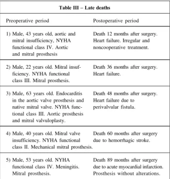
Table III – Late deaths
Preoperative period Postoperative period
1) Male, 43 years old, aortic and Death 12 months after surgery. mitral insufficiency, NYHA Heart failure. Irregular and functional class IV. Aortic noncooperative treatment. and mitral prosthesis
2) Male, 22 years old. Mitral insuf- Death 36 months after surgery. ficiency. NYHA functional Heart failure.
class III. Mitral prosthesis.
3) Male, 63 years old. Endocarditis Death 48 months after surgery. in the aortic valve prosthesis and Heart failure due to native mitral valve. NYHA func- perivalvular fistula. tional class III. Aortic prosthesis
and mitral valvuloplasty.
4) Male, 40 years old. Mitral valve Death 60 months after surgery insufficiency. NYHA functional due to hemorrhagic stroke. class II. Mechanical mitral prosthesis.
sophageal echocardiography, which have been used in our unit since 1991, were definitive for making surgical deci-sions. Transesophageal echocardiography has been used during surgery to search for residual fistulas while the extra-corporeal circulation cannula is still installed in the patient. The causal agent is identified through blood cultures. A positive blood culture is important for the diagnosis and management of the case, directly influencing the morbidity and mortality of these patients. In patients who had not previously taken antibiotics, positivity of the blood cultu-res should be expected in more than 95%. However, the
Fig. 4 – Patients’ survival according to the Kaplan-Meier curve. Of the 26 patients under regular clinical follow-up, 5 died 12, 36, 48, 60, and 89 months after surgery.
0 10 20 30 40 50 60 70 80 90 100 110 120 130 140 150 160 170 180 Months
1.0
0.9
0.8
0.7
0.6
0.5
0.4
0.3
0.2
0.1
0.0
P
ro
b
a
b
il
it
y
CLASS IV CLASS III CLASS II
A
B
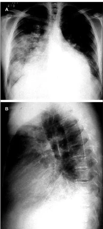
Fig. 3 – 3a, 3b) Active endocarditis in native aortic valve. Aortic insufficiency.
indiscriminate use of antibiotics in the initial phase of symp-toms is the major cause of lack of growth of microorganisms in culture in approximately 35% of the patients 19. This is a
common practice, and, in addition, several laboratories experience technical difficulties with cultures in an anaero-bic medium. These facts may have accounted for the low rate of positivity in the cultures (32.3%) in our case series. The list of microorganisms causing infective endocarditis has increased due to the great number of uncommon patho-gens that continue to be identified. Gram-positive microor-ganisms, particularly Streptococcus and Staphylococcus, still predominate. In patients using intravenous drugs and in immunocompromised patients, gram-negative and fungal infections have been reported. Streptococcus viridans
accounts for 30 to 65% of the cases of endocarditis in native valves. Enterococci are responsible for 10 to 20% of the ca-ses of infective endocarditis and may infect normal valvular tissue. Staphylococcus aureus, a common cause of infective endocarditis, is a highly virulent microorganism that usually has severe consequences for the patient. When present in the left cardiac chambers, Staphylococcus aureus is asso-ciated with a mortality of 35 to 40%, probably due to its abili-ty to destroy normal and abnormal valvular tissue 20-22,
cau-sing frequent embolisms and metastatic infection. Staphy-lococcus aureus was identified in only 3 patients (32.3% of the positive cultures), 2 of whom died at the hospital, and, in the third patient, Staphylococcus aureus was the initial cause of infective endocarditis and 3 interventions were required because of the recurrence of the disease. In deve-loped countries, epidemiological studies suggest that
pros-Fig. 5 – Evolution of pre- and postoperative functional classes.
NYHA Evolution
Number of patients
Pré-op e rator
y
Death s
Loss of follow
thetic valve endocarditis (PVE) represents 10 to 30% of the cases of infective endocarditis 23. The major risk of
develo-ping infective endocarditis occurs in the first 6 months after surgery, mainly within the 5 and 6 initial weeks; the risk then declines, although some persists 13,23. Patients with
pre-vious endocarditis in the native valves, especially when operated upon in the active phase of the disease, are more prone to develop PVE 13,24,25. Prosthetic valve endocarditis
occurs more often in the aortic position, and no statistical difference exists whether it is biological or mechanical. When the infected prosthesis is of the mechanical type, perivalvular destruction and presence of fistulas and abs-cesses are the most frequent findings, because no leaflet of organic material exists to be destroyed by the microorga-nisms. However, if the infection is early, the prosthesis may still be sterilized with early antibiotic therapy, which will avoid the need for a new surgery in the acute phase. Eight (23.5%) patients who underwent surgery here developed endocarditis in an implanted prosthesis; 7 (87.5%), like the report in the literature, were prostheses in the aortic posi-tion. One of the 4 in-hospital deaths occurred in this group with a biological aortic prosthesis, with extensive perivalvu-lar necrosis and clinical findings of sepsis. General consen-sus exists about the unsatisfactory long-term results in PVE, despite adequate surgical techniques and good clinical management. Lytle et al 26 reported the recurrence of
infec-tion in 25% of their patients in a 5-year follow-up. In that study, 7 patients discharged from hospital after intervention for PVE were followed up. Two (28%) patients required new surgical interventions as follows: the first patient, after the second and third years, died from the recurrence of infec-tion in the following year; the second patient, after the first and fifth years, had a small periaortic fistula without clinical repercussions and free of infection, and 9 years have alrea-dy passed since the last surgical intervention. The microor-ganisms most frequently found in PVE are of the nosoco-mial and even community types. In the first 60 days after surgery, Staphylococcus epidermidis, Staphylococcus aureus, the gram-negative bacilli, diphtheroids, and fungi are the most common causes of PVE 26.
The indications for the surgical treatment of infective endocarditis reported in the literature 27,28 are similar to those
in our series and may be grouped as follows: severe and pro-gressive heart failure refractory to clinical treatment; infec-tion that could not be controlled with antimicrobial treat-ment; uncontrolled infection in a cardiac prosthesis, and systemic arterial embolisms. In regard to functional class, as reported by other authors 29-31, most patients were in NYHA
functional classes III and IV. Of the indications for surgical treatment, heart failure and infection not controlled with adequate antibiotics are the most common 30,31. In our case
series, those were responsible for approximately 91% of the surgical indications. Patients with mechanical prostheses under continuous use of coumarin anticoagulants (4) had their medication replaced by intravenous heparin, infusion of fresh plasma, and interruption of the infusion of heparin 4 hours prior to the surgical procedure.
The most common surgical procedure was valvular replacement with a biological or metallic prosthesis and homograft. Valvuloplasty was also possible with recons-truction techniques in select cases 8-10.
The presence of total atrioventricular block in the clinical presentation of infective endocarditis and the need to implant a definitive pacemaker showed the severity and the extension of paravalvular tissue necrosis. The choice of endomyocardial electrodes will depend on the phase and the clinical situation in which these devices are implanted. Of the 3 patients receiving definitive pacemakers, the epicardial electrode was chosen for 2 patients, because definitive implantation was indicated early.
Perivalvular involvement with the formation of abs-cesses and fistulas represents a risk factor for recurrence of the disease 32. Despite the extensive débridement and
closure of the cavities and fistulas by using autologous or bovine pericardium, 5 patients required more than 1 surgical intervention due to a relapse of the endocarditis or its sequelae. Danchin et al 33 and Renzulli et al 34 did not find
differences between the patients with and without perival-vular involvement, and, in a 10-year follow-up, reported a 58% rate of no relapse.
The in-hospital mortality of 11.7% (4 patients) here reported resulted from the delay in indicating surgery and consequent severe myocardial failure, to the present sepsis, and to the anatomic propagation with formation of intraca-vitary abscesses. Although 3 of the patients were young, which would be a factor favorable to survival, because they only experienced intervention in the final phase of the disease, they did not survive. A preoccupying fact in this and other surgical heart pathologies is the critical state at which surgery is considered for many of these patients. In this situation, integrity of the central nervous system was the major feature to be considered. If the central nervous system is considered suitable, with no irreversible injury, surgery is usually the option. However, 1 patient in our series was in a critical clinical situation and died in the opera-ting room, although her neurologic condition was suitable when surgery was indicated. All 5 (14.7%) deaths after hos-pital discharge were the consequence of heart failure, except for 1 patient operated upon in class II, who died due to hemorrhagic stroke while using coumarin anticoagulants. This confirms the fact that even with the infection under control, ventricular dysfunction is a preponderant factor in the postoperative evolution of these patients. For us, the difficulty in the clinical follow-up of patients, who usually come from regions far from specialized centers, in addition to the inaccuracy in controlling the coumarin anticoagulants, is a reality that should be emphasized.
References
1. Ruiz E, Schirmbeck T, Figueredo LTM. Estudo sobre endocardite infecciosa em Ribeirão Preto, SP, Brasil. Análise de casos ocorridos entre 1992 e 1997. Arq Bras Cardiol 2000; 74: 217-24.
2. Arnoni AS, Castro Neto J, Arnoni RT, et al. Endocardite infecciosa: 12 anos de tratamento cirúrgico. Rev Bras Cir Cardiovasc 2000; 15: 308-19.
3. Bayer AS, Bolger AF, Taubert KA, et al. Diagnosis and management of infective endocarditis and its complications. Circulation 1998; 98: 2936-48. 4. Sandra RM, Shafran SD. Infective endocarditis: review of 135 cases over 9 years.
Clin Infect Dis 1996; 22: 276-86.
5. Mansur AJ. Endocardite infecciosa. In: Barreto ACP, Souza AGMR, (eds). Cardiologia: Atualização e Reciclagem. Rio de Janeiro: SOCESP, 1994: 455-65. 6. Arvay A, Lengyel M. Incidence and risk factors of prosthetic valve endocarditis.
Eur J Cardiothorac Surg 1988; 2: 340-6.
7. Cardewood SB, Swinski LA, Waternaux CM, et al. Risk factor for the development of prosthetic valve endocarditis. Circulation 1985; 72: 31-7.
8. Pomerantzeff PMA, Brandão CMA, Mansur AJ, et al. Tratamento cirúrgico do abscesso de anel valvar associado a endocardite bacteriana: resultados imediatos e tardios. Rev Bras Cir Cardiovasc 1996; 11: 259-62.
9. Ergin MA, Raissi S, Follis F, et al. Annular destruction in acute bacterial endocar-ditis: surgical techniques to meet the challenge. J Thorac Cardiovasc Surg 1989; 97: 755-63.
10. David TF, Feindel CM. Reconstruction of the mitral annulus. Circulation 1987; 76(suppl 3): 102-7.
11. Hogevik H, Olaison L, Anderson R, et al. Epidemiologic aspects of infective endocarditis in an urban population: a 5-year prospective study. Medicine 1995; 74: 324-39.
12. Berlin JA, Abrutyn E, Strom BL, et al. Incidence of infective endocarditis in the Delaware Valley, 1088-1990. Am J Cardiol 1995; 76: 933-36.
13. Arvay A, Lengyel M. Incidence and risk factors of prosthetic valve endocarditis. Eur J Cardiothorac Surg 1988; 2: 340.
14. Kazanjian P. Infective endocarditis: Review of 60 cases treated in community hospitals. Infect Dis Clin Pract 1993; 2: 41.
15. Durack DT, Lukes AS, Bright DK. New criteria for diagnosis of infective endocar-ditis: utilization of specific echocardiographic findings. Am J Med 1994; 96: 200. 16. Mugge A. Echocardiographic detection of cardiac valve vegetations and
prog-nostic implications. Infect Dis Clin North Am 1993; 7: 877.
17. Sochowski RA, Chan KL. Implication of negative results on a monoplane tran-sesophageal echocardiographic study in patients with suspected infective endocarditis. J Am Coll Cardiol 1993; 21: 216.
18. Daniel WG, Mugge A. Transesophageal echocardiography. N Engl J Med 1995; 332: 1268.
19. Hoen B, Selton-Suty C, Lacassin F, et al. Infective endocarditis in patients with negative blood cultures: analysis of 88 cases from a one-year nationwide survey in France. Clin Infect Dis 1995; 20: 501.
20. Benn M, Hagelskjaer LH, Tvede M. Infective endocarditis, 1984 through 1993: a clinical and microbiological survey. J Intern Med 1997; 242: 15-22. 21. Roder BL, Wandall DA, Espersen F, Frimodt-Moller N, Skinhoj P, Rosdahl VT.
Neurological manifestations in Staphylococcus aureus endocarditis: a review of 260 bacteremic cases in nondrug addicts. Am J Med 1997; 102: 379-86. 22. Roder BL, Wandall DA, Frimodt-Moller N, Espersen F, Skinhoj P, Rosdahl VT.
Clinical features of Staphylococcus aureus endocarditis: a 10-year experience in Denmark. Arch Intern Med 1999; 159: 462-9.
23. Horskotte D, Piper C, Niehues R, et al. Late prosthetic valve endocarditis. Eur Heart J 1995; 16(suppl B); 1995: 39.
24. Agnihotri AK, McGiffin DC, Galbraith AJ, O’Brien MF. Surgery for acquired heart disease. J Thorac Cardiovasc Surg 1995; 110: 1708-24.
25. Ivert TSA, Dismukes WE, Cobbs CG, et al. Prosthetic valve endocarditis. Circulation 1984; 69: 223.
26. Lytle BW, Priest BP, Taylor PC, et al. Surgical treatment of prosthetic valve endocarditis. J Thorac Cardiovasc Surg 1996; 111: 198-207.
27. Al Jubair K, Al Fagih M, Ashmeg A, et al. Cardiac operations during active endocarditis. J Thorac Cardiovasc Surg 1992; 104: 487.
28. ACC/AHA guidelines for the management of patients with valvular heart disease. A report of the American College of Cardiology/American Heart Asso-ciation. Task force on Practice Guidelines (Committee on Management of Patients with Valvular Heart Disease). J Am Coll Cardiol 1998; 32: 1486-588. 29. Haydock D, Barrat-Boyes B, Macedo T, Kirklin JW, Borst HG. Aortic valve replacement for active infectious endocarditis in 108 patients. J Thorac Cardio-vasc Surg 1992; 103: 130-9.
30. Jault F, Gandjbakhck I, Chastre JC, et al. Prosthetic valve endocarditis with ring abscesses. J Thorac Cardiovasc Surg 1993; 105: 1106-13.
31. Watanabe G, Haverich A, Speier R, Dresler C, Borst HG. Surgical treatment of active endocarditis with paravalvular involvement. J Thorac Cardiovasc Surg 1994; 107: 171-7.
32. McGiffin DC, Galbraith AJ, McLachlan GJ, et al. Aortic valve infection: risk factors for death and recurrent endocarditis after aortic valve replacement. J Thorac Cardiovasc Surg 1992; 104: 511-520.
33. Danchin N, Retournay G, Stchepinsky O, et al. Comparison of long-term outcome in patients with or without aortic ring abscess treated surgically for aortic valve infective endocarditis. Heart 1999; 81: 177-81.
34. Renzulli A, Carozza A, Romano G, et al. Recurrent infective endocarditis: a mul-tivariate analysis of 21 years of experience. Ann Thorac Surg 2001; 72: 39-43.
of the antibiotics in acting in the presence of an infected foreign body is well known.
Recent advances in the diagnosis of infective endo-carditis, mainly transesophageal echocardiography, new antibiotic regimens, and adequate surgical techniques have provided early therapy and consequent improvement in the results. However, morbidity and mortality of infective endocarditis caused by microorganisms like Staphylococ-cus aureus and PVE still remain high in these patients. This study carried out in a regional cardiology center in the Brazi-lian state of Rio Grande do Sul shows that, in addition to those principles already internationally diffused, the early
diagnosis of infective endocarditis should be emphasized for the obtainment of greater positivity in blood cultures, that clinical and surgical treatment should be instituted as soon as possible and not when cardiogenic shock and sep-sis had already occurred. On the other hand, new prospec-tive research is required to answer several pending ques-tions, ie, how to improve the management of complications and the indications of therapies in infective endocarditis.