Arq Bras Cardiol 2003; 80: 355-8.
Póvoa et al Electrocardiographic abnormalities in neurological diseases
3 5 5
Hospital do Servidor Público Estadual.
Mailing address: João Pimenta - Rua das Camélias, 357 - 04048-060 - São Paulo, SP – Brazil - E-mail:[email protected]
Arq Bras Cardiol, volume 80 (nº 4), 355-8, 2003
Rui Póvoa, Luciano Cavichio, Ana Lúcia de Almeida, Danielle Viotti, Celso Ferreira, Luciane Galvão, João Pimenta
São Paulo, SP - Brazil
Electrocardiographic Abnormalities in Neurological Diseases
Original Article
Central nervous system disorders are known to be res-ponsible for the onset of cardiac alterations, frequently observed in the electrocardiogram 1,2. Although serum potassium disturbance has been suggested as an important factor in these alterations, this finding was not confirmed in several studies 3,4. The majority of studies suggest that these ECG abnormalities result from disturbances in the autonomous system, due to neurologic disease, promoting a local excess of catecholamines, associated with enhanced adrenal production, and activation of the calcium channels, leading to an increase in cytosolic and mitochondrial cal-cium, as well as release of free radicals, causing contraction band necrosis and reflected in ECG alterations. Although the causes of cardiac alterations are a matter of scientific speculation, evidence exists that myocardial lesion is media-ted by catecholamines, and an important left ventricle dys-function with the liberation of cardiac enzymes, such as tro-ponin-I, occurs in some cases 5. The increase in intracranial pressure is also considered a factor in these alterations 6.
Data described by Goldstein 7 in a certain population demonstrated a high incidence (up to 92%) of electrocardio-gram alteration in neurological disorders. This result was not observed in clinical practice. In our study, we observed a frequency of electrocardiogram alterations in pathological brain processes contrary to that observed in the literature.
Methods
We carried out a study for 6 years of 1,590 patients admitted because of neurological diseases, such as brain tumor, stroke, subarachnoid hemorrhage, subdural hemorr-hage, brain aneurysm, and head injury, with ages ranging from 10 to 60 years. Patients with concomitant diseases that could lead to electrocardiogram alterations, such as syste-mic hypertension, cardiac valve abnormalities, coronary diseases, congenital cardiopathies, endocrine diseases, and others were excluded. Those patients who did not have a complete clinical history or an electrocardiogram per-formed at the time of admittance were also excluded. A cardiologist unaware of the neurological diseases and any other clinical data regarding the patient interpreted the
elec-Objective – To evaluate electrocardiographic
ab-normalities in patients with neurologic diseases.
Methods – We studied 161 patients with neurologic disorders by analyzing the 12-lead electrocardiogram during the pathological process. An expert who did not know anything about the patients evaluated the traces.
Results – Neurological process included brain tumor (41%), stroke (27.3%), cerebral aneurysm (15.5%), sub-arachnoid hemorrhage (6.8%), subdural hemorrhage (5%), and head injury (4.4%). Electrocardiograms were normal in 61% of cases, and the most frequent abnorma-lity was ventricular repolarization (23.7%). The presence of T waves (4.6%) and prolonged QT intervals (8.8%) was the most characteristic of brain injuries.
Conclusion – We observed a lower incidence of elec-trocardiographic abnormalities than that described in the literature.
3 5 6
Póvoa et al
Electrocardiographic abnormalities in neurological diseases
Arq Bras Cardiol 2003; 80: 355-8.
regarding the incidence of electrocardiogram alterations in neurological diseases. Hersch 3 described 20 cases, with a pattern of subepicardial ischemia in 15% and ST segment elevation in 8% of the patients, without prolonged QT interval. In the present study, we verified in the 66 cases of brain tumor, a pattern of subepicardial ischemia in only 7.5% of the cases, and ST segment elevation in 3% of the cases. Goldstein 7 evaluated 150 cases of patients with acute stroke and 150 controlled cases paired by sex and ages to determine the frequency of abnormalities in the electrocar-diogram in the several groups, observing alterations in 92% of cases, characteristic of neurological diseases, prolonged QT interval in 45% of the cases, and cerebral T wave in 29% of cases (fig. 1). Burch et al 8, in a report of 17 cases, found prolonged QT intervals in all patients and U wave in 64.7%. In our cohort, in 44 cases of acute stroke, the incidence of abnormalities in the electrocardiogram was 29.6%. The alterations considered as indicative of cerebral lesions were not frequent, considering that in all cases the cerebral T wave appeared in 2.5% (4/161), prolonged QT interval in 5.6% (9/161), and subepicardial ischemia (less specific) in 4.9% (8/161). In Goldstein’s study 7, an incidence of 25% of new arrhythmias was observed compared with 3% of the control group. Among these, atrial fibrillation was the most common, present in 14% of the cases, followed by ventri-cular arrhythmia in 5%, with ventriventri-cular extrasystoles in the majority of cases. Although the analysis of any arrhythmic event by using only conventional electrocardiography is not accurate, atrial fibrillation occurred in 2.3% of cases and was the only arrhythmia found in this group of patients.
In the studies on subarachnoid hemorrhage, Byer et al 9 assessed 29 patients, observing that 67% of the electro-trocardiograms. In electrocardiogram evaluation rhythm,
QRS complex, T wave, ST segment, PR interval, QT interval corrected using Bazett’s formula and the presence of bundle branch blocks or fascicular blocks were analyzed.
Results
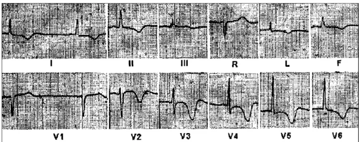
We evaluated 1590 charts, and 1,429 were excluded because they did not meet inclusion criteria. We included 161 patients, 87 women (54%) with a mean age of 43.8 years. Neurological diseases found included 66 cases (41%) of brain tumor, 44 cases (27.3%) of stroke, 25 cases (15.5%) of brain aneurysm, 11 cases (6.8%) of subarachnoid hemorrha-ge, 8 cases (5%) of subdural hemorrhahemorrha-ge, and 7 cases (4.4%) of head injury. The electrocardiograms analyzed were abnormal in 35.4% (57/161) and normal in 104 of the remaining patients (64.5%). Frequency of alterations of QRS complex, ST segment, and T wave in neurological diseases are found in table I, and the presence of alterations of the rhythm is found in table II. Figure 1 presents the electrocar-diogram of a patient with stroke, with suggestive T waves (cerebral T wave), with significant increase in T wave dura-tion, as well as ST segment and, consequently prolonged QT intervals. Figure 2 presents the electrocardiogram of a patient with subarachnoid hemorrhage, with alterations suggestive of subepicardial ischemia in the anterior wall (fig. 2-A). These alterations disappeared after proper surgical treatment (fig. 2-B).
Discussion
We noticed a discrepancy in the findings described
Table I - Electrocardiographic abnormalities (QRS, ST, and T) in 161 patients with neurological diseases (percentage in parentheses)
Non-specific ST-T Subepicardiol Subendocerdiol Brain Prolonged U Bundle ST abnormalities ischemia ischemia T wave QT interval wave branch block elevation
Tumor (n=66) 16 (24) 0 6 (7.5) 0 2 (3) 0 2 (3) 2 (3)
Stroke (n=44) 06 (13.5) 0 0 1 (2.3) 3 (6.8) 1 (2.3) 3 (6.8) 1 (2.3)
Aneurysm (n=25) 06 (20) 2 (8) 1 (4) 1 (4) 0 1 (4) 1 (4) 0
SAH (n=11) 02 (18.2) 0 1 (9) 1 (9) 2 (18) 1 (9) 0 0
SDH (n=8) 03 (37.5) 0 0 1 (12.5) 2 (25) 0 0 0
Head Injury (n=7) 02 (28.6) 0 1 (14.3) 0 0 0 0 1 (14.3)
Total (n=161) 35 (21.7) 2 (1.2) 9 (5.6) 4 (2.5) 9 (5.6) 3 (1.9) 6 (3.8) 4 (2.5)
SAH- subarachnoid hemorrhage; SDH- subdural hemorrhage.
Table II - Main arrhythmias in 12-lead electrocardiograms found in neurologic diseases (percentage in parentheses)
Sinus Sinus AV Atrial Supraventricular Ventricular Junctional bradycardia tachycardia block fibrillation ectopic beats ectopic beats rhythm
Tumor (n=66) 4 (6.1) 0 2 (3) 0 3 (4.5) 0 0
Stroke (n=44) 0 0 0 1 (2.3) 0 0 0
Aneurysm (n=25) 0 1 (4) 1 (4) 0 1 (4) 1 (4) 0
SAH (n=11) 1 (9.1) 0 1 (9.1) 0 0 0 1 (9.1)
SDH (n=8) 0 0 0 0 1 (12.5) 0 0
Head Injury (n=7) 0 0 0 0 0 0 0
Arq Bras Cardiol 2003; 80: 355-8.
Póvoa et al Electrocardiographic abnormalities in neurological diseases
3 5 7
Fig. 1 - Electrocardiogram of patient with stroke. Observe sinus bradycardia, inverted T waves with wide bases (cerebral T wave), and prolonged QT interval.
Fig. 2 - Electrocardiogram of patient with subarachnoid hemorrhage. In A, primary and diffuse alterations of T wave, suggesting myocardial ischemia. In B, after surgical therapy, electrocardiogram is normal.
cardiograms suggested a pattern of ischemia associated with a prolonged QT interval. Croop (Check the spelling of Croop. It is different in the reference list.) and Manning 10
3 5 8
Póvoa et al
Electrocardiographic abnormalities in neurological diseases
Arq Bras Cardiol 2003; 80: 355-8.
References
1. Calvo-Romero JM, Soria-Pantoja RF, Garcia JDA, et al. Electrocardiographic abnormalities in subarachnoid hemorrage. Rev Neurol 2001; 6: 536-7. 2. Hirashima Y, Takashima S, Matsumura N, et al. Right sylvian fissure subarachnoid
hemorrage has electrocardiographic consequences. Stroke 2001; 32: 2278-81. 3. Hersch C. Electrocardiographic changes in subarachnoid hemorrhage, meningitis and intracranial space-occupying lesions. Brain Heart J 1964; 26: 785-93.
4. Wasserman F, Choquette G, Cassinelli R. The eletrocardiographic observations in patients with cerebrovascular accidents. Am J Med Sci 1956; 231: 502-10. 5. Parekh N, Venkatesh B, Cross D, et al. Cardiac troponin I predicts myocardial
dysfunction in aneurysmal subarachnoid hemorrhage. J Am Coll Cardiol 2000; 36: 1328-35.
6. Levine H. Non-specificity of the electrocardiogram associated with coronary artery disease. Am J Med 1953; 15: 344.
7. Goldstein DS. The electrocardiogram in stroke: relationship to pathophysiolo-gical type and comparison with prior tracings. Stroke 1979; 10: 253-9.
8. Burch GE, Meyers R, Abildskov A. A new electrocardiographic pattern observed in cerebrovascular accidents. Circulation 1954; 9: 719-23. 9. Byer E, Ashman R., Toth LA. Electrocardiogram with large upright T waves and
long QT intervals. Am Heart J 1947; 33:796-806.
10. Cropp GJ, Manning G. Electrocardiographic changes simulating myocardial ischemia and infarction associated with spontaneous intracranial hemorrhage. Circulation 1960; 22: 25-38.
11. Rudehill A, Gordon E, Sylven C. A study of ECG abnormalities and myocardial specific enzymes in patient with subarachnoid hemorrhage. Acta Anaesth Scand 1982; 26: 344-50.
12. Brouwers PJ, Wijdicks EFM, Hasan D, et al.. Serial electrocardiographic recording in aneurysmal subarachnoid hemorrhage. Stroke 1989; 20:1162-7. 13. Stober T. Cardiac arrhythmias in subarachnoid hemorrhage. Acta Neurochir
1998; 93: 37-44.
14. Kantor HL, Krishman SC. Cardiac problems in patients with neurologic disease. Cardiol Clin 1995; 13: 179-208.
abnormalities were prolonged QT intervals (75%), promi-nent U waves (45%), and cerebral T waves (15%). Brouwers et al 12 also demonstrated, in a study of 61 patients, a pattern of ischemia in 50.8%, U waves in 44.2%, and prolonged QT intervals in 39.3%. However, in our cohort, in the 11 cases of subarachnoid hemorrhage, only 36.4% were abnormal. Stober et al 13 described sinus bradycardia in 23%, multifo-cal ventricular ectopic beats in 54%, asystolic intervals in 27%, and atrial fibrillation in 4% of the cases with conventio-nal electrocardiography. In the present study, sinus brady-cardia, atrioventricular block, and the junctional rhythm were the only arrhythmias found (9.1% for each). Kantor and Krishnan 14 discussed in a literature review the high inciden-ce of alterations in electrocardiograms in cases of head injury, in contrast with 57.1% of the normal electrocardio-grams reported in their cohort. Likewise, of the 8 cases of subdural hemorrhage of in the present study, 50% of the
electrocardiograms were normal, and the most frequent alte-ration observed was the nonspecific altealte-ration of repolariza-tion, corresponding to 37.5% of cases.
These differences in the prevalence of electrocardio-graphic alterations discussed in the literature probably occurred because of the presence, in these patients, of diseases that may alter the electrocardiogram, especially hypertension, which is frequently associated with left ventricular hypertrophy. In the present study, we tried to minimize this problem, excluding patients with diseases that may lead to alterations in the electrocardiogram.