Cells involved in extracellular matrix remodeling after
acute myocardial infarction
Células envolvidas no remodelamento da matriz extracelular após infarto agudo do miocárdio
Larissa Ferraz Garcia1, Fábio D’Aguiar Mataveli2, Ana Maria Amaral Antônio Mader1,
Thérèse Rachell Theodoro1, Giselle Zenker Justo2, Maria Aparecida da Silva Pinhal2
1 Faculdade de Medicina do ABC, Santo André, SP, Brazil. 2 Universidade Federal de São Paulo, São Paulo, SP, Brazil.
Corresponding author: Larissa Ferraz Garcia − Avenida Príncipe de Gales, 821 − Príncipe de Gales − Santo André, SP, Brazil – Zip code: 09060-650 − Phone: (55 11) 4993-5400 – E-mail: [email protected] Received on: Sep 16, 2013 – Accepted on: Jan 10, 2015
Conflict of interest: none. DOI: 10.1590/S1679-45082015AO2970
ABSTRACT
Objective: Evaluate the effects of VEGF165 gene transfer in the process of remodeling of the extracellular matrix after an acute myocardial infarct. Methods: Wistar rats were submitted to myocardial infarction, after the ligation of the left descending artery, and the left ventricle ejection fraction was used to classify the infarcts into large and small. The animals were divided into groups of ten, according to the size of infarcted area (large or small), and received or not VEGF165 treatment. Evaluation of different markers was performed using immunohistochemistry and digital quantification. The primary antibodies used in the analysis were anti-fibronectin, anti-vimentin, CD44, E-cadherin, CD24, alpha-1-actin, and anti-PCNA. The results were expressed as mean and standard error, and
analyzed by ANOVA, considering statistically significant if p≤0.05.
Results: There was a significant increase in the expression of undifferentiated cell markers, such as fibronectin (protein present in the extracellular matrix) and CD44 (glycoprotein present in the endothelial cells). However, there was decreased expression of vimentin and PCNA, indicating a possible decrease in the process of cell proliferation after treatment with VEGF165. Markers of differentiated cells, E-cadherin (adhesion protein between myocardial cells), CD24 (protein present in the blood vessels), and alpha-1-actin (specific myocyte marker), showed higher expression in the groups submitted to gene therapy, compared to non-treated group. The value obtained by the relation between alpha-1-actin and vimentin was approximately three times higher in the groups treated with VEGF165, suggesting greater tissue differentiation. Conclusion: The results demonstrated the important role of myocytes in the process of tissue remodeling, confirming that VEGF165 seems to provide a protective effect in the treatment of acute myocardial infarct.
Keywords: Myocardial infarction; Genetic therapy; Angiogenesis inducing agents; Neovascularization physiologic; Vascular endothelial growth factors
RESUMO
Objetivo: Avaliar os efeitos da transferência gênica do VEGF165 no processo de remodelamento da matriz extracelular após infarto agudo do miocárdio. Métodos: Ratos Wistar foram submetidos ao infarto do miocárdio por ligação da artéria coronária descendente esquerda, e a fração de ejeção de ventrículo esquerdo foi utilizada para classificar os infartos em grandes e pequenos. Os animais foram divididos em grupos de dez animais, de acordo com o tamanho do infarto (grande ou pequeno), e receberam ou não tratamento com o VEGF165. A avaliação dos diferentes marcadores foi realizada por imuno-histoquímica e quantificação digital. Os anticorpos primários utilizados foram antifibronectina, antivimentina, anti- CD44, anti-E-caderina, anti-CD24, anti-alfa-1-actina e anti-PCNA. Os resultados foram representados como média e erro padrão, e analisados por ANOVA, sendo
considerado estatisticamente significativo se p≤0,05. Resultados: Houve aumento significativo da expressão de marcadores de células indiferenciadas, como fibronectina (proteína presente na matriz extracelular) e CD44 (glicoproteína presente nas células endoteliais). Entretanto, houve diminuição da expressão de vimentina e PCNA, indicando possível diminuição do processo de proliferação celular após o tratamento com VEGF165. Os marcadores de células diferenciadas, E-caderina (proteína de adesão entre as células do miocárdio), CD24 (proteína presente nos vasos sanguíneos) e alfa-1-actina (marcador especifico de miócitos) também apresentaram maior expressão nos grupos submetidos à terapia gênica, comparativamente com o grupo não tratado. O valor obtido pela relação entre alfa-1-actina e vimentina foi aproximadamente três vezes maior nos grupos tratados com VEGF165, indicando maior diferenciação tecidual. Conclusão: O papel dos miócitos se mostrou importante no processo de remodelamento tecidual, confirmando que o VEGF165 parece conferir um efeito protetor no tratamento do infarto agudo do miocárdio.
INTRODUCTION
Cardiovascular disease represents one of the main causes of mortality in the world.(1) Unfortunately, current
pharmacological treatment for acute myocardial infarction is limited as to the process of ventricle remodeling, besides not preventing progression to cardiac failure.(2-4)
Gene therapy represents an alternative modality of treatment for cardiac diseases, since it intensifies the action of exogenous angiogenic factors, therefore inducing the formation of new arterial capillaries
and promoting remodeling of existing vessels.(5) The
possible benefits of gene therapy within the context of acute myocardial infarct include, besides intensification of the action of angiogenic factors, the promotion of myocyte protection (by inhibiting cellular processes such as apoptosis and guaranteeing cell survival) and the alteration of the expression of components responsible for the integrity of the ventricular extracellular matrix.(6-9)
The vascular endothelial growth factor (VEGF) protein shows molecular heterogeneity and has five different isoforms with 121, 145, 165, 189, and 206 amino acids. Its most common form is a polypeptide
homodimer with 165 or 121 amino acids (VEGF165
and VEGF121), and both are capable of increasing the
collateral blood flow in experimental models. VEGF121
regulates vascular permeability, while VEGF165 regulates
angiogenesis.(10)
VEGF165 is a potent mitogen of endothelial cells
of arteries, veins, and lymphatic vessels, since besides stimulating the proliferation and migration of endothelial cells, it promotes the formation of new vessels and
increases vascular permeability.(10-12) The expansion
of collateral vessels in the infarcted myocardial tissue, triggers invasion and proliferation of endothelial cells and smooth muscle cells, inducing responses such as mitosis, migration, cell differentiation, degradation, and remodeling of the extracellular matrix. In the heart of an adult individual, the genes that encode induction factors for formation of new vessels, as well as their receptors, are expressed at levels apparently insufficient in response to a chronic ischemia.(13,14)
VEGF165 presents hypoxia-induced expression and
promotes the differentiation of mesenchymal stem-cells in various types of specialized cells, including myocytes, therefore allowing greater cardiac protection, followed by
formation of new vessels in the ischemic myocardium.(15)
The overexpression of VEGF165 during survival
of the myocytes under conditions of hypoxia, besides stimulating the formation of new blood vessels, increases the vascular density and the blood flow in the infarcted
zone. Neovascularization determined by VEGF165 is
responsible for the decrease in apoptosis of myocytes and for the increased contractility of the cardiac
ventricles.(15,16) In this way, gene therapy with VEGF
165
can affect the fibrous remodeling in the ischemic area
and impede the expansion of the scar.(17)
Recent studies have suggested a broader role for
VEGF165 in the maintenance of the ventricular function,
since the drop in number of myocytes associated with myocardial infarct leads to a relative state of deficiency of
VEGF165 and this fact contributes towards an alteration
of normal cardiac function and of the ventricular structure. Consequently, such discoveries make evident
an autocrine and paracrine role of VEGF165 that seems
to be able to confer a protective effect on myocardium and extracellular heart matrix, which extends to its role in the formation of new vessels.(18,19)
Alterations of molecules of the extracellular matrix and of the surface of myocytes after an acute myocardial infarction were evaluated by the antibodies that identify cell differentiation and tissue proliferation, such as an adhesive glycoprotein present in the soluble form in the plasma and insoluble in the extracellular matrix of most tissues responsible for the interaction between the cell surface and the component of the extracellular matrix (anti-fibronectin), cell surface receptor present in monocytes and vascular endothelium cells (anti-CD44), intermediate filament of fibroblasts and mesenchymal cells (anti-vimentin), adhesion protein present in cardiac cells that guarantee adhesion during systole and diastole (anti-E-cadherin), blood vessel marker (anti-CD24), myocyte-specific market (anti-alpha-1-actin), and the marker that identifies the process of cell proliferation, defined as proliferating cell nuclear
antigen − anti-PCNA.(19)
OBJECTIVE
Evaluate the effects of VEGF165 gene transfer in the
process of remodeling of the extracellular matrix after an acute myocardial infarct.
METHODS
Animal Model
Wistar rates (adult females) were anesthetized and submitted to ligation of the left descending coronary artery to provoke an infarction, with subsequent gene transfer to the treated animal group, as previously described by Mataveli et al.(13) Next, 250µg of recombinant
injected in 150µl of 0.1 M phosphate buffer immediately after the induced infarct. The intramyocardial injection was done at three equidistant points, around the area irrigated by the left portion of the descending coronary artery.
Six weeks after induction of acute myocardial infarction, all animals were submitted to echocardiography before being euthanized. The test had the objective of calculating the size of the infarction on the anterior wall of the left ventricle (LV) and measuring the ejection fraction (EF) as a dimension of cardiac function, as described in detail by Mataveli et al.(13)
The groups of animals were divided according to the size of the infarction, that is, infarctions that reached less than 30% of the anterior LV wall were considered small (SAMI), and infarcts that reached more than 30% of the anterior wall of the LV were considered large (LAMI). The group of animals that suffered infarction (LAMI or SAMI) received no treatment with
pVEGF165, but served as controls. Each group was made
up of ten animals.
This project was approved by the Ethics in Animal
Research Committee of the Universidade Federal de
São Paulo (UNIFESP), process number 0933/07. Infarct induction in animals was conducted at the animal house of the National Institute of Pharmacology of UNIFESP, during the period between 2008 and 2010. Nevertheless, the immunohistochemical analyses of the different markers were done at the Biochemistry Department
Laboratory, at the Faculdade de Medicina do ABC,
during the period between 2009 and 2012.
Immunohistochemistry reactions
The primary antibodies used were: anti-fibronectin (sc-6953, Santa Cruz Biotechnology, California, United States),
anti-CD-44 (AM310-5M, BioGenex®, Andhra Pradesh,
India), anti-vimentin (sc-7558, Santa Cruz Biotechnology, California, United States), anti-E-cadherin (number 4,065,
Cell Signaling Technology Inc®, Beverly Hills, United
States), anti-CD-24 (sc-7036, Santa Cruz Biotechnology, California, United States), anti-alpha-1-actin (M0851,
Dako®, Glostrup, Denmark), and anti-PCNA (sc-56,
Santa Cruz Biotechnology, California, United States). The secondary antibody used was LSAB-HRP (Large Streptavidin-Avidin-Biotin-; System Peroxidase; k-0690;
Dako®, Glostrup, Denmark). The slides were developed
with a chromogene (3-3’-diaminobenzidine (DAB),
obtained from Sigma Diagnostics®, St. Louis, United
States, and counterstained with Harris’s hematoxylin (Sigma Diagnostics®, St. Louis, United States).
Digital quantification of the immunohistochemistry
reactions
For the quantitative analysis of the immunohistochemistry reactions, digital image processing software was
used (ImageLab®, Rio de Janeiro, Brazil), following
the instructions described by Matos et al.(20) Digital
quantification of each immunohistochemical reaction was determined as an expression index (EI), indicated in optic units/mm2 (ou/mm2).
Statistical analyses
The results were expressed as means and standard deviation or median and interquartile variation, assessed by Analysis of Variance (ANOVA), and was considered
statistically significant if p≤0.05. For the statistical
analyses, the Statistical Package for Social Science (SPSS) software was used, version 13.0 (SPSS Inc., Illinois, United States).
RESULTS
The results were presented according to the group of markers that determined cell undifferentiation, differentiation, and proliferation.
Evaluation of the cell undifferentiation markers
The gene transfer of VEGF165 increased the expression
of specific markers that identified undifferentiated cells, such as fibronectin, CD44, and vimentin, as is shown in figure 1.
An increase in the expression of fibronectin and of CD44 was noted in the infarcted tissues treated with
VEGF165 comparatively with the respective tissues
of infarcted groups and not treated with VEGF165.
However, vimentin, showed a decrease in expression in the infarcted groups, after treatment with gene therapy, as can be observed in figure 1.
Figure 1 also shows that there was a significant increase in the expression of fibronectin for the
group of animals with SAMI, treated with VEGF165,
comparatively with the non-treated group – respectively
21.74±6.00ou/µm2 and 4.62±1.43ou/µm2, with p=0.0125.
Nevertheless, there was no statistically significant difference in the expression of fibronectin when comparing the group of animals who suffered a treated or
non-treated LAMI: 18.2±4.72ou/µm2 and 11.45±2.61ou/µm2,
respectively, with p=0.2108.
The significant increase of CD44 for the groups of animals treated with gene transfer, containing
Values obtained for the non-treated and treated
SAMI groups were, respectively, 0.0023±0.01ou/µm2
and 0.82±0.27ou/µm2, with p=0.0358). In the LAMI
group, the values obtained for CD44 expression were
2.57±0.91ou/µm2 for the group without treatment
and 10.83±2.76ou/µm2 for the treated with VEGF
165
(p=0.0299), as is shown in figure 1.
There was greater expression of vimentin in the
groups of animals not treated with VEGF165, as can be
observed in figure 1, both for LAMI and for SAMI. One can clearly see the significant decrease in the expression of vimentin, when comparing, respectively, the SAMI groups non-treated and treated with
VEGF165: 34.18±6.81ou/µm2 and 17.11±2.02ou/µm2, with
p=0.0120. However, there was no statistical difference
for the non-treated LAMI group (34.00±3.45ou/µm2)
and that treated with VEGF165 (25.58±3.025ou/µm2),
with p=0.1122.
Evaluation of the cell differentiation markers
The markers of cellular differentiation were increased in the infarcted tissues that received the recombinant
plasmid containing VEGF165, compared to the tissues
from the non-treated group.
The E-cadherin showed a significant increase in expression in the LAMI group that received gene therapy, in comparison with the non-treated group
(respectively, 34.08±5.03ou/µm2 and 12.40±6.37ou/µm2;
p=0.0331), as shown in figure 2. However, no significant difference was seen between the treated group of
SAMI and the non-treated with VEGF165 (respectively,
26.87±5.52ou/µm2 and 25.35±4.93ou/µm2; p=0.8419),
as can be seen in figure 2.
Marking of differentiated cells with specific antibodies targeted at proteins present in blood vessels (CD24) and in myocytes (alpha-1-actin) was also analyzed.
The number of vessels, identified by antibody anti-CD24, showed a significant increase (p=0.0018) in the tissues collected from the SAMI group that
received treatment with VEGF165 relative to the
non-treated animals (respectively, 10.04±1.69ou/µm2 and
0.46±0.44ou/µm2), as can be seen in figure 2.
No significant difference was observed in the expression of CD24 (p=0.1626), when compared to the
LAMI group, submitted or not to transfer with VEGF165
(respectively, 5.56±0.91ou/µm2 and 7.45±0.86ou/µm2).
The anti-alpha-1-actin antibody also showed an expressive increase (p=0.0490) in the tissues of animals
that presented with SAMI treated with VEGF165
(16.26±1.99ou/µm2) compared with tissues of non-treated
animals (10.20±1.69ou/µm2), as can be seen in figure 2.
Figure 1. Expression of fibronectin, CD44, and vimentin in infarcted animals, submitted or not to treatment with VEGF165. The values indicate the median
and the interquartile variation, obtained by statistical analysis after determining the expression index (EI), by digital quantification of the immunohistochemical reactions, as was described in details in Methods. There were ten (n=10) animals in each group. Asterisks indicate the p-values obtained for each marker, comparing the results obtidos from animals treated with VEGF
There was no significant difference (p=0.5534) in the expression of alpha-1-actin among the groups of
animals affected with LAMI, treated with VEGF165
(27.54±4.38ou/µm2) and non-treated (32.02±5.70ou/µm2),
as is shown in figure 2.
Relation between differentiation and undifferentiation
markerstion
The mean values of EI, obtained by digital quantification of the immunohistochemical reactions, utilizing the different antibodies that mark undifferentiated cells (fibronectin, vimentin, and CD44) and differentiated cells (E-cadherin, CD24, and alpha-1-actin), are summarized on table 1.
Figure 2. Expression of E-cadherin, CD24, and alpha-1-actin in infarcted animals submitted or not to treatment with VEGF165. The values indicate the median and interquartile range, obtained by statistical analysis after obtaining the expression index (EI), by digital quantification of the immunohistochemistry reactions, as described in detail in Methods. The number of animals in each group was ten (n=10). Asterisks (*) indicate the p-values obtained for each marker, comparing the results acquired of animals treated with VEGF versus
the non-treated group. *p=0.0331; **p=0.0018; ***p=0.0490. SAMI: group of animals submitted to acute myocardial infarction (small infarction) without treatment; SAMI + VEGF: group of animals submitted to acute myocardial infarction (small infarction) treated with gene transfer containing VEGF165; LAMI: group of animals submitted to acute myocardial infarction (large infarction) without treatment; LAMI + VEGF: group of animals submitted to acute myocardial infarction (large infarction) treated with gene transfer containing VEGF165.
Table 1. Expression index of markers of differentiated and undifferentiated cells
Fibronectin Vimentin CD44 E-cadherin CD24 Alpha-1-actin
SAMI 4.62 34.18 0 26.87 0.46 10.20
SAMI + VEGF 21.74 17.10 0.82 25.35 10.04 16.26
LAMI 11.45 34.00 2.57 12.40 7.45 27.54
LAMI + VEGF 18.12 25.58 10.83 34.80 5.56 32.02
SAMI: small infarciont; LAMI: large infarction; VEGF: animals treated with injection of the recombinant plasmid containing VEGF165.
Table 2 shows the value that expresses the relation between the EI of alpha-1-actin (differentiation marker) and vimentin (undifferentiation marker). The greatest value obtained by the alpha-1-actin/vimentin relation indicated that there was a greater degree of cellular differentiation of the tissue analyzed. The choice of alpha-1-actin and vimentin as markers of cell differentiation and undifferentiation was due to the fact that both markers are present in the interior of differentiated (myocytes) and undifferentiated cells, as was mentioned before.
Table 2. Alpha-1-actin and vimentin relation. Note the evidence of the myocardial differentiation level
Groups Alpha-1-actin/vimentin
SAMI 0.3
SAMI + VEGF 0.95
LAMI 0.81
LAMI + VEGF 1.03
SAMI: small infarction; LAMI: large infarction; VEGF: animals treated with injection of recombinant plasmid containing VEGF165.
Tissues of animals from the group submitted to
three times greater proportion of cell differentiation markers, comparatively with their respective non-treated groups. This relation shows the increase in
proportion of myocytes, after treatment with VEGF165,
which suggests that such differentiated cells are important in tissue remodeling.
Evaluation of cell proliferation
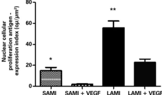
PCNA was significantly diminished in the animals
submitted to treatment with VEGF165. Relative to the
group that suffered a small infarction, the result was,
respectively, for those treated with VEGF165 and not
treated: 1.92±0.66ou/µm2 and 14.75±3.36ou/µm2, with
p=0.0078. The group that suffered LAMI presented
with the result 22.82±3.23ou/µm2 for the treated group
and 55.70±6.54ou/µm2 for the non-treated (p=0.0006),
as can be seen in figure 3.
Literature data suggest that the increase in expression of molecules that determine undifferentiation might be
related to the increased degree in cell proliferation.(10)
Results obtained from the present study suggest that the increased expression of fibronectin in the infarcted area is related to greater cell proliferation, which
occurs in the myocardium after treatment with VEGF165
(specifically in the areas of small infarction).
The significant increase of CD44 after treatment
with VEGF165 indicated the formation and regeneration
of blood vessels in the myocardium after the infarct, regardless of the size of the infarct.
The decrease in expression of vimentin, which corresponds to an intermediate filament of the cytoskeleton, characteristic of fibroblasts and mesenchymal cells, demonstrated that possibly such cells were not responsible for the processes of cardiac tissue remodeling after the myocardial infarction.
It is important to point out that E-cadherin represents an adhesion protein present in cardiac cells
and serves to maintain systole and diastole.(16,19) The
increased expression of E-cadherin after treatment with
VEGF165 indicates that it is likely that this marker has
a fundamental role in the protection and maintenance of cardiac contractility, stimulated by gene transfer with
VEGF165, mainly in areas of a large infarction.
The increased expression of CD24 in the SAMI group
treated with gene transfer suggested that VEGF165 could
stimulate the proliferation and migration of endothelial cells, hence promoting the formation of new vessels and greater ease in myocardial tissue regeneration.
An increase in number of blood vessels would
be expected after treatment with VEGF165 in both
groups of animals, with a small or large infarcted area. However, no significant increase was observed in CD24
in the areas of large infarction. Possibly, the VEGF165
demonstrated a more effective role in treating small myocardial lesions.
The increased expression of the antibody anti-alpha-1-actin in the groups submitted to gene therapy indicated that such a growth factor likely promoted the induction of myocyte proliferation. Therefore, the data obtained demonstrated that myocytes were possibly involved in the process of myocardial extracellular matrix remodeling after the infarction.
The fact that no difference was observed in marking of the myocytes in the tissues of animals of the large infarct group made evident, once again, that possibly
VEGF165 was not effective in inducing the proliferation
of myocytes, when the area affected by the infarction was very large.
Figure 3. Expression of the nuclear cellular proliferation antigen in infarcted animals submitted or not to treatment with VEGF165. The values indicate
the mean and standard deviation by analysis of the expression index, after digital quantification of the immunohistochemical reactions, as is described in methods. The number of animals in each group was 10 (n=10). Asterisks (*) indicated the p values obtained for each marker, comparing the results acquired of animals treated with VEGF versus the non-treated group. *p=0.0078; **p=0.0006. SAMI: group of animals submitted to acute myocardial infarction (small infarction) without treatment; SAMI + VEGF: group of animals submitted to acute myocardial infarction (small infarction) treated with gene transfer containing VEGF165; LAMI: group of animals submitted to acute myocardial infarction (large infarction) without treatment; LAMI + VEGF: group of animals submitted to acute myocardial infarction (large infarction) treated with gene transfer containing VEGF165.
DISCUSSION
In results previously published by our group, it was
described that the maximal expression of VEGF165
occurs 14 days after gene transfer. Consequently, the evaluation of the samples was done within the period of 6 weeks after induction of the infarction and posterior treatment with gene transfer. Therefore, the late response of the effect promoted by treatment
with VEGF165 was evaluated, as had been previously
The groups that received treatment with VEGF165 presented with lower expression of PCNA and, therefore, less cell proliferation. The decrease in rate
of tissue proliferation after gene transfer of VEGF165
corroborates the data already mentioned that evidenced an intense process of cell differentiation.
Therefore, six weeks after the treatment with
VEGF165, it was possible to demonstrate that VEGF165
was related to alterations in the infarction area, responsible for the increased expression of markers specific for cell differentiation. Additionally, the results demonstrated that myocytes were the differentiated cells responsible for such a process. Data obtained in the present study clarified that the damaged tissue after infarction was under intense remodeling and did not present with only scar tissue, as suggested in literature.
CONCLUSIONS
Treatment with VEGF165 represented a fundamental
alternative for extracellular matrix remodeling after acute myocardial infarction, promoting stimulation of cell differentiation. Myocytes actively participated in the remodeling process of the extracellular matrix after an acute myocardial infarction.
ACKNOWLEDGMENTS
We wish to thank the financing obtained for conducting this
study from the Fundação de Amparo à Pesquisa do Estado
de São Paulo (FAPESP) [State of São Paulo Research Foundation] with Regular Research Grant, Process
number 2011/188638-300, of the Conselho Nacional de
Desenvolvimento Científico e Tecnológico (CNPq) [National Council of Scientific and Technological Development],
Process number 127212/2010-8, and of Coordenação de
Aperfeiçoamento de Pessoal de Nível Superior (CAPES) [Coordination for the Improvement of Higher Education Personnel].
REFERENCES
1. Sanderson JE, Mayosi B, Yusuf S, Reddy S, Hu S, Chen Z, et al. Global burden of cardiovascular disease. Heart. 2007;93(10):1175
2. McGinn AN, Nam HY, Ou M, Hu N, Straub CM, Yockman JW, et al. Bioreducible polymer-transfected skeletal myoblasts for VEGF delivery to acutely ischemic myocardium. Biomaterials. 2011;32(3):942-9.
3. Katare RG, Kakinuma Y, Arikawa M, Yamasaki F, Sato T. Chronic intermittent fasting improves the survival following large myocardial ischemia by activation of BDNF/VEGF/PI3K signaling pathway. J Mol Cell Cardiol. 2009;46(3):405-12. 4. Novotny NM, Ray R, Markel TA, Crisostomo PR, Wang M, Wang Y, et al.
Stem cell therapy in myocardial repair and remodeling. J Am Coll Surg. 2008; 207(3):423-34. Review.
5. Furlani AP, Kalil RA, Castro I, Cañedo-Delgado A, Barra M, Prates PR, et al. Effects of therapeutic angiogenesis with plasmid VEGF165 on ventricular function in a canine model of chronic myocardial infarction. Rev Bras Cir Cardiovasc. 2009;24(2):143-9.
6. Wang R, Crystal RG, Hackett NR. Identification of an exonic splicing silencer in exon 6A of the human VEGF gene. BMC Mol Biol. 2009;10:103.
7. Ward MR, Stewart DJ. Progenitor cell therapy for cardiac regeneration following acute myocardial infarction: So far, so good? Can J Cardiol. 2008;24(Suppl C): 5C-10C.
8. Payne TR, Oshima H, Okada M, Momoi N, Tobita K, Keller BB, et al. A relationship between vascular endothelial growth factor, angiogenesis, and cardiac repair after muscle stem cell transplantation into ischemic hearts. J Am Coll Cardiol. 2007;50(17):1677-84.
9. Kivelä R, Bry M, Robciuc MR, Räsänen M, Taavitsainen M, Silvola JM, et al. VEGF-B-induced vascular growth leads to metabolic reprogramming and ischemia resistance in the heart. EMBO Mol Med. 2014 Mar;6(3):307-21. 10. Levashova Z, Backer M, Backer JM, Blankenberg FG. Imaging vascular
endothelial growth factor (VEGF) receptors in turpentine-induced sterile thigh abscesses with radiolabeled single-chain VEGF. J Nucl Med. 2009;50(12):2058-63. 11. Yan D, Wang X, Li D, Liu W, Li M, Qu Z, et al. Macrophages overexpressing
VEGF target to infarcted myocardium and improve neovascularization and cardiac function. Int J Cardiol. 2013;164(3):334-8.
12. Sato D, Otani H, Enoki C, Fujita M, Minato N, Iwasaka T. Phenotypic modulation and turnover of bone marrow-derived cells after myocardial infarction in rats. Cardiovasc Pathol. 2011;20(3):146-55.
13. Mataveli FD, Han SW, Nader HB, Mendes A, Kanishiro R, Tucci P, et al. Long-term effects for acute phase myocardial infarct VEGF165 gene transfer cardiac extracellular matrix remodeling. Growth Factors. 2009;27(1):22-31. 14. Jujo K, Li M, Losordo DW. Endothelial progenitor cells in neovascularization of
infarcted myocardium. J Mol Cell Cardiol. 2008;45(4):530-44. Review. 15. Ye L, Zhang W, Su LP, Haider HK, Poh KK, Galupo MJ, et al. Nanoparticle based
delivery of hypoxia-regulated VEGF transgene system combined with myoblast engraftment for myocardial repair. Biomaterials. 2011;32(9):2424-31. 16. Zhang Y, Furumura M, Morita E. Distinct signaling pathways confer different
vascular responses to VEGF 121 and VEGF 165. Growth Factors. 2008;26(3):125-31. 17. Hagikura K, Fukuda N, Yokoyama S, Yuxin L, Kusumi Y, Matsumoto T, et al.
Low invasive angiogenic therapy for myocardial infarction by retrograde transplantation of mononuclear cells expressing the VEGF gene. Int J Cardiol. 2010;142(1):56-64.
18. Vilahur G, Juan-Babot O, Peña E, Oñate B, Casaní L, Badimon L. Molecular and cellular mechanisms involved in cardiac remodeling after acute myocardial infarction. J Mol Cell Cardiol. 2011;50(3):522-33.
19. Xu H, Shi BM, Lu XF, Liang F, Jin X, Wu TH, et al. Vascular endothelial growth factor attenuates hepatic sinusoidal capillarization in thioacetamide-induced cirrhotic rats. World J Gastroenterol. 2008;14(15):2349-57.