Radiofrequency Catheter Ablation of Atrial Fibrillation Guided by
Spectral Mapping of Atrial Fibrillation Nests in Sinus Rhythm
José Carlos Pachón Mateos, Enrique I. Pachón Mateos, Tasso J. Lobo, Maria Zélia C. Pachón, Juán Carlos Pachón Mateos, Denilda Queiroz V. Pachón, Remy Nelson A. Vargas, Leopoldo S. Piegas, Adib D. Jatene
Hospital do Coração (HCor) and Instituto Dante Pazzanese de Cardiologia - São Paulo, SP - Brazil
Summary
Background: Two types of myocardia can be observed through the endocardial spectral mapping (SM) in sinus rhythm: the compact type with a smooth spectrum and the fibrillar type with a segmented spectrum (atrial fibrillation nests). During the atrial fibrillation (AF), the compact type has an organized activation and low frequency (passive), whereas the fibrillar type has a rather disorganized activation and high frequency (active/resonant), with both being activated by high-frequency sustained tachycardia - the background tachycardia (BT).
Objective: To describe the treatment of AF by the ablation of the AF nests and BT.
Methods: 1) Catheter ablation of the AF nests with RF [4/8mm-60º/30-40J/30s] guided by SM in sinus rhythm, outside the pulmonary vein; 2) atrial stimulation -300ppm; 3)Additional ablation of the AF nests if AF is induced; 4) Focal ablation if BT and/or Flutter is induced; 5)Clinical follow-up+ ECG+ Holter.
Results: A total of 50±18 AF nests/patient were treated. After 11.3±8m, 81 patients (88%) did not present AF (28.3% with antiarrhythmic drugs). After the ablation of the AF nests, AF was not reinduced in 61 patients (71%) and BT was induced and treated in 24 patients (26%). There were two episodes of pericardial bleeding (1 treated clinically and 1 surgically), caused by sheaths that are no longer used.
Conclusion: The SM in sinus rhythm can be used in the ablation of AF nests. During the AF, the AF nests present a reactive-resonant pattern and the compact myocardium is passive, stimulated by the high frequency of the BT. After the ablation of the AF nests and the BT, it was not possible to reinduce the sustained AF. The Ablation of AF nests outside the pulmonary veins showed to be safe and highly effective in the cure and/or clinical control of the AF. (Arq Bras Cardiol 2007;89(3):124-134)
Key words: Catheter ablation; atrial fibrillation; spectrum analysis; heart rate.
Mailing address: José Carlos Pachón Mateos •
Av. Açocê, 515/31 - 04075-000 - São Paulo, SP - Brazil E-mail: [email protected]
Manuscript received February 25, 2007; revised manuscript received March 1, 2007; accepted March 7, 2007.
Introduction
The conventional electrophysiological study is based on the analysis of amplitude variations over time (time domain). However, the spectral analysis of a wave correlates amplitude and/or potency with the intrinsic signal frequencies (frequency domain), supplying a large amount of additional information. The heart rate is measured in beats per minute (extrinsic signal frequency), i.e., time domain, whereas the normal R wave peak, for instance, changes voltage abruptly due to the fact that it has high intrinsic spectral frequencies (frequency domain), measured in hertz (Hz, cycles per second). Therefore, the term frequency can be related to temporal or spectral values. Using real-time spectral analysis in sinus rhythm, it is observed that the atrial walls are constituted by two distinct types of myocardia, the compact and the fibrillar types (Fig. 1). The first
type presents a continuous and organized spectrum, with most of the potency grouped to the left (low frequency). The second type presents a segmented spectrum, with a large amount of the segments shifted to the right (high frequencies, > 80 Hz)1. The fibrillar myocardium is usually
grouped in small areas, which are more or less extensive. During the atrial fibrillation (AF), the compact myocardium presents an organized behavior and high-amplitude electrical activation, low frequency, slightly irregular, suggesting a passive behavior. On the other hand, the fibrillar myocardium presents low-amplitude electrical activity, which is highly disorganized and has high frequency, showing, from an electrical point of view, a resonant behavior.
of AF, 56 cases (61%) presented the paroxysmal type (3.6 ± 1.3, with 2 to 7 crises per year), 25 (27%) presented the persistent type (2.6 ± 1.2, with 1 to 6 crises per year) and 11 (12%) presented the permanent type. Very frequent episodes of non-sustained or incessant AF were observed in 9 patients. Most of the patients did not present any significant cardiopathy. The left atrium diameter was normal or moderately increased (41.9 ± 5 mm). The mean ejection fraction was 0.61 ± 0.8. The most relevant etiologies were: idiopathic, 42%; hypertension, 22%; congenital cardiopathy, 11%; rheumatic cardiopathy, 10%; coronaropathy, 9%; and others, 6%.
Methods
The patients using oral anticoagulants were admitted at the hospital 24 or 48 to have the International Normalized Ratio
(INR) adjusted for 1.4. All cases, especially those with a history of thromboembolism, were treated only after the confirmation of the absence of cardiac thrombi through transesophageal echocardiography3-5. The conventional monitoring with surface
electrocardiography (ECG) radiotransparent defibrillation adhesive plaques, orotracheal intubation with mechanical ventilation and inhalation anesthesia was used, as well as the passing of a transesophageal echocardiography transducer. After asepsis, five venipunctures were performed (one subclavian and four femoral ones). A decapolar catheter was introduced through the subclavian vein to catheterize the coronary sinus. Two quadripolar catheters were introduced through the femoral vein into the right atrium and ventricle, an octapolar catheter was positioned at the His and a transeptal punction was performed (five patients had a patent foramen ovale). The following equipment were utilized: Cícero It has been observed that the elimination of the fibrillar
myocardium makes it progressively more difficult to reinduce AF. Additionally, these areas present he highest frequencies during this type of arrhythmia. Therefore, the places with high density of fibrillar myocardium were called AF “nests”1,2.
Moreover, after the elimination of the AF nests, it has been possible to induce very rapid atrial tachycardia that is highly protected from reversion. Some evidence has demonstrated that this tachycardia occurs secondarily during the AF (background tachycardia), being its maintenance element. The present study describes the mapping and the ablation of the AF nests, followed by the induction and ablation of the background tachycardia.
Hypothesis - The AF nests can be easily identified through spectral mapping during the sinus rhythm and eliminated with ablation by radiofrequency. This could weaken one of the mechanisms of AF maintenance, contributing to its ultimate treatment. Additionally, it would allow the visualization of background tachycardia2, permitting its mapping and
respective ablation.
Objectives - To propose a new alternative for the treatment of AF based on a spectral mapping during sinus rhythm and describe new findings, aiming at a better understanding of the physiopathology of this arrhythmia.
Patients - A total of 92 patients, 76 males, whose mean age was 52.4 ± 11 yrs were included in the study. All patients had frequent AF that was refractory to at least two antiarrhythmic drugs and 51 (55.4%) reported at least one electrical cardioversion. In this group, all the antiarrhythmic drugs available in our country, as well as flecainamide, showed to be ineffective in one or more patients. Regarding the type
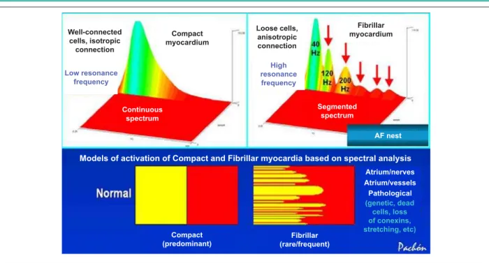
Fig. 1 -The compact myocardium presents a continuous spectrum (left), whereas the fibrillar myocardium presents a typically segmented spectrum. These very distinct spectral behaviors result in a significant difference in the electrophysiological properties, with the first being very resistant to and the last being highly susceptible to AF.
Well-connected cells, isotropic
connection
Low resonance frequency
Compact myocardium
Continuous spectrum
Loose cells, anisotropic connection
High resonance frequency
Fibrillar myocardium
Segmented spectrum
AF nest
Models of activation of Compact and Fibrillar myocardia based on spectral analysis
Atrium/nerves Atrium/vessels Pathological
(genetic, dead cells, loss of conexins, stretching, etc)
Compact (predominant)
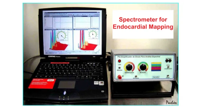
EM anesthesia equipment (Dräger); HP/Philips M1026A multiparametric monitor; HP/Philips echocardiography equipment model Sonos 2500; 32-channel TEB polygraph with modified software for real-time spectral post-analysis (TEB-Pachón); computerized spectrometer (Pachón®) for
real-time spectral analysis (Fig. 2); Digital Siemens radioscopy; Biotronik Abcontrol MDS; radiofrequency generator; Philips Heartstart XL biphasic defibrillator with transcutaneous pacemaker; brain-activity spectrometer-BIS (in cases with higher thromboembolic risk). The ablations were carried out with a Blazer EPT 4-mm or 8-mm catheter or 4-mm or 8-mm Medtronic Conductor. All patients received information about the methods and potential complications and agreed with the
procedures by signing the informed consent form.
Through a transeptal punction a spiral catheter (Supreme St. Jude or Biotronik Lexx) and the ablation catheter were introduced. Heparin was administered IV to maintain the activated coagulation time between 300 and 400 s. The atrial endocardia were scanned with real-time spectral analysis. All AF nests found were eliminated by applying radiofrequency (30-40 W/30 s/60oC) until the fibrillar pattern disappeared
(Fig. 3). No complete intentional isolation of the pulmonary veins or blockage lines was carried out, according to the existing conventional techniques6. The spiral catheter was
used to safely identify the atrial limits and detect AF nests, which are less evident with an 8-mm catheter.
Fig. 2 - Spectrometer developed by Pachón & Pachón, consisting of a high-precision amplifier with specific filters and IBM-PC compatible software to obtain two-dimensional as well as three-two-dimensional real-time spectral analysis of endocardial signals. The connection to the patient is made by photo-coupler.
Spectrometer for
Endocardial Mapping
30-40 W / 30s / 60°C
Fig. 3 -Radiofrequency ablation guided by spectral mapping during sinus rhythm. The radiofrequency catheter connected to the spectrometer scans the entire atrial endocardium. All points that present fibrillar spectrum in sinus rhythm are submitted to ablation with 30-40 W/30 s/60ºC. In the present study, the antrum of the pulmonary veins was treated only at the points that presented AF nests.
“AF nest”
Post-ablation re-study - After the ablation of the AF nests, programmed (up to three extra-stimuli) as well as progressive (up to 300 ppm) atrial stimulation were performed. If AF were induced, new AF nests would be sought and treated. If residual tachycardia were induced (background tachycardia2), it would
be mapped and treated at its focus of origin. The authors of the present study chose to use the term “background tachycardia”, instead of secondary tachycardia, as the term “background” has been incorporated into the Brazilian Portuguese language, according to version 5.11 of Novo Dicionário Aurélio. In case of history or induction of flutter, the specific ablation was carried out. The criteria to finish the ablation were: 1. Impossibility to re-induce AF or tachycardias; 2. Induction only of non-sustained tachycardia/AF < 10 sec; and 3. Impossibility to eliminate residual tachycardia or prolonged procedure (more than five hrs); in these cases, the external thoracic cardioversion was carried out.
Post-ablation medication - Even being ineffective or little effective in the pre-ablation phase, the antiarrhythmic medication was used for two months to reduce the atrial excitability caused by the cicatricial process. Warfarin anticoagulants were administered for three months post-ablation, keeping the prothrombin time between 2 and 3 INR. After this period, several anti-aggregants were used in cases that presented thromboembolism risk (age, heart failure, mitral dysfunction, dilated left atrium, etc), including those patients with sustained or non-sustained AF recurrence and adequate pharmacological response. The antiarrhythmic medication was withdrawn, except in 23 cases in whom it was necessary to maintain the sinus rhythm.
Post-ablation follow-up - The follow-up consisted of clinical control and ECG (at hospital discharge, after one, three and six months and after one year) and Holter (one month, six months and one year) or whenever there were symptoms. Additionally, a continuous control was kept through telephone calls.
Results
On average, 50 ± 18 AF nests were treated per patient. After the ablation, it was not possible to induce sustained AF in 65 (71%) cases. In 27 cases (29%), it was possible to induce a non-sustained episode of atrial tachycardia or AF (< 10 s). In 24 patients (26%), it was possible to induce atrial tachycardia with special characteristics (induced by programmed or progressive high-frequency atrial stimulation, highly protected by inflow blockage, difficult to induce and very difficult reversion through atrial stimulation), which was called “background tachycardia”2. Eighteen patients (19%)
presented a history of common flutter or it was induced, being treated with the bidirectional blockage of the cavotricuspid isthmus. After a mean follow-up of 11.3 ± 8 months, 81 patients (88%) were in sinus rhythm, 23 cases (28%) using previously ineffective antiarrhythmic drugs at reduced doses (amiodarone, 100 mg/day to 300 mg/day; sotalol, 60 mg/day to 160 mg/day; propafenone, 150 mg/day to 600 mg/day; quinidina, 200 mg/day to 400 mg/day). Nine patients (9.8%) underwent re-ablation due to the difficulty in the clinical control of AF or atrial tachycardia in the late phase post-ablation (more than six months of evolution).
At the initial phase of this method, two episodes of pericardial bleeding occurred (one that resolved clinically and another that had to be surgically drained), probably related to the simultaneous manipulation of multiple sheaths inside the left atrium, as the tip of the involved electrodes (EPT 4 mm large), differently from the sheaths, is very malleable, being unlikely that it would have caused the lesion. Additionally, this complication did not occur after the sheaths started to be removed from the left atrium during the ablation, even when more rigid 8-mm electrodes were used. No other complication was observed.
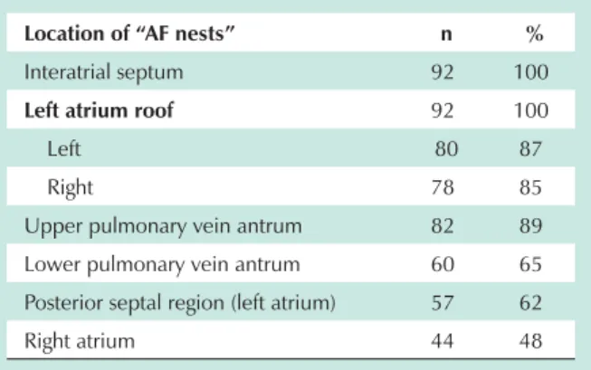
The most common locations of “AF nests” are shown in Table 1.
In 11 patients (46%) of the 24 cases in whom background tachycardia was induced, the focus was well defined by mapping, with tachycardia reversion following the use of radiofrequency at the place of better mapping (Table 2). In the other 13 cases (54%), the external thoracic cardioversion (10) or reversion by overdrive (3) was carried out due to inconsistent mappings, very high tachycardia frequency, more than one focus or prolonged procedures.
The mean procedure and radioscopy duration were, respectively: 4.3 ± 1.2 hrs and 53.8 ± 19 minutes.
Discussion
This study demonstrates the use of real-time spectral
Table 1 - Frequency of the locations of “AF nests”
Location of “AF nests” n %
Interatrial septum 92 100
Left atrium roof 92 100
Left 80 87
Right 78 85
Upper pulmonary vein antrum 82 89
Lower pulmonary vein antrum 60 65
Posterior septal region (left atrium) 57 62
Right atrium 44 48
n - number of patients.
Table 2 - Location of the focus of the background tachycardias reversed during the application of radiofrequency
Location of the focus n %
Interatrial septum 4 36
Right atrium 3 27
Coronary sinus 2 18
Left upper pulmonary vein 2 18
mapping, in sinus rhythm, in the identification and ablation of AF nests. It was possible to observe that the more extensive the ablation of the AF nests, the lower the possibility of inducing this arrhythmia through stimulation. Additionally, a large number of these patients (88%) remained without AF or had controlled AF during follow-up, despite the 39% of cases with persistent and permanent AF, types that are normally associated to poorer outcomes7. These data suggest that,
apparently, the lower the number of AF nests, more stable the atrial walls become, from an electrical point of view. Additionally, their elimination allowed the identification of tachycardia with very specific characteristics, likely constituting an essential connection in the physiopathology of AF.
AF substrate - Large contributions have been made in the identification of AF triggers8-10 as well as in the treatment of
this arrhythmia by the ablation or isolation of these triggers11,12.
However, the identification of the substrate has advanced very little. It is relatively easy to understand an arrhythmia such as the AF in a dilated atrium with significant fibrosis and degeneration; however, how does one explain this arrhythmia in apparently normal hearts? The initial observations of the present study indicate the AF nests and the background tachycardias. When comparing normal patients with and without paroxysmal AF, it was observed that the number of AF nests is significantly higher in cases with spontaneous or laboratory-induced AF1,13.
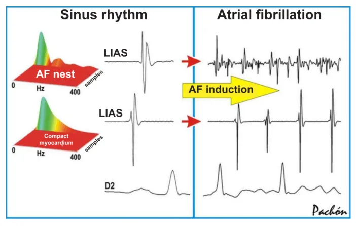
Fibrillar myocardium and AF nests: The different spectrums ofThe different spectrums of compact and fibrillar myocardia suggest that the great difference between the two types lies in the degree of connection among the cells, which gives those very distinct electrical resonance frequencies. The compact myocardium is very well connected and functions as a “large cell”, whereas the fibrillar type behaves as a set of bundles with varied velocities of conduction and refractoriness. From the spectral point of view, the fibrillar and compact myocardia have high and low resonance frequencies, respectively. Therefore, when the atrial walls are stimulated with progressive frequencies, it is observed that the fibrillar myocardium presents autolimited repetitive responses at a much lower frequency than the compact myocardium (234 ± 28 x > 300 ppm; p = 0.018), i.e., the compact myocardium is much more resistant to rapid atrial stimulation. Due to this property, during the AF the fibrillar myocardium constantly maintains a very high, irregular and disorganized frequency (multiplying element), justifying the name “AF nests”, whereas the compact myocardium maintains an organized depolarization, much lower frequency and no repetitive responses (passive element) (Fig. 4).
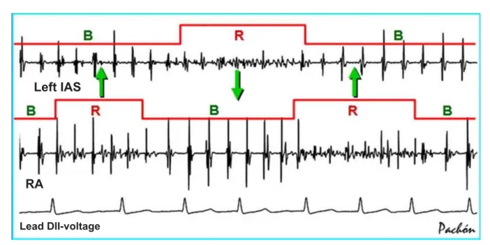
Resonance of the AF nests - During the AF, the AF nests frequently alternate periods of low amplitude and disorganized high frequency with high amplitude and organized low frequency (Fig. 4). This happens due to a periodical electrical instability that promotes the alternateness of high and low frequencies. In this case, AF nests that are close and at different phases
Fig. 4 - The AF nests identified during the sinus rhythm by spectral mapping show a very different behavior when compared to the compact myocardium during AF. Characteristically, the more segmented the spectrum during the sinus rhythm, the higher the frequency of activation after the AF induction. AF - atrial fibrillation; Compact M. - compact myocardium; Hz - hertz; LIAS - left interatrium septum; RA - right atrium; DII - lead DII.
Sinus rhythm
$WULDO¿EULOODWLRQ
LIAS
LIAS
AF nest
Compact myocardium
AF induction
sam ples sam
can feed again, favoring the maintenance of AF (Fig. 5). This periodicity of electrical organization/disorganization is called electrical resonance (a known biological property observed in some cells of the mammalian and amphibian nervous system), as its behavior is comparable to that of an electronic resonator14,
capable of multiplying the initial stimulation. The microreentry and/or reflection between close cell bundles, as well as periods of autolimited triggered activity due to a transitory calcium phenomenon, can explain this property of AF nests15,16.
Probable focus of the AF nests - In the group of patients assessed in the present study, it was possible to associate the AF nests with the following conditions:
1. Congenital: innervation entry (fiber interpolation)20.
2. Constitutional: reduction of conexins, with a predominance of termino-terminal connections (crista terminalis)21.
3. Atriovenous transition (interpolation of tissues at the insertion of the pulmonary veins).
4. Mechanical stretching of the atrial myocardium (separation of cells due to pathological conditions, such as interatrial septum aneurysm, etc).
5. Decreased expression of conexins (congestive heart failure, aging, congenital conditions).
6. Pathological: degeneration, ischemia, inflammation or infiltrative disease com remodeling and reduction of conexins.
Recently it was observed that the elimination of the AF nests makes the response to atropine negative in patients who presented a previously normal response, showing a close association between AF nests and vagal innervation20 (Fig. 6).
Treating atrial fibrillation in sinus rhythm through ablation with radiofrequency of AF nests - The advantage of the concept of AF nests was to allow the ablation by catheter during sinus rhythm1 (Fig. 4) without performing the classical blockage lines.
It has been observed that the higher the number of AF nests, more frequent and prolonged is the AF, whether spontaneous or induced. Patients that present a very reduced number of AF nests are typically resistant to arrhythmia induction by atrial stimulation. Additionally, when induced, the AF tends to be short and present spontaneous reversion. In the present study, we observed that as the number of AF nests was reduced through ablation with radiofrequency, it became more difficult to re-induce arrhythmia.
Background Tachycardia - An extremely interesting phenomenon observed during the spectral ablation of AF nests was the background tachycardia2. The latter is an atrial
tachycardia observed only after the ablation of AF nests and attained at the following conditions: during AF, by progressive ablation of AF nests or during sinus rhythm, through programmed and/or progressive atrial stimulation (up to 300 ppm) after most of the AF nests have been submitted to ablation.
Main characteristics of background tachycardia:
1. Regular or slightly irregular atrial tachycardia. 2. Very high frequency (256 ± 34 bpm).
3. Originated by reentry (generally microreentry induced by rapid and/or programmed atrial stimulation and reversed by overdrive).
4. Highly protected focus (very difficult to be reversed by overdrive) (Fig. 7).
Fig. 5 -Simultaneous tracings showing the dynamic behavior of the AF nests. During the AF, a periodical electrical instability occurs, which promotes the alternateness of high and low frequencies – the resonant and bystander states. Close AF nests and those at different phases favor the refeeding and maintenance of the AF (arrows). In this sense, the techniques that create lines of blockage on the atrial wall favor the electrical stability17-19. R - resonant; B - bystander; Left IAS - left interatrial sinus; RA - right atrium; DII – N - lead DII - voltage.
Left IAS
RA
5. It can be identified by the digital subtraction of the QRS at the surface ECG and spectral analysis of the tracing of the fast Fourier transform (FFT), before the ablation.
Association between background tachycardia and atrial fibrillation - In the present study it was possible to visualize the background tachycardia after the ablation of the AF nests. It was observed that the AF starts to organize and becomes background tachycardia with the progressive elimination of the AF nests. These data suggest that this tachycardia is a key element in the physiopathology of AF. It is a regular tachycardia (originated by reentry), which, apparently exists at a second plane (background) during the AF. Its very high frequency stimulates the AF nests, promoting its disorganization, its resonance and refeeding. The presence of an entry blockage protects the background tachycardia focus, so that it is not reversed by the stimuli that originate from the activation of the AF nests. The larger the entry blockage is (more difficult to reverse with atrial stimulation), the more prolonged the AF. The combination of background tachycardia with very high and disorganized frequency of AF nests constitutes the apparently chaotic electrical activity that characterizes the classical morphology of f waves at the ECG of frank AF. Many times there is a periodical alternateness of resonance of the AF nests, making the background tachycardia to appear more or less cyclically organized. This constitutes a condition that is frequently known as “fibrillo-flutter”. From an electrical point
of view, this tachycardia is the main oscillator of the AF2. Ablation of the background tachycardia - After the ablation of the AF nests, the focus of the background tachycardia induced by atrial stimulation was sought and its ablation was attempted (Fig. 8). Eventually, the focus of this tachycardia might have been eliminated indirectly during the ablation of the AF nests. In this study, the evolution of the patients who had the background tachycardia treated or those in whom it was impossible to induce it when compared to the group who remained untreated was very different. In the first group, the success rate was 88.9%; in the second group, however, the AF was eliminated in only 73% (p=0.008). Nevertheless, due to mechanisms of protection, many times the background tachycardia could not be induced. Additionally, its mapping is hindered by the high frequency.
Physiopathology of atrial fibrillation based on AF nests - This study suggests that the AF results from the fusion of arrhythmias constituted essentially by a protected atrial tachycardia (background tachycardia), which stimulates the nests and maintain them under a constant state of repetitive responses (Fig. 8). The observation of this group of patients, of the respective ablations and the clinical history of each case allows us to establish a specific physiopathology for AF based on the following components:
1. Modulator: autonomic nervous system (extensively
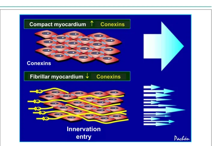
Fig. 6 - Based on the spectral characteristics, the location and association with nerves, veins and myocardial stretching, the differences between the compact and fibrillar myocardia seem to be closely related with the amount of intercellular connections. The lower the number of connections, the more segmented is the spectrum. Naturally, the large amount of conexins among the cells is reduced in regions of great interpolation of nervous fibers (neuromyocardial interface). This characteristic, as well as the mix of different cells (such as in fibrosis or at the transition to the veins) can modify the spectrum.
Compact myocardium
Conexins
Fibrillar myocardium
Conexins
Innervation
entry
Fig. 7 - Schematic representation showing the behavior of fibrillar and compact myocardia activated by background tachycardia, which behaves as a protected microreentry with high frequency, induced by the triggers. Due to its high frequency, the compact myocardium has a limited response during the refractory period (lower frequency and higher regularity), whereas the fibrillar one has a high and irregular response. The sum of these rhythms results in the electrocardiographic expression known as atrial fibrillation. AF - atrial fibrillation.
Triggers
AF nests
“Background tachycardia”
Compact myocardium
Sustained reentry
Fibrillar myocardium
AF
Fig. 8 - Tracings from the same patient, before and after the ablation of the AF nests; in this case, during AF. To the right, the organization of the rhythm can be observed after the removal of the AF nests, resulting in background tachycardia. The observation of this group of patients suggests that this type of tachycardia exists during AF and functions as an element of arrhythmia maintenance. RF - radiofrequency; AF - atrial fibrillation; BKGT - background tachycardia; AF nests - atrial fibrillation nests; PCS - Proximal Coronary Sinus; LIAS - left interatrium septum; LRA - lateral wall of right atrium; V2 - lead V2.
Pre-RF
AF
Post-RF
BKGT
AF nest ablation
PCS
LIAS
LRA
modified by the ablation of the “AF nests”20).
2. Trigger: extra-systoles and non-sustained tachycardias of the pulmonary veins9,19,22-24.
3. Substrate (multiplier element): AF nests that respond with high frequency to the rapid atrial stimulation, due to the resonant properties (microreentry and/or triggered activity). The capacity to multiply the stimuli is one of the factors that sustain the AF.
4. Maintainer: background tachycardia, which works as an asynchronic atrial pacemaker with a very high frequency, capable of activating the AF nests, maintaining them within the range of repetitive responses. It is the analogue of the artificial pacemaker used by Wijffels et al25 and Moreira et al26
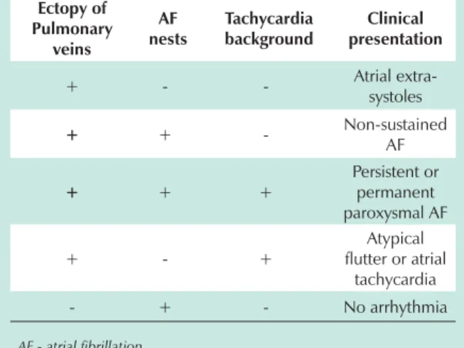
to produce AF, stimulating with very high frequencies. This physiopathology explains the several classically known clinical pictures (Table 3).
large amounts of energy. The exclusive elimination of the high frequencies of the spectrum allows the preservation of the more compact myocardium of the atrial wall, preventing deep lesions and reducing the risk of perforation, of atrioesophageal fistulas29,32 and of stenosis of the pulmonary veins33,34, a fact
that also differentiates the technique from other methods based on extensive and deep lines of blockage19.
As well as in the techniques that directly eliminate the paracardiac ganglia35,36, the ablation of the AF nests, in a
simplified form, promotes a significant parasympathetic denervation through the direct elimination of intramural neurons and the indirect elimination of the paracardiac ganglia. Therefore, in addition to acting directly on the autonomous nervous system, it eliminates a large part of the neuromyocardial interface, which has resonant properties (due to its microstructure) and specific spectral characteristics20
(interpolation of fibers). The elimination of the intramural neuron and the fibrosis of the neuromyocardial junction significantly reduces the extension of the reinnervation.
The method developed by Nademanee et al37, also
published in 2004, describes the ablation during the AF oriented by complex fractionated electrograms (CFAEs), which are areas of highly fractioned electrical activity and directly identified by the time domain. It is known, however, that during the AF, due to the large number of wavefront collisions, many virtual areas of complex potentials are formed, a problem that does not exist during the sinus rhythm and, therefore, during the ablation of AF nests.
Likely due to this fact, it is well-known the difficulty to reverse a persistent or chronic AF with the ablation of CFAES. Only the paroxysmal AF can be reversed more easily and the reversion, in this case, can be occasional and not necessarily electrophysiological.
In fact, most of the methods have developed in the sense of causing progressively more extensive intramural lesions on the atrial wall. Clearly, the larger the area destroyed, the larger the number of eliminated AF nests, which can influence the results38, regardless of the method. Additionally, it has been
observed that the origin of the background tachycardia can inadvertently eliminated during these extensive ablations, favoring the long-term outcome. This aspect is highly desirable; however, considering the spectral mapping as the basis, it is understood that it is preferable to direct the ablations rationally into the substrate, eliminate only the resonant frequencies (fibrillar myocardium), preserve the compact muscle, apply the least amount of energy possible to prevent complications and try to induce the background tachycardia with the subsequent ablation of its focus of origin.
Conclusions
The AF nests were easily found in sinus rhythm through the spectral mapping, allowing them to be treated by ablation with conventional radiofrequency, regardless of the isolation of the pulmonary veins. During the AF, this type of myocardium (fibrillar type) presents a reactive-resonant high-frequency pattern, which refeeds the arrhythmia. Differently, the compact myocardium responds in an organized form and with low frequency. After the ablation of the AF nests,
Table 3 – Physiopathology of the atrial fibrillation according to the observations of spectral ablation of the AF nests (the several combinations of the basic elements of AF determine the appearance of different clinical pictures, and have been long known by cardiologists)
Ectopy of Pulmonary veins AF nests Tachycardia background Clinical presentation
+ - - Atrial
extra-systoles
+ + - Non-sustained
AF + + + Persistent or permanent paroxysmal AF + - + Atypical flutter or atrial
tachycardia
- + - No arrhythmia
AF - atrial fibrillation.
Comparison with other ablation methods of atrial fibrillation - The cure of AF is a great challenge for all the available technique27. Although it was published in 20041, the ablation
of AF nests was developed in the end of the 90’s, without being based on previous techniques. The initial proposal of the authors of the present study was to use spectral mapping to search for the AF substrate even in an apparently normal heart, and including patients who had presented a history of AF and, certainly, without atrial fibrosis. The AF nests are frequent in the antrum of pulmonary veins. During their elimination, the electrical isolation of these structures can occur. However, as the spectrum-oriented isolation searches for the frequencies > 80 Hz, most of the times the pulmonary veins are not completely isolated by the ablation of the AF nests, resulting in the electrical conduction of potentials < 80 Hz. This fact differentiates this technique from the methods developed by Haissaguerre et al11, Natale et al28, Pappone et al19,29 and Oral et al30,31, among
it became progressively more difficult the reinduction of the AF, showing that they are directly associated with the physiopathology of arrhythmia. Additionally, the removal of the AF nests allowed the visualization of a residual tachycardia with entry blockage and high frequency, the background tachycardia. The treatment of this condition was followed by significant outcome improvement. These observations, in addition to contributing to the better understanding of the AF physiopathology, allow us to conclude that the ablation of the AF nests followed by the treatment of the background tachycardia is a safe, feasible and highly efficient method to cure or clinically control the AF.
Potential Conflict of Interest
No potential conflict of interest relevant to this article was reported.
Sources of Funding
There were no external funding sources for this study.
Study Association
This article is part of the thesis associate professor submitted by José Carlos Pachón Mateos, from Universidade de São Paulo.
References
1. Pachón M JC, Pachón M EI, Pachón M JC, Lobo TJ, Pachón MZ, Vargas RN, et al. A new treatment for atrial fibrillation based on spectral analysis to guide the catheter RF-ablation. Europace. 2004; 6 (6): 590-601. Erratum in: Europace. 2005; 7 (1): 92-3.
2. Pachón M JC, Pachón M EI, Lobo TJ, Pachón JC, Pachon MZ, Piegas LS, et al. Atrial fibrillation RF catheter ablation based on the spectral mapping of the AF-nests: the Second ISHNE AF World-Wide Internet Symposium. [Acesso em 2007 February 10]. Disponível em: url:http://www.af-symposium. org/2007/@lectures.php>.
3. Maltagliati A, Galli CA, Tamborini G, Calligaris A, Doria E, Salehi R, et al. Usefulness of transesophageal echocardiography before cardioversion in patients with atrial fibrillation and different anticoagulant regimens. Heart. 2006; 92 (7): 933-8.
4. Tatani SB, Fukujima MM, Lima JA, Ferreira LD, Ghefter CG, Prado GF, et al. Clinical impact of transesophageal echocardiography in patients with stroke without clinical evidence of cardiovascular sources of emboli. Arq Bras Cardiol. 2001; 76 (6): 453-61.
5. Zabalgoitia M, Halperin JL, Pearce LA, Blackshear JL, Asinger RW, Hart RG. Transesophageal echocardiographic correlates of clinical risk of thromboembolism in nonvalvular atrial fibrillation. Stroke Prevention in Atrial Fibrillation III Investigators. J Am Coll Cardiol. 1998; 31 (7): 1622-6.
6. Sanders P, Morton JB, Deen VR, Davidson NC, Sparks PB, Vohra JK, et al. Immediate and long-term results of radiofrequency ablation of pulmonary vein ectopy for cure of paroxysmal atrial fibrillation using a focal approach. Intern Med J. 2002; 32: 202-7.
7. Mehta N, Tavora MZ, Takeschita N, Figueiredo E, Lourenco RM, Germiniani H, et al. Useful clinical features for the selection of ideal patients with atrial fibrillation for mapping and catheter ablation. Arq Bras Cardiol. 2002; 78 (1): 1-16.
8. Jais P, Haissaguerre M, Shah DC, Chouairi S, Gencel L, Hocini M, et al. A focal source of atrial fibrillation treated by discrete radiofrequency ablation. Circulation. 1997; 95 (3): 572-6.
9. Haissaguerre M, Jais P, Shah DC, Takahashi A, Hocini M, Quiniou G, et al. Spontaneous initiation of atrial fibrillation by ectopic beats originating in the pulmonary veins. N Engl J Med. 1998; 339 (10): 659-66.
10. Rocha Neto AC, Farias RL, de Paola AA. Treatment of atrial fibrillation with radiofrequency ablation and simultaneous multipolar mapping of the pulmonary veins. Arq Bras Cardiol. 2001; 77 (5): 407-28.
11. Haissaguerre M, Jais P, Shah DC, Garrigue S, Takahashi A, Lavergne T, et al. Electrophysiological end point for catheter ablation of atrial fibrillation initiated from multiple pulmonary venous foci. Circulation. 2000;101:1409-17.
12. Morady F. Treatment of paroxysmal atrial fibrillation by pulmonary vein isolation. Circ J. 2003; 67: 567-71.
13. Pachón M JC, Pachón M EI, Pachón M J, Lobo TJ, Pachón MZC, Vargas RNA, et al. “AF-Nests” electrical resonance: is it a new atrial fibrillation physiopathology? Heart Rhythm. 2005; 2 (5): S149.
14. Armstrong CE, Roberts WM. Electrical properties of frog saccular hair cells: distortion by enzymatic dissociation. J Neurosci. 1998; 18 (8): 2962-73.
15. Patterson E, Po SS, Scherlag BJ, Lazzara R. Triggered firing in pulmonary veins initiated by in vitro autonomic nerve stimulation. Heart Rhythm. 2005; 2 (6): 624-31.
16. Roberts WM. Localization of calcium signals by a mobile calcium buffer in frog saccular hair cells. J Neurosci. 1994; 14 (5 Pt 2): 3246-62.
17. Cox JL, Schuessler RB, Cain ME, Corr PB, Stone CM, D’Agostino HJ Jr, et al. Surgery for atrial fibrillation. Semin Thorac Cardiovasc Surg. 1989; 1 (1): 67-73.
18. Kalil RA, Albrecht A, Lima GG, Vasconcellos D, Cunha B, Hatem D, et al. Results of the surgical treatment of chronic atrial fibrillation. Arq Bras Cardiol. 1999; 73 (2): 139-48.
19. Pappone C, Rosanio S, Oreto G, Tocchi M, Gugliotta F, Vicedomini G, et al. Circumferential radiofrequency ablation of pulmonary vein ostia: a new anatomic approach for curing atrial fibrillation. Circulation. 2000; 102: 2619-28.
20. Pachón JC, Pachón EI, Pachón JC, Lobo TJ, Pachón MZ, Jatene AD. “Cardioneuroablation” – new treatment for neurocardiogenic syncope, functional AV block and sinus dysfunction using catheter RF-ablation. Europace. 2005; 7 (1): 1-13.
21. Saffitz JE, Kanter HL, Green KG, Tolley TK, Beyer EC. Tissue-specific determinants of anisotropic conduction velocity in canine atrial and ventricular myocardium. Circ Res. 1994; 74 (6): 1065-70.
22. Chen SA, Tai CT, Tsai CF, Hsieh MH, Ding YA, Chang MS. Radiofrequency catheter ablation of atrial fibrillation initiated by pulmonary vein ectopic beats. J Cardiovasc Electrophysiol. 2000; 11 (2): 218-27.
23. Jais P, Haissaguerre M, Shah DC, Chouairi S, Gencel L, Hocini M, et al. A focal source of atrial fibrillation treated by discrete radiofrequency ablation. Circulation. 1997; 95 (3): 572-6.
24. Seshadri N, Marrouche NF, Wilber D, Packer D, Natale A. Pulmonary vein isolation for treatment of atrial fibrillation: recent updates. Pacing Clin Electrophysiol. 2003; 26: 1636-40.
25. Wijffels MC, Kirchhof CJ, Dorland R, Allessie MA. Atrial fibrillation begets atrial fibrillation: a study in awake chronically instrumented goats. Circulation. 1995; 92 (7): 1954-68.
27. Scanavacca MI, Sosa E. Catheter ablation of atrial fibrillation: techniques and results. Arq Bras Cardiol. 2005; 85 (4): 295-301.
28. Natale A, Pisano E, Shewchik J, Bash D, Fanelli R, Potenza D, et al. First human experience with pulmonary vein isolation using a through-the-balloon circumferential ultrasound ablation system for recurrent atrial fibrillation. Circulation. 2000; 102 (16): 1879-82.
29. Pappone C, Oral H, Santinelli V, Vicedomini G, Lang CC, Manguso F, et al. Atrio-esophageal fistula as a complication of percutaneous transcatheter ablation of atrial fibrillation. Circulation. 2004; 109 (22): 2724-6. Epub 2004 May 24.
30. Oral H, Pappone C, Chugh A, Good E, Bogun F, Pelosi F Jr, et al. Circumferential pulmonary-vein ablation for chronic atrial fibrillation. N Engl J Med. 2006; 354 (9): 934-41.
31. Oral H, Scharf C, Chugh A, Hall B, Cheung P, Good E, et al. Catheter ablation for paroxysmal atrial fibrillation: segmental pulmonary vein ostial ablation versus left atrial ablation. Circulation. 2003; 108 (19): 2355-60.
32. Scanavacca MI, D’Avila A, Parga J, Sosa E. Left atrial-esophageal fistula following radiofrequency catheter ablation of atrial fibrillation. J Cardiovasc Electrophysiol. 2004; 15 (8): 960-2.
33. Saad EB, Rossillo A, Saad CP, Martin DO, Bhargava M, Erciyes D, et al.
Pulmonary vein stenosis after radiofrequency ablation of atrial fibrillation: functional characterization, evolution, and influence of the ablation strategy. Circulation. 2003; 108 (25):3102-7.
34. Dong J, Vasamreddy CR, Jayam V, Dalal D, Dickfeld T, Eldadah Z, et al. Incidence and predictors of pulmonary vein stenosis following catheter ablation of atrial fibrillation using the anatomic pulmonary vein ablation approach: results from paired magnetic resonance imaging. J Cardiovasc Electrophysiol. 2005; 16 (8): 845-52.
35. Scherlag BJ, Nakagawa H, Jackman WM, Yamanashi WS, Patterson E, Po S, et al. Electrical stimulation to identify neural elements on the heart: their role in atrial fibrillation. J Interv Card Electrophysiol. 2005 (Suppl 1): 37-42.
36. Scanavacca M, Pisani CF, Hachul D, Lara S, Hardy C, Darrieux F, et al. Selective atrial vagal denervation guided by evoked vagal reflex to treat patients with paroxysmal atrial fibrillation. Circulation. 2006; 114 (9): 876-85.
37. Nademanee K, McKenzie J, Kosar E, Schwab M, Sunsaneewitayakul B, Vasavakul T, et al. A new approach for catheter ablation of atrial fibrillation: mapping of the electrophysiologic substrate. J Am Coll Cardiol. 2004; 43 (11): 2044-53.