Original Article
9 2 Arq Bras Oftalmol. 2016;79(2):92-5 http://dx.doi.org/10.5935/0004-2749.20160028
IntroductIon
Intraocular pressure (IOP) and central corneal thickness (CCT) have been measured in term and pre-term newborns in previous studies, and are well-documented(1-5).However, no data are available on the efect of the mode of delivery on IOP and CCT or on the changes in these in the irst hours of life. Birth itself, and particularly the duration of active labor until vaginal delivery (VD), is the main stressor for the infant during the peripartum period(6). This stress results in higher plasma catecholamine levels, grades of excitability, and heart rates in vaginally delivered infants(7-9). In previous adult and animal studies, physical and emotional stress have also been shown to induce eleva-tion in IOP(10-11). Thus, IOP in vaginally-delivered infants might also be afected by delivery stress.
CCT in newborns decreases during the irst days of life(1,2), and the corneal endothelium is primarily responsible for the pump function.
Previous studies of mouse corneas have demonstrated that the pump function of the corneal endothelium can be enhanced by dexametha-sone(12,13). During VD, stress increases the plasma levels of catechola-mines(14-16).This increment may also activate the corneal endothelial pump system and result in thinner corneas in vaginally-delivered infants. The efect of gender on CCT in newborns has been evaluated previously, and corneas in male newborns have been found to be thicker than corneas in female newborns(1). However, the efect of gender on IOP and CCT in the irst hours of life has not been analyzed.
The gold standard for IOP measurement is Goldmann applana-tion tonometry, and in many studies the measured IOP has been de-monstrated to vary with CCT(17-19). In recent studies, Tono-Pen XL and iCare have been recommended for obtaining accurate IOP measure-ments of edematous corneas(20). These methods were presumed to be less dependent on CCT due to the smaller area of applanation(21).
The effects of delivery type and gender on intraocular pressure
and central corneal thickness in newborns
Os efeitos do tipo de parto e sexo sobre a pressão intraocular e espessura corneana central em
recém-nascidos
Zeynep Gursel OZkurt1, selahattin Balsak2, Berrin Balsak3, hande Guclu4, MuhaMMed sahin1, harun yuksel1, Fatih M. turkcu1
Submitted for publication: August 24, 2015 Accepted for publication: January 14, 2016
1 Ophthalmology Department, Dicle University of Medicine, Diyarbakir, Turkey. 2 Diyarbakir Education and Research Hospital, Diyarbakir, Turkey.
3 Obstetrics and Gynecology Department, Diyarbakir Women’s and Children’s Diseases Hospital, Diyarbakir, Turkey.
4 Ophthalmology Department, Trakya University of Medicine, Edirne, Turkey.
Funding: No specific financial support was available for this study.
Disclosure of potential conflicts of interest: None of the authors have any potential conflicts of interest to disclose.
Corresponding author: Zeynep Gursel Ozkur. E-mail: [email protected]
Approved by the following research ethics committee: Dicle University Medical Faculty Ethics Committee for Non-Interventional Studies (August 27, 2013 #474).
ABStrAct
Purpose: To analyze intraocular pressure (IOP) and central corneal thickness (CCT ) in newborns during the first 12 h of life.
Methods: Forty-three newborns born by vaginal delivery (VD) and 30 newborns born by cesarean section (CS) were evaluated. IOP and CCT were measured using Tono-Pen and handheld pachymeter, respectively, at both the 5th minute after
delivery and at the 12th h of life.
results: The mean IOP for the VD group was significantly higher than that of the CS group at both the 5th minute and 12th h (p=0.042 and p=0.018, respectively). In
both groups, the IOP decreased by the 12th h, but the decrease was only significant
for the CS group (p=0.020). The decrease in CCT over the 12 h was significant for both groups (p<0.001). In the VD and CS groups, the IOP values of the males were significantly higher than those of the females at the fifth minute only (p=0.024 and p=0.043, respectively). No other values were significantly different between the genders.
conclusions: Newborn IOP is affected by the mode of delivery and gender. A higher IOP was found in vaginally delivered newborns than in CS newborns for at least 12 h postpartum. CCT showed a significant decline within 12 h. Male newborns have significantly higher IOP values in the first minutes of life.
Keywords: Cesarean section; Delivery, obstetric; Intraocular pressure; Cornea/ana-tomy & histology; Infant; Newborn
RESUMO
Objetivos: Analisar a pressão intraocular (IOP) e a espessura corneana central (CCT) em recém-nascidos durante as primeiras 12 horas de vida.
Método: Quarenta e três recém-nascidos nascidos por parto vaginal (VD) e 30 re-cém-nascidos nascidos após cesariana (CS) foram avaliados. IOP e CCT foram medidos com Tono-Pen e Handheld Pachymeter no quinto minuto após o parto e na décima segunda hora de vida.
Resultados: A média de IOP para o grupo VD foi significativamente maior do que o grupo CS tanto no quinto minuto quanto na décima segunda hora (p=0,042, p=0,018, respectivamente). Em ambos os grupos, a IOP diminuiu na décima segunda hora, mas a redução foi significativa apenas para o grupo CS (p=0,020). A diminuição da CCT nas doze horas foi significativa para ambos os grupos (p<0,001). Nos grupos VD e CS os valores de IOP dos homens foram significativamente maiores do que das mulheres apenas no quinto minuto (p=0,024 e p=0,043, respectivamente). Outros valores não foram significativamente diferentes entre os sexos.
Conclusões: A IOP em recém-nascidos é afetada pela via de parto e pelo sexo. A IOP é maior em recém-nascidos de parto normal durante pelo menos 12 horas. A CCT mostra queda significativa no prazo de 12 horas. Recém-nascidos do sexo masculino têm valores de IOP significativamente mais elevados nos primeiros minutos de vida.
Ozkurt ZG, et al.
9 3
Arq Bras Oftalmol. 2016;79(2):92-5
In newborns, corneas are edematous, so the Tono-Pen is a suitable instrument for measuring IOP.
In this study, we prospectively studied the behavior of IOP and CCT in newborns in terms of their delivery type and gender. Our aim was to establish a standard of normality of IOP and CCT of newborns in the irst 12 h of their lives.
MethodS
This study was approved by the institutional review board of the hospital, and adhered to the tenets of the Declaration of Helsinki. In this prospective study, 43 right eyes of 43 infants born by VD and 30 right eyes of 30 infants born after elective cesarean section (CS) by epidural anesthesia at Diyarbakir Women’s and Children’s Diseases Hospital, were evaluated. Parental consent was obtained after the purpose and risks of the study had been fully explained.
The inclusion criteria included: infants with a gestational age between 37 and 41 weeks according to Dubowitz assessment (a method of clinical assessment in newborns from birth until ive days old that includes neurological criteria for the infant’s maturity and other physical criteria to determine gestational age), and with a birth weight between 2500 g and 4000 g. Further, in the VD group, infants presented cephalically, and in the CS group, infants born after electi-ve operations, were included. The exclusion criteria were as follows: ocular abnormalities resulting in a negative red relex; corneal and iris alterations; familial congenital glaucoma; maternal major organ dysfunction or a syndrome that can afect IOP; and complicated or assisted deliveries, general anesthesia, and the mother being given anesthetic drugs in addition to epidural anesthesia.
The ophthalmological examination included an external exami-nation, testing of pupil reactivity, and visualization of the red relex. Measurements were performed using the smallest wire lid speculum under topical anesthesia (proparacaine hydrochloride 0.5%) in a supi-ne position. The right eyes of all the supi-newborns were measured by the same operator. The newborn was given time to become accustomed to the speculum (quiet and still) to avoid a Valsalva-like efect. IOP and CCT were measured centrally immediately after the delivery at the ifth minute and at the 12th h of the postpartum period. IOP was deter-mined using the Tono-Pen AVIA (TPA; Reichert Inc., Depew, NY, USA). The TPA displays the mean of 10 independent readings, along with a statistical conidence index. Each series of measurements was per-formed three times, and the mean was taken into account. The mean of the measurements was accepted with a conidence index of 95%, which represents very reliable measurements. CCT was determined using the Palm Scan AP2000 Handheld Pachymeter (Micromedical Devices, Inc., Englewood, CO, USA), by determining the mean of four independent readings.
Statistical calculations were performed using the SPSS 15.0 sta-tistical package (SPSS for Windows, Chicago, IL, USA). All data are presented as the mean ± standard deviation. The distribution of the data was analyzed using the Kolmogorov-Smirnov test, and the distri-butions of IOP and CCT were found to be normal; therefore, pa rametric tests were used. Qualitative data were evaluated using the paired
samples t-test and the independent samples t-test. The Pearson test was used for correlation analysis, and a p-value of <0.05 was regarded as statistically signiicant.
reSultS
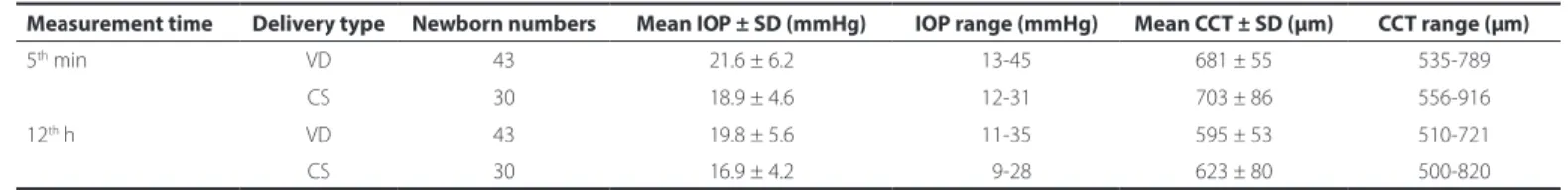
In this study, 43 right eyes of 43 infants (28 girls, 15 boys, ratio: 1.86) born by VD and 30 right eyes of 30 infants (18 girls, 12 boys, ratio: 1.5) born by CS, were evaluated. In the VD group, the mean gestational age at birth was 38.4 ± 0.63 weeks (range: 37-41 weeks), and the mean birth weight was 3199 ± 329 g (range: 2700-3900 g). In the CS group, the mean gestational age at birth was 38.4 ± 0.62 weeks (range: 37-40 weeks), and the mean birth weight was 3290 ± 349 g (range: 2770-4000 g). Diferences between the two groups in terms of gestational age, birth weight, and gender were not statistically sig-niicant (p=0.957, p=0.388, and p=0.656, respectively). The mean IOP and CCT values for both groups at the ifth minute and at the 12th h of the postpartum period are shown in table 1. The independent samples t-test was used to compare the IOP and CCT values between the VD and CS groups. The IOP values of the VD group were signii-cantly higher than those of the CS group, both at the ifth minute and at the 12th h of the postpartum period (p=0.042 and p=0.018, respectively). In the CS group, the IOPs of ive newborns were above 21 mmHg at the ifth minute (range: 31-23 mmHg), and the IOP of two of these infants (40%) remained above 21 mmHg at the 12th h (23 and 28 mmHg). In the VD group, the IOPs of 16 newborns were above 21 mmHg at the ifth minute (range: 43-22 mmHg), and the IOP of ive of these infants (31.25%) remained above 21 mmHg at the 12th h (range: 35-22 mmHg). The CCT diferences between the two groups were not statistically signiicant (p=0.188 and p=0.075, respectively). We analyzed the changes in IOP and CCT between the ifth minute and 12th h in both groups, using the paired samples t-test. The CCT values for both the VD and CS groups declined signiicantly (p<0.001 for both groups). In the VD group, the IOP appeared to be decreased, but the diference was not statistically signiicant (p=0.120). Con-versely, IOP decreased signiicantly in the CS group (p=0.020). We analyzed the correlations between the IOP and CCT values in both delivery groups at both measurement times. In the VD group, a weak correlation was found at the ifth minute and 12th h (p=0.006, r=0.416 and p=0.000, r=0.538, respectively). However, in the CS group, no correlation was found at the ifth minute and 12th h (p=0.870, r=-0.031 and p=0.665, r=0.082, respectively).
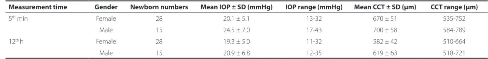
In the VD and CS groups, we also evaluated the efect of gender on IOP and CCT both at the ifth minute and at the 12th h of the postpar-tum period. The VD group values are shown in table 2, while the CS group values are shown in table 3. In the VD and CS groups, the IOP values of the males were signiicantly higher than those of the fema-les at the ifth minute (p=0.024 and p=0.043, respectively). When the delivery type was not taken into account, at the ifth minute all of the males’ IOP was again signiicantly higher than that of the females (p=0.003). In the VD and CS groups, at the ifth minute, the CCT values of the males were not signiicantly diferent to those of the females (p=0.091 and p=0.841, respectively). Further, when the delivery
table 1. IoP and cct values of the Vd- and cS-delivery groups at the ifth minute and at the 12th hour of the postpartum period
Measurement time delivery type newborn numbers Mean IoP ± Sd (mmhg) IoP range (mmhg) Mean cct ± Sd (µm) cct range (µm)
5th min VD 43 21.6 ± 6.2 13-45 681 ± 55 535-789
CS 30 18.9 ± 4.6 12-31 703 ± 86 556-916
12th h VD 43 19.8 ± 5.6 11-35 595 ± 53 510-721
CS 30 16.9 ± 4.2 09-28 623 ± 80 500-820
The effects of delivery type and gender on intraocular pressure and central corneal thickness in newborns
9 4 Arq Bras Oftalmol. 2016;79(2):92-5
type was not taken into account, there was no signiicant diferen-ce of the CCT values between the genders (p=0.360). At the 12th h, IOP values between the genders were not signiicantly diferent in the VD group, CS group, or in all of the studied newborns (p=0.386,
p=0.107, and p=0.155, respectively). Similarly, at the 12th h, the CCT values between the genders were not signiicantly diferent in the VD group, CS group, or all of the studied newborns (p=0.055, p=0.811, and p=0.225, respectively).
dIScuSSIon
In recent studies, IOP and CCT have been evaluated in pre-term and full-term newborns. However, to the best of our knowledge, CCT and IOP values immediately after birth and after 12 h have not been studied. Therefore, the current study is the irst to report the efect of the mode of delivery and gender on IOP and CCT in the irst 12 h of life.
Active labor is the main cause of stress for infants during the pe ripartum period(6). The inal efectors of physical and emotional stresses during birth are glucocorticoids and the main actors are ca techolamines such as adrenaline and noradrenaline. Previously published studies have shown that plasma catecholamine levels of infants born by CS are lower than those of infants born by VD(6,7). Another study identiied a signiicant correlation between a lower plasma level of noradrenaline and poor muscle tone and/or lower grade of excitability in infants born by CS compared to infants born by VD(8). In the irst minutes of life, higher heart rates have also been reported in infants born by VD compared to infants born by CS(9). Psychological stress is known to induce elevated IOP in adults(10). In our study, IOP values were signiicantly higher in the VD group than in the CS group at the ifth minute. The psychological stress of VD may be the cause of the higher IOP.
In the present study, IOP was also measured at the 12th h of the postpartum period. Although the IOP had decreased by the 12th h in both groups, the IOP values of the VD group were still signiicantly higher than those of the CS group. In a previous study, infant saliva cortisol levels were examined 72 h after birth, and higher levels were observed in the VD group than in the CS group(6). The study demons-trated that the efects of delivery stress continue for at least 72 h
postpartum. Therefore, the higher IOP in the VD group at the 12th h may have been the result of sustained higher cortisol levels.
Fetuses delivered vaginally are at greater risk of head compres-sion than those delivered by CS. The direct physical efect of vaginal birth may be the reason for this increment in IOP. Further, the in tra-cranial major venous system may be afected by the increased pres-sure around it due to the thin wall structure of the venous vessels. The efect of the mode of delivery on cerebral hemodynamics has been discussed in previous studies(22,23). Moreover, similar changes of cerebral hemodynamics have been observed in newborns delivered by VD and CS(24). Additionally, no correlation was found between the duration of labor and venous and arterial Doppler indices and ve-locities(25). These data support our theory of hormonal efects on the IOP of newborns.
In our study, at the ifth minute, IOP of males was found to be sig-niicantly higher than that of females in both delivery groups. In a pre-vious study, IOP was measured in 150 newborns between 12 and 24 h after birth, and again no signiicant diference was found between the genders(4). This result is similar to the results of our study, because in our study the diference between the genders was only signiicant at the ifth minute. At the 12th h, however, while the males’ mean IOP values appeared to be higher than those of the females in both de-livery groups, the diferences were not statistically signiicant. As far as we know, none of the studies researching delivery stress, cerebral hemodynamics, and the IOP of newborns have evaluated the efect of gender. Further investigation is necessary to understand the reason for the higher IOP values in males.
Newborn CCT has also been evaluated in recent studies. The earliest measurements in the literature were performed at 6 h postpartum without grouping the newborns according to delivery type. The mean CCT was 647 ± 61 μm at 0-6 h postpartum and 611 ± 72 μm at 7-12 h postpartum(1). For both groups, our results were higher than those of the previous study at 0-6 h postpartum, most likely due to our earlier measurement time. The efect of gender on the CCT of newborns was evaluated in the same study, at six days postpartum. In males, the mean CCT was 631 ± 67 μm in the right eyes and 630 ± 65 μm in the left eyes, while in females, these values were 601 ± 52 μm in the right eyes and 559 ± 49 μm in the left eyes; the diference between the genders was statistically signiicant(1). In another study, table 2. IoP and cct values of both genders at the ifth minute and at the 12th h of the postpartum period in the Vd group
Measurement time Gender newborn numbers Mean IoP ± Sd (mmhg) IoP range (mmhg) Mean cct ± Sd (µm) cct range (µm)
5th min Female 28 20.1 ± 5.1 13-32 670 ± 51 535-752
Male 15 24.5 ± 7.0 17-43 700 ± 58 584-789
12th h Female 28 19.3 ± 5.0 11-32 582 ± 42 510-664
Male 15 20.9 ± 6.8 12-35 619 ± 63 518-721
IOP= intraocular pressure; CCT= central corneal thickness; SD= standard deviation; VD= vaginal delivery.
table 3. IoP and cct values of both genders at the ifth minute and at the 12th h of the postpartum period in the cS group
Measurement time Gender newborn numbers Mean IoP ± Sd (mmhg) IoP range (mmhg) Mean cct ± Sd (µm) cct range (µm)
5th min Female 18 17.3 ± 3.3 12-25 699 ± 75 556-916
Male 12 21.1 ± 5.4 16-31 705 ± 94 559-864
12th h Female 18 15.9 ± 3.6 12-23 626 ± 81 513-820
Male 12 18.4 ± 4.7 09-28 618 ± 82 500-817
Ozkurt ZG, et al.
9 5
Arq Bras Oftalmol. 2016;79(2):92-5
the efect of gender was evaluated again at six days postpartum. The mean CCT of the males was again found to be higher than that of the females, but the diference was not signiicant(2). In our study, although the diferences did not reach statistical signiicance, in the VD group the males’ mean CCT at both times was higher than that of the females. In the CS group, the diferences between the genders were negligible. In the previous studies mentioned above, the de-livery type was not taken into account. If the newborns had been grouped according to delivery type, the results could have been more signiicant in the VD groups.
Previous studies have reported a decline in CCT in newborns during the irst days of life(1,2). In our study, this decrease was highly signiicant in both the VD and CS groups. This decline is thought to be due to improving control of corneal hydration, evaporation, and cor-neal remodeling over time (2,5). Prolonged eye closure may explain the thicker corneas during the irst days of life. Infants are known to have physiologic intracellular luid excess and, after birth, sudden luid elux occurs from the intracellular compartment to the extracellular compartment(26). In addition to prolonged eye closure, physiologic in-tracellular luid excess might be the cause of the edematous cornea. The respiratory outcome in newborns is linked to fetal maturity and the mode of delivery. Infants delivered by CS have a higher inci-dence of respiratory distress than vaginally delivered infants. Active Na+ transport across the pulmonary epithelium drives liquid from the lung lumen to the interstitium, and labor is critical for the activation of the Na+ pump system. There is considerable evidence that high levels of endogenous catecholamines at birth may be important for accelerating alveolar luid clearance by increasing its activity(27). In our study, although the diferences were not statistically signiicant, the mean CCT was higher for the CS group than for the VD group both at the ifth minute and at the 12th h. The Na+- and K+-dependent ATPase (Na+/K+-ATPase) expressed in the basolateral membrane of corneal endothelial cells is primarily responsible for the pump function of the corneal endothelium. In two diferent studies of mouse corneal en-dothelial cells, dexamethasone was shown to increase Na+/K+-ATPase activity and pump function(12,13). We suggest that, like the pulmonary epithelium Na+ transport system, the corneal endothelial pump func-tion may be induced by higher endogenous plasma catecholamine levels in vaginally-delivered infants, causing thinner CCT values in the VD group than in the CS group.
To measure IOP and CCT in newborns, a protocol based on the experiences documented in previous studies was used. A previous study reported that the use of the Alfonso eyelid speculum in chil-dren can falsely elevate IOP by 4 mmHg(28). Further, topical anesthe-tics have been found to afect IOP and CCT readings(29,30). Therefore, the use of a speculum and topical anesthetic drops are limitations of our study. The small number of cases involved in our study is another limitation. Since the international gold standard for IOP measure-ment is Goldmann applanation tonometry, the use of a Tono-Pen in our study could be considered a limitation. However, Tono-Pen XL and iCare have recently been recommended for obtaining accurate IOP measurements of edematous corneas(20). These methods were presumed to be less dependent on CCT due to the smaller area of applanation(21). The duration of active labor of the deliveries was not taken into account because, in our region, most pregnant women arrive at hospital after the beginning of active labor, which is another limitation of our study.
In conclusion, IOP remains higher in vaginally-delivered infants than in infants delivered by CS for at least 12 h postpartum. Infant CCT decreases signiicantly over the 12 h postpartum. Vaginally-delivered infants have thinner CCT values than infants delivered by CS. The present study establishes reference values for IOP and CCT in newborns, which could be used in the early diagnosis of congenital eye disor-ders. Additionally, these data may be useful for understanding the ocular physiology of newborns. Further studies are needed to clarify the possible efects on IOP and CCT in newborns.
referenceS
1. Rushood AA, Zahrani MH, Khamis A, Rushood AA. Central corneal thickness in full term Saudi newborns. Acta Ophthalmol. 2012;90(5):355-8.
2. Remón L, Cristóbal JA, Castillo J, Palomar T, Palomar A, Pérez J. Central and peripheral corneal thickness in full-term newborns by ultrasonic pachymetry. Invest Ophthalmol Vis Sci. 1992;33(11):3080-3.
3. Uva MG, Reibaldi M, Longo A, Avitabile T, Gangliano C, Scollo D, et al. Intraocular pres-sure and central corneal thickness in premature and full-term newborns. J AAPOS. 2011;15(4):367-9.
4. Reddy SC, Alias R. Tono-pen measurement of intraocular pressure under topical anaesthesia in full term normal newborns. Int J Ophthalmol. 2014;7(1):92-4. 5. Kirwan C, O’Keefe M, Fitzsimon S. Central corneal thickness and corneal diameter in
premature infants. Acta Ophthalmol Scand. 2005;83(6):751-3.
6. Schuller C, Känel N, Müller O, Kind AB, Tinner EM, Hösli I, et al. Stress and pain response of neonates after spontaneous birth and vacuum-assisted and cesarean delivery. Am J Obstet Gynecol. 2012;207(5):416-22.
7. Faxelius G, Lagercrantz H, Yao A. Sympathoadrenal activity and peripheral blood low after birth: comparison in infants delivered vaginally and by caesarean section. J Pediatr. 1984;105(1):144-8.
8. Otamiri G, Berg G, Ledin T, Leijon I, Lagercrantz H. Delayed neurological adaptation in infants delivered by elective caesarean section and the relation to catecholamine levels. Early Hum Dev. 1991;26(1):51-60.
9. Dawson JA, Kamlin CO, Wong C, te Pas AB, Vento M, Cole TJ, et al. Changes in heart rate in the first minutes after birth. Arch Dis Child Fetal Neonatal Ed. 2010;95(3): 177-81.
10. Brody S, Erb C, Veit R, Rau H. Intraocular pressure changes: the inluence of psycho-logical stress and the Valsalva maneuver. Biol Psychol. 1999;51(1):43-57.
11. Miyazaki Y, Matsuo T, Kurabayashi Y. Immobilization stress induces elevation of intraocu-lar pressure in rabbits. Ophthalmic Res. 2000;32(6):270-7.
12. Hatou S. Hormonal regulation of Na+/K+-dependent ATPase activity and pump function in corneal endothelial cells. Cornea. 2011;30(10):60-6.
13. Hatou S, Yamada M, Mochizuki H, Shiraishi A, Joko T, Nishida T. The efects of dexa-methasone on the Na,K-ATPase activity and pump function of corneal endothelial cells. Curr Eye Res. 2009;34(5):347-54.
14. Vogl SE, Worda C, Egarter C, Bieglmayer C, Szekeres T, Huber J, et al. Mode of delivery is associated with maternal and fetal endocrine stress response. BJOG. 2006;113(4):441-5. Comment in: BJOG.2007;114(1):120-1.
15. Miller NM, Fisk NM, Modi N, Glover V. Stress responses at birth: determinants of cord arterial cortisol and links with cortisol response in infancy. BJOG. 2005;112(7):921-6. 16. Bergqvist LL, Katz-Salamon M, Hertegard S, Anand KJ, Lagercrantz H. Mode of delivery
modulates physiological and behavioral responses to neonatal pain. J Perinatol. 2009; 29(1):44-50.
17. Li J, Herndon LW, Asrani SG, Stinnett S, Allingham RR. Clinical comparison of the proview eye pressure monitor with the Goldmann applanation tonometer and the Tonopen. Arch Ophthalmol. 2004;122(8):1117-21.
18. Lleo A, Marcos A, Calatayud M, Alonso L, Rahhal SM, Sanchis-Gimeno JA. The rela-tionship between central corneal thickness and Goldmann applanation tonometry. Clin Exp Optom. 2003;86(2):104-8.
19. Shimmyo M, Ross AJ, Moy A, Mostafavi R. Intraocular pressure, Goldmann applanation tension, corneal thickness, and corneal curvature in Caucasians, Asians, Hispanics, and African Americans. Am J Ophthalmol. 2003;136(4):603-13.
20. Neuburger M, Maier P, Böhringer D, Reinhard T, F Jordan J. The impact of corneal edema on intraocular pressure measurements using goldmann applanation tonometry, Tono- Pen XL, iCare, and ORA: an in vitro model. J Glaucoma. 2013;22(7):584-90. 21. Hager A, Wiegand W. [Methods of measuring intraocular pressure independently of
central corneal thickness]. Ophthalmologe. 2008;105(9):840-4. German.
22. Dubowitz LM, Dubowitz V, Palmer P, Verghote M. A new approach to the neurological assessment of the preterm and full-term newborn infant. Brain Dev 1980;2(1):3-14. 23. Baytur YB, Tarhan S, Uyar Y, Ozcakir HT, Lacin S, Coban B, et al. Assessment of fetal
cerebral arterial and venous blood low before and after vaginal delivery or Cesarean section. Ultrasound Obstet Gynecol. 2004;24(5):522-8.
24. Dani C, Martelli E, Bertini G, Pezzati M, Rubaltelli FF. Haemodynamic changes in the brain after vaginal delivery and caesarean section in healthy term infants. BJOG. 2002; 109(2):202-6.
25. Sihota R, Tuli D, Dada T, Gupta V, Sachdeva MM. Distribution and determinents of in-traocular pressure in a normal pediatric population. Pediatr Ophthalmol Strabismus. 2006;43(1):14-8.
26. Aggarwal R, Deorari AK, Paul VK. Fluid and electrolyte management in term and preterm neonates. Indian J Pediatr. 2001;68(12):1139-42.
27. Jain L, Eaton DC. Physiology of fetal lung luid clearance and the efect of labor. Semin Perinatol. 2006;30(1):34-43.
28. Epley KD, Tychsen L, Lueder GT. The efect of an eyelid speculum on intraocular pressure measurement in children. Am J Ophthalmol. 2002;134(6):926-7.
29. Almubrad TM, Ogbuehi KC. Clinical investigation of the efect of topical anesthesia on intraocular pressure. Clin Ophthalmol. 2007;1(3):305-9.