Case Report
Percutaneous Mitral Valvuloplastry in a Pregnant Woman Guided
only by the Transesophageal Echocardiography
José A. Mangione, Salvador André Bavaresco Cristovão, Gustavo Ithamar Souto Maior, Henry Abensur
Departamento de Hemodinâmica e Cardiologia Intervencionista do Hospital Beneficência Portuguesa - São Paulo, SP - BrazilMailing address: Gustavo Ithamar Souto Maior •
R. Maestro Cardim, 769 – 1º SS – SL 71 – BL 1 - 01323-900 – São Paulo/SP - Brazil
E-mail: [email protected]
Manuscript received May 3, 2006; revised manuscript received June 1, 2006; accepted June 1, 2006.
Introduction
Fluoroscopy-guided balloon valvuloplasty is known to be safe and effective in pregnant patients with symptomatic mitral stenosis, and its role is well established in the literature worldwide1-3. However, the fetus is exposed to the harmful
effects of ionizing radiation. The lack of case reports in the literature on the use of transesophageal echocardiography as a guide to valvuloplasty in pregnant patients, and the elimination of fetal exposure to the harmful effects of ionizing radiation encouraged us to publish this article.
Case Report
Patient, 24 years old, Caucasian, married, primigravida, born and residing in Porto Velho – State of Rondônia, Brazil, was admitted in our service in October, 2005 with rheumatic mitral stenosis and NYHA (New York Heart Association) functional class III, despite the use of propranolol (120mg/day) and hydrochlorothiazide (25mg/day).
Her physical examination revealed good general conditions: weight - 64Kg, height – 1.65 m, heart rate – 68 bpm, regular heart rhythm with no extra sounds, accentuated S1, grade 4/6 diastolic murmur in the mitral area and the presence of thrill. She had vesicular breath sounds and a pregnant abdomen.
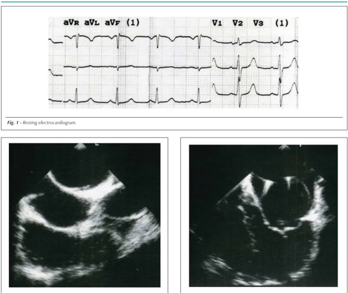
The electrocardiogram showed p-wave duration at the upper limit of normal (Figure 1).
The transthoracic echocardiogram at admission revealed a mitral valve area of 0.9 cm2with a mean gradient of 15
mmHg and a Wilkins and Block score4 of 9 points (3 mobility, 3
thickening, 1 calcification, and 2 subvalvular apparatus); mild mitral regurgitation; left ventricular (LV) diastolic diameter of 48 mm; LV ejection fraction of 0.81; left atrium (LA) with 49 mm (significant enlargement); mild tricuspid regurgitation; and systolic pulmonary artery pressure estimated at 50 mmHg, without further significant alterations.
Based on the patient’s clinical status and on the favorable morphology of the mitral valve apparatus as described in the echocardiographic study, the decision was to treat the mitral stenosis percutaneously using the Inoue technique5,
guided only by transesophageal echocardiography, so as to prevent fetal exposure to the potentially harmful effects of ionizing radiation6.
A gynecological and obstetric evaluation was performed prior to the procedure. The ultrasonography revealed a single fetus, fetal biometry estimated at 24 weeks and two days, adequate development and fetal heart rate of 154 bpm, anterior grade 0 placenta, cephalic presentation, longitudinal lie, normal amniotic fluid volume, and a three-vessel umbilical chord.
The procedure was carried out in the presence of the obstetric and echocardiography teams in the catheterization laboratory. The patient underwent conscious sedation and analgesia with intravenous midazolam (6mg) and meperidine (15mg), not requiring orotracheal intubation. The right femoral artery and vein were punctured and cannulated with 6F and 7F introducers, respectively.
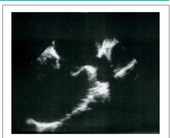
We positioned the guidewire at the superior vena cava using the subcostal bicaval longitudinal view (90º). Transeptal puncture was performed with the Brockenbrough technique7
utilizing a Mullins catheter after visualization of the interatrial septum using the same view (Figure 2). After puncture, IV heparin was given at 70U/kg. The guidewire was positioned in the left atrium (LA) (Figure 3), and the dilation of the interatrial septum was performed with a 14F dilator. A 26-mm Inoue balloon (calculated with the formula: balloon diameter = [height (cm) /10] + 10) was introduced up to the left ventricle (LV). After making sure that the balloon was free in the left ventricular chamber, it was inflated in its distal portion and anchored at the mitral annulus, and valve dilation was performed with a 25-mm-diameter balloon for approximately 5 seconds (Figure 4).
Key words
Mitral valve stenosis; balloon dilatation; pregnancy. Mitral valve stenosis is the most common valve lesion in pregnancy. In spite of an optimized clinical treatment and a favorable valve anatomy according to Wilkins and Block score, in symptomatic patients, percutaneous intervention is shown to be of great importance. In these patients, avoiding x-ray exposure as much as possible is recommended so as to protect the fetus from the deleterious effects of ionizing radiation. In this case report, a 24-year old pregnant patient with severe mitral stenosis (valve area of 0.9 cm²) was successfully submitted to a TEE-guided percutaneous treatment, without the use of x-ray.
Case Report
Mangione et al Percutaneous mitral valvuloplastry in a pregnant woman by the transesophageal ECHOArq Bras Cardiol 2007; 88(3) : e60-e63
volume and heart rate) worsen the hemodynamic status resulting from the obstruction during left atrial emptying, which causes a pressure increase in the LA and in the pulmonary region.
In addition to the repercussions for the mother, mitral stenosis is related to a significant increase in prematurity, fetal growth retardation and low birth weight11,12.
Clinical stabilization of pregnant women with mitral stenosis can be obtained, in most cases, with heart rate control and left atrial pressure reduction, by means of limited physical activities and the use of beta-blockers and diuretics. Nevertheless, the latter two must be used with great caution so as to avoid a significant reduction in placental blood flow. In patients who persist with significant symptoms despite adequate clinical therapy, percutaneous or invasive surgical treatment is indicated.
It is important to note that even functional class I and II patients may have an unfavorable outcome because they may present an obstructive valve disease. Functional class III and After the first inflation, the echocardiographic result
obtained was not considered satisfactory (valve area of 1.6 cm2), so we decided to perform a second inflation, with a
26-mm-diameter balloon, with a resulting valve area of 2.2 cm2 and mild mitral regurgitation. The Inoue balloon was
removed, and no left-to-right shunt was detected at the interatrial septum. (Figure5).
The procedure was uneventful and had a total duration of 25 minutes as of the femoral puncture.
The patient remained in the Intensive Care Unit for 24 hours under observation, after which she was discharged.
Discussion
Mitral valve stenosis is the most common valve disease in pregnancy8,9. In Brazil, it accounts for 50% of all heart
valve diseases in pregnant women10. During pregnancy, most
patients with moderate to severe mitral stenosis progress with a deterioration of one or two NYHA functional classes8,11.
The physiological alterations (increased cardiac output, blood Fig. 1 -Resting electrocardiogram.
Fig. 3 -Four-chamber view – lower esophagus: guidewire placement inside the left atrium.
Fig. 2 -Bicaval longitudinal view (90º): Puncture of interatrial septum with a Brockenbrough needle.
Case Report
Mangione et alPercutaneous mitral valvuloplastry in a pregnant woman by the transesophageal ECHO
Arq Bras Cardiol 2007; 88(3) : e60-e63
IV patients usually have an unfavorable outcome. Therefore, invasive therapies have proven to be of great value for the adequate clinical control of these patients.
However, open or closed surgery are related to high fetal and maternal mortality and morbidity rates, which limit their use13,14.
Several studies1-15 have shown favorable immediate results
with percutaneous treatment in these patients, with a significant increase in valve area and low complication rates.
So far, the children’s clinical follow-up has been favorable, with normal development and no adverse effects resulting from ionizing radiation3,16. However, these patients are advised
to avoid x-ray exposure as much as possible, particularly in the first trimester of pregnancy, when fetal organogenesis occurs. Although after this period the risks are minimized, radiation exposure is associated with harmful effects. Intrauterine growth retardation, central nervous system abnormalities and
increased incidence of childhood cancer, such as leukemia, are reported6.
The most commonly used form of fetal protection during PMV includes the shortest possible time of fluoroscopy and the placement of a lead apron on the mother’s abdominal and pelvic regions.
In an attempt to completely eliminate the possible risks associated with the use of ionizing radiation, valvuloplasty was performed using only transesophageal echocardiography, which made the procedure easy and safe. This option should be encouraged in pregnant patients, particularly with the use of the Inoue technique, because of its greater simplicity, and performed by teams experienced in the percutaneous treatment of mitral stenosis.
Potential Conflict of Interest
No potential conflict of interest relevant to this article was reported.
Fig. 5 -Longitudinal two-chamber view: mitral valve with good opening at the end of the procedure.
Fig. 4 -Longitudinal two-chamber view: Inoue balloon inflation with mitral valve opening.
References
1. Mangione JA, Zuliani MFM, DelCastillo JM, Nogueira EA, Arie S. Percutaneous double balloon mitral valvuloplasty in pregnant women. Am J Cardiol. 1989; 64: 99-102.
2. Safian RD, Berman A, Sachs B, Diver DJ, Come PC, Baim DS, et al. Percutaneous balloon mitral valvuloplasty in a pregnant woman with mitral stenosis. Cathet Cardiovasc Diagn. 1988; 15: 103-8.
3. Farhat MB, Gama H, Betbout F, Maatouk F, Jarrar M, Addad F, et al. Percutaneous balloon mitral comissurotomy during pregnancy. Heart. 1997; 77: 564-7.
4. Block PC. Who is suitable for percutaneous balloon mitral valvotomy? Int J Cardiol. 1988; 20: 9-14.
5. Inoue K, Owaki T, Nakamura T, Kitamura F, Miyamoto N. Clinical applications of transvenous mitral comissurotomy by a new balloon catheter. J Thorac Cardiovasc Surg. 1984; 87: 394-402.
6. Debakan AS. Abnormality in children exposed to X-radiation during various stages of gestation: tentative timetable of radiation injury to the human fetus.
J Nucl Med. 1968; 9: 471-7.
7. Brockenbrough EZ, Braunwald EA. A new technic for left ventricular angiocardiography and transseptal left heart catheterization. Am J Cardiol. 1960; 6: 1062-4.
8. Essop MR, Sareli P. Rheumatic valvular disease and pregnancy. In Elkayam U, Gleicher N (eds). Cardiac problems in pregnancy. 3rd ed. New York: Wily-Liss; 1998. p. 55-60.
9. Reimold SC, Rutherford JD: Valvular heart disease in pregnancy. N Eng J Med. 2003; 349: 52-9.
10. AÁvila WS, Rossi EG, Ramires JF, Grinberg M, Bortolotto MR, Zugaib M, et al. Pregnancy and heart disease: experience with 1000 cases. Clin Cardiol. 2003; 26: 135-42.
11. Hameed AB, Karaalp IS, Tummala PP, Wani OR, Canetti M, Akhter MW, et al. The effect of valvular heart disease on maternal and fetal outcome in pregnancy. J Am Coll Cardiol. 2001; 37: 893-9.
Case Report
Mangione et al Percutaneous mitral valvuloplastry in a pregnant woman by the transesophageal ECHOArq Bras Cardiol 2007; 88(3) : e60-e63
12. Naidoo DP, Desai DK, Moodley J. Maternal death to pre-existing cardiac disease. Cardiovasc J S Afr. 2002; 13: 17-20.
13. Vosloo S, Reichart B. The feasibility of closed mitral valvotomy in pregnancy. J Thorac Cardiovasc Surg. 1987; 93: 675-9.
14. Bernal JM, Mirales PJ. Cardiac surgery with cardiopulmonary bypass during
pregnancy. Obstet Gynecol Surv. 1986; 41: 1-6.
15. Esteves CA, Ramos AIO, Braga SLN, Harrisson JK, Sousa JEMR. Effectiveness of percutaneous balloon mitral valvotomy during pregnancy. Am J Cardiol. 1991; 68: 930-4.
16. Kinsara AJ, Ismail O, Fawzi ME. Effect of ballon mitral valvuloplasty during pregnancy on childhood development. Cardiology. 2002; 97: 155-8.